 E-submission
Search
- Page Path
- HOME > Search
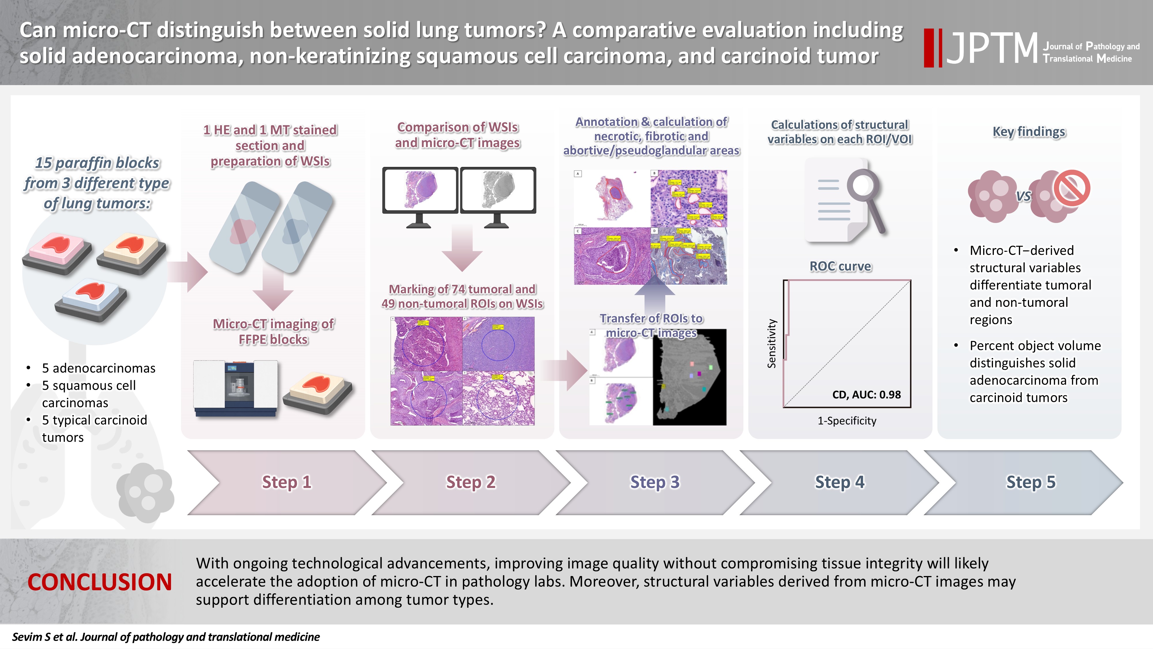
- Can micro-CT distinguish between solid lung tumors? A comparative evaluation including solid adenocarcinoma, non-keratinizing squamous cell carcinoma, and carcinoid tumor
- Selim Sevim, Serpil Dizbay Sak, Kaan Orhan, Arda Buyuksungur, Duru Karasoy, Hilal Ozakinci, Ayten Kayi Cangir
- J Pathol Transl Med. 2026;60(2):231-245. Published online March 10, 2026
- DOI: https://doi.org/10.4132/jptm.2025.12.16
- 1,827 View
- 122 Download
-
 Abstract
 PDF
PDF Supplementary Material
 - Background
Some pulmonary carcinomas display a solid pattern, and immunohistochemistry is commonly used for tumor differentiation. Micro–computed tomography (micro-CT), with its ability to produce detailed three-dimensional images using small voxel sizes, may offer additional insights. This study investigates whether three solid tumor types, solid adenocarcinoma (sAC), non-keratinizing squamous cell carcinoma, and carcinoid tumor (CaT), can be differentiated using micro-CT. Methods: Fifteen paraffin blocks, five for each type, were scanned with micro-CT (Skyscan 1275, Bruker). These images were compared to whole slide images (WSIs) of the same tumors. Consequently, tumoral (n = 74) and non-tumoral (n = 49) regions of interest (tumor ROIs [tROIs] and non-tumor ROIs [ntROIs]) were selected on the micro-CT images and evaluated in terms of certain structural variables (percent object volume, structure model index, structure thickness, structure linear density, connectivity, connectivity density, open porosity, closed porosity) to investigate whether tumors can be differentiated from normal parenchyma and from each other. Results: Although detailed images comparable to WSIs could not be obtained, it was considered an important advantage to be able to examine the entire depth of the paraffin blocks. tROIs and ntROIs could be distinguished based on all variables (p < .001). Additionally, sAC showed a notable difference from CaT in “percent object volume” (p = .011). Conclusions: With ongoing technological advancements, improving image quality without compromising tissue integrity will likely accelerate the adoption of micro-CT in pathology labs. Moreover, structural variables derived from micro-CT images may support differentiation among tumor types.
- TRPS1 expression in non-melanocytic cutaneous neoplasms: an immunohistochemical analysis of 200 cases
- Yi A. Liu, Phyu P. Aung, Yunyi Wang, Jing Ning, Priyadharsini Nagarajan, Jonathan L. Curry, Carlos A. Torres-Cabala, Doina Ivan, Victor G. Prieto, Qingqing Ding, Woo Cheal Cho
- J Pathol Transl Med. 2024;58(2):72-80. Published online February 26, 2024
- DOI: https://doi.org/10.4132/jptm.2024.01.23

- 9,346 View
- 416 Download
- 15 Web of Science
- 15 Crossref
-
Abstract
PDFSupplementary Material
- Background
Although trichorhinophalangeal syndrome type 1 (TRPS1) was initially thought to be highly sensitive and specific for carcinomas and mesenchymal tumors of mammary origin, more recent data suggest its expression is not limited to breast neoplasms but also can be seen in other cutaneous neoplasms, such as extramammary Paget disease and squamous cell carcinoma (SCC) in situ.
Methods
Two-hundred cases of non-melanocytic cutaneous neoplasm, including basal cell carcinomas (BCCs) (n = 41), SCCs (n = 35), Merkel cell carcinomas (MCCs) (n = 25), and adnexal neoplasms (n = 99), were tested for TRPS1 expression using a monoclonal anti- TRPS1 rabbit anti-human antibody.
Results
TRPS1 expression was present in almost all cases of SCC (94%), with a median H-score of 200, while it was either absent or only focally present in most BCCs (90%), with a median H-score of 5. The difference between BCCs and SCCs in H-score was significant (p < .001). All MCCs (100%) lacked TRPS1 expression. TRPS1 expression was frequently seen in most adnexal neoplasms, benign and malignant, in variable intensity and proportion but was consistently absent in apocrine carcinomas. All endocrine mucin-producing sweat gland carcinomas (EMPSGCs) (100%, 6/6) showed diffuse and strong TRPS1 immunoreactivity, with a median H-score of 300, which was significantly different (p < .001) than that of BCCs.
Conclusions
Our study shows that TRPS1 may be an effective discriminatory marker for BCCs and SCCs. It also has a role in distinguishing BCCs from EMPSGCs. -
Citations
Citations to this article as recorded by
- Metastatic Vulvar Paget's Disease Presenting in a Supraclavicular Lymph Node: A Diagnostic Challenge on Fine Needle Aspiration Cytology
Thiri Htoo Aung, Neha Seth, Anam Khan, Kasturi Das
Diagnostic Cytopathology.2026;[Epub] CrossRef - The evolving role of TRPS1 in dermatopathology: insights from the past 4 years
Mokhtar H. Abdelhammed, Woo Cheal Cho
Journal of Pathology and Translational Medicine.2026; 60(2): 129. CrossRef - Correspondence: Primary Cutaneous NUT Adnexal Carcinoma: A Case Report With Novel Clinical and Pathological Observations
Woo Cheal Cho
Journal of Cutaneous Pathology.2026; 53(7): 622. CrossRef - Trichorhinophalangeal syndrome type 1 (TRPS1) in breast pathology: diagnostic utility and pitfalls
Atif Ali Hashmi, Edi Brogi, Hannah Y. Wen
Diagnostic Pathology.2025;[Epub] CrossRef - Refining NTRK Fusion Detection in Papillary Thyroid Carcinoma Through Pan-TRK Immunohistochemistry and Histopathologic Features
Hyun Lee, Sue Youn Kim, Ji Min Park, Seung-Hyun Jung, Ozgur Mete, Chan Kwon Jung
Endocrine Pathology.2025;[Epub] CrossRef - Endocrine mucin-producing sweat gland carcinoma: Case report and literature review
Nan Guo, Zhenlin Fan, Yitong Chen, Qian Li, Limin Guo
European Journal of Ophthalmology.2025;[Epub] CrossRef - Updates on utility of immunohistochemistry in diagnosis of metastatic breast cancer
Hongxia Sun, Aysegul A. Sahin, Qingqing Ding
Human Pathology.2025; 162: 105821. CrossRef - Primary Cutaneous NUT Adnexal Carcinoma With BRD4::NUTM1 Fusion: A 19-Year Follow-Up
Elsayed Ibrahim, Richard K. Yang, Maria A. Gubbiotti, Victor G. Prieto, Woo Cheal Cho
The American Journal of Dermatopathology.2025; 47(9): 731. CrossRef - Primary mucinous carcinoma of the skin with co-expression of TRPS1 and GATA3: a case report
Liling Song, Ning Zhu, Lei Jiang, Dong Gao, Guohua Yu
Frontiers in Oncology.2025;[Epub] CrossRef - Diagnostic Algorithm for Secondary Extramammary Paget Disease from Institutional Cases and Literature Review
Salin Kiratikanon, Ayaka Fukui, Masahiro Hirata, Jakob M. T. Moran, Masakazu Fujimoto, Mai P. Hoang
Cancers.2025; 17(24): 4014. CrossRef - TRPS1 Expression Is Frequently Seen in a Subset of Cutaneous Mesenchymal Neoplasms and Tumors of Uncertain Differentiation: A Potential Diagnostic Pitfall
Moon Joo Kim, Yi A. Liu, Yunyi Wang, Jing Ning, Woo Cheal Cho
Dermatopathology.2024; 11(3): 200. CrossRef - TRPS1 expression in MPNST is correlated with PRC2 inactivation and loss of H3K27me3
Rossana Lazcano, Davis R. Ingram, Gauri Panse, Alexander J. Lazar, Wei-Lien Wang, Jeffrey M. Cloutier
Human Pathology.2024; 151: 105632. CrossRef - Syringocystadenoma Papilliferum-Like Features in Poroma: An Unusual Morphologic Pattern of Poroma or True Synchronous Occurrence of 2 Distinct Neoplasms?
Mouaz Alsawas, Fiorinda F. Muhaj, Phyu P. Aung, Priyadharsini Nagarajan, Woo Cheal Cho
The American Journal of Dermatopathology.2024; 46(12): 871. CrossRef - A Comprehensive Review of TRPS1 as a Diagnostic Immunohistochemical Marker for Primary Breast Carcinoma: Latest Insights and Diagnostic Pitfalls
Antonia-Carmen Georgescu, Tiberiu-Augustin Georgescu, Simona-Alina Duca-Barbu, Lucian Gheorghe Pop, Daniela Oana Toader, Nicolae Suciu, Dragos Cretoiu
Cancers.2024; 16(21): 3568. CrossRef - Expression of TRPS1 in Metastatic Tumors of the Skin: An Immunohistochemical Study of 72 Cases
Kassiani Boulogeorgou, Christos Topalidis, Triantafyllia Koletsa, Georgia Karayannopoulou, Jean Kanitakis
Dermatopathology.2024; 11(4): 293. CrossRef
- Metastatic Vulvar Paget's Disease Presenting in a Supraclavicular Lymph Node: A Diagnostic Challenge on Fine Needle Aspiration Cytology
- Intrathyroidal metastasis of tonsillar squamous cell carcinoma masquerading as a primary thyroid tumor
- Jai-Hyang Go
- J Pathol Transl Med. 2023;57(4):242-245. Published online July 11, 2023
- DOI: https://doi.org/10.4132/jptm.2023.06.16

- 5,630 View
- 118 Download
- 2 Web of Science
- 2 Crossref
-
Abstract
PDF
- Intrathyroidal metastasis of tonsillar squamous cell carcinoma is rare. To date, only six cases have been reported in the literature. This case was unusual and presented with thyromegaly before the diagnosis of the primary tumor. A 55-year-old male patient was suspected to have a primary thyroid tumor with nodal metastasis. The thyroid gland was diffusely enlarged, with no discernible mass. Histologically, the thyroid parenchyma revealed extensive endolymphatic tumor emboli, which were positive for p40 and p16 in a background of chronic lymphocytic thyroiditis. Positron emission tomography–computed tomography revealed hypermetabolic activity in the right tonsillar region. Tonsillar biopsy revealed human papillomavirus–positive squamous cell carcinoma. The present case is the first reported case of intrathyroidal metastasis of tonsillar squamous cell carcinoma with an initial clinical presentation of thyroid enlargement before the primary tumor of tonsillar cancer was diagnosed.
-
Citations
Citations to this article as recorded by- Metastasis to Thyroid from Recurrent Head and Neck Squamous Cell Carcinoma: A Case Series and Review of Literature
Avneet Kaur, Rohit Nayyar, Harit Kumar Chaturvedi, Akshat Malik
Indian Journal of Surgical Oncology.2025; 16(1): 122. CrossRef - Metastatic oropharyngeal squamous cell carcinoma to the thyroid: A case report and review of literature
Hannah Walker, Jed Speers, Milena Fabry, Sameep Kadakia
American Journal of Otolaryngology.2024; 45(4): 104306. CrossRef
- Metastasis to Thyroid from Recurrent Head and Neck Squamous Cell Carcinoma: A Case Series and Review of Literature
- Evaluation of the characteristics of multiple human papillomavirus (HPV) infections identified using the BD Onclarity HPV assay and comparison with those of single HPV infection
- Jinhee Kim, Moonsik Kim, Ji Young Park
- J Pathol Transl Med. 2022;56(5):289-293. Published online September 13, 2022
- DOI: https://doi.org/10.4132/jptm.2022.08.02

- 9,814 View
- 141 Download
- 12 Web of Science
- 9 Crossref
-
Abstract
PDFSupplementary Material
- Background
Human papillomavirus (HPV) infection is a major cause of cervical cancer and associated precursor lesions. Multiple HPV genotype infections have been reported. However, their clinicopathological characteristics still remain elusive.
Methods
For this study, 814 consecutive patients who had undergone colposcopy and HPV genotyping test using BD Onclarity HPV assay were retrospectively selected. Clinicopathological parameters of multiple HPV infections were compared with those of single HPV infection.
Results
Multiple HPV infections were found in 110 out of 814 cases (13.5%). Multiple HPV infections were associated with a significantly higher incidence of high-grade intraepithelial lesions (HSILs) compared with single HPV infection. Other high-risk HPV genotypes, in addition to HPV 16, were found more frequently in the multiple HPV infections group; these included HPV 51, 52, 33/58, 56/59/66, and 35/39/68. No specific coinfection pattern was not identified. Additionally, the number of HPV genotypes in multiple HPV infections was not associated with the progression to HSIL or squamous cell carcinoma.
Conclusions
Multiple HPV infections have distinct clinicopathological characteristics (compared with single HPV infection). As their biological behavior is uncertain, close and frequent follow-up is warranted. -
Citations
Citations to this article as recorded by- Informative HPV testing after conization and its impact on time-varying estimates: a GAMM-based cohort study
Jie Zhou, Jian hong Liao, Lin Jie Su, Yan Chen, Hong bo Hu
Frontiers in Public Health.2026;[Epub] CrossRef - The Prevalence of Multi-Type Infections Among Human Papillomavirus Types in Korean Women
Jang Mook Kim, Hee Seung Song, Jieun Hwang, Jae Kyung Kim
Pathogens.2025; 14(4): 369. CrossRef - Multiple high-risk human papillomavirus infections exacerbate cervical lesion risk: epidemiological evidence from suining, Sichuan
Yaling Jing, Jianhui Chen, Fang Lin, Xiaonan Huang, Yulin Liu, Mingcai Zhao, Chuan Ye, Lianfang Zhao, Xiaofang Liu, Jiayan Yang
Virology Journal.2025;[Epub] CrossRef - The cervical cancer related distribution, coinfection and risk of 15 HPV types in Baoan, Shenzhen, in 2017–2023
Rukai Li, Weiwei Meng, Yunhai Zuo, Yanli Xu, Shaonan Wu
Virology Journal.2024;[Epub] CrossRef - Molecular findings and virological assessment of bladder papillomavirus infection in cattle
Francesca De Falco, Anna Cutarelli, Francesca Luisa Fedele, Cornel Catoi, Sante Roperto
Veterinary Quarterly.2024; 44(1): 1. CrossRef - Patterns of single and multiple HPV infections in female: A systematic review and meta-analysis
Dan Zhou, Jing Xue, Yaqiong Sun, Liling Zhu, Ming Zhao, Meimei Cui, Min Zhang, Jingjing Jia, Limei Luo
Heliyon.2024; 10(17): e35736. CrossRef - Age distribution of patients with multiple High-Risk Human Papilloma Virus (HR-HPV) genotypes and HPV vaccine recommendations by age
Gülçin Çetin Uysal, Nil Tekin
Family Practice and Palliative Care.2024; 9(3): 80. CrossRef - Relative distribution of HPV genotypes in histological cervical samples and associated grade lesion in a women population over the last 16 years in Burgundy, France
Christelle Auvray, Serge Douvier, Odile Caritey, Jean-Baptiste Bour, Catherine Manoha
Frontiers in Medicine.2023;[Epub] CrossRef - Epidemiologic characteristics of high-risk HPV and the correlation between multiple infections and cervical lesions
Qinli Luo, Xianghua Zeng, Hanyi Luo, Ling Pan, Ying Huang, Haiyan Zhang, Na Han
BMC Infectious Diseases.2023;[Epub] CrossRef
- Informative HPV testing after conization and its impact on time-varying estimates: a GAMM-based cohort study
- Frequency of PIK3CA mutations in different subsites of head and neck squamous cell carcinoma in southern Thailand
- Arunee Dechaphunkul, Phatcharaporn Thongwatchara, Paramee Thongsuksai, Tanadech Dechaphunkul, Sarayut Lucien Geater
- J Pathol Transl Med. 2022;56(3):126-133. Published online February 28, 2022
- DOI: https://doi.org/10.4132/jptm.2022.01.04

- 9,255 View
- 195 Download
- 4 Web of Science
- 4 Crossref
-
Abstract
PDF
- Background
Phosphatidylinositol-4,5-bisphosphate 3-kinase catalytic subunit alpha (PIK3CA) mutations have been reported in many cancers, including head and neck squamous cell carcinoma (HNSCC). The frequency of these mutations varies among tumor locations and might be relevant to treatment outcomes among HNSCC. In this study, we examined the frequency of PIK3CA mutations in the different subsites of HNSCC.
Methods
Ninety-six fresh biopsy specimens were investigated for mutations in PIK3CA exons 4, 9, and 20 using allele-specific real-time polymerase chain reaction. Patient characteristics and survival were analyzed and compared between specimens with or without PIK3CA mutations.
Results
The study included primary tumors originating from the oral cavity (n=63), hypopharynx (n=23), and oropharynx (n=10). We identified mutations in 10.4% of patients (10 of 96 specimens). The overall mutational frequency was 17.4% (4/23) and 9.5% (6/63) in the hypopharynx and oral cavity, respectively. No patients with oropharyngeal carcinoma had mutations. Among the 10 mutant specimens, five were missense mutations (exon 9 [E545K] in two samples and exon 20 [H1047R] in three samples) and five were silent mutations in exon 20 (T1025T). Mutations were not found in exon 4. Among 84 patients with available clinical data, we found no significant differences in clinical characteristics and survival based on the presence or absence of PIK3CA mutations.
Conclusions
The results indicate that PIK3CA mutations are involved in HNSCC carcinogenesis, and the hypopharynx should be considered a primary site of interest for future studies, particularly in Southeast Asian populations. -
Citations
Citations to this article as recorded by- Comprehensive Genomic Profiling of Sinonasal Carcinomas: Identification of Common Mutations and Potential Targets for Therapy
Gabriel Bitar, Beau Hsia, Saif Alshaka, Bastien A. Valencia, Jeeho Kim, Mariko Sato, John Crawford, Michael L. Levy, Sean Polster, Vijay A. Patel
Journal of Neurological Surgery Part B: Skull Base.2026; 87(03): 337. CrossRef - Anti-breast cancer effects of Pterocarpus soyauxii Taub aqueous extract and its compounds by integrating ADMET, network pharmacology, molecular docking, dynamic simulation, CLC-Pred and pdCSM-Cancer/PPI approaches, and in vitro validation
Owona Pascal Emmanuel, Mengue Ngadena Yolande Sandrine, Bilanda Danielle Claude, Ayissi Mbomo Rigobert-Espoir, Oluwafemi Adeleke Ojo, Ella Armand Fils, Bidingha A Goufani Ronald, Bindzi Georges Michel, Dzeufiet Djomeni Paul Désiré, Tariq Aziz, Abdulhakeem
Journal of Ethnopharmacology.2025; 353: 120407. CrossRef - A retrospective study of laryngeal squamous cell carcinoma and the significance of the PIK3CA mutation for survival
Akinobu Kubota, Nobuyuki Bandoh, Takashi Goto, Michihisa Kono, Ryosuke Sato, Shiori Suzuki, Shota Sakaue, Ryuhei Takeda, Shuto Hayashi, Misaki Hayashi, Daisuke Araki, Shogo Baba, Yasutaka Kato, Miki Takahara, Hiroshi Nishihara, Hajime Kamada
Molecular and Clinical Oncology.2025; 23(4): 1. CrossRef - An empirical review on the resistance mechanisms of epidermal growth factor receptor inhibitors and predictive molecular biomarkers in colorectal cancer
Sankha Bhattacharya
Critical Reviews in Oncology/Hematology.2023; 183: 103916. CrossRef
- Comprehensive Genomic Profiling of Sinonasal Carcinomas: Identification of Common Mutations and Potential Targets for Therapy
- Primary squamous cell carcinoma of the salivary gland: immunohistochemical analysis and comparison with metastatic squamous cell carcinoma
- Uiree Jo, Joon Seon Song, Seung-Ho Choi, Soon Yuhl Nam, Sang Yoon Kim, Kyung-Ja Cho
- J Pathol Transl Med. 2020;54(6):489-496. Published online August 31, 2020
- DOI: https://doi.org/10.4132/jptm.2020.07.19

- 12,165 View
- 220 Download
- 19 Web of Science
- 20 Crossref
-
Abstract
PDFSupplementary Material
- Background
Primary squamous cell carcinoma (SCC) of the salivary gland is a rare disease, and distinguishing primary SCC from metastatic SCC is difficult. This study investigated the histological and immunohistochemical differences between primary and metastatic salivary gland SCC to improve the accuracy of diagnosis and to explore the pathogenesis of this disease.
Methods
Data of 16 patients who underwent surgery for SCC of salivary glands between 2000 and 2018 at Asan Medical Center were retrieved. Eight patients had a history of SCC at other sites, and eight patients had only salivary gland SCC. Immunostaining for p16, p53, androgen receptor (AR), gross cystic disease fluid protein 15 (GCDFP-15), and c-erbB2, as well as mucicarmine staining, were compared between the two groups.
Results
Most tumors were located in the center of the salivary glands with extraparenchymal extension. The histology of primary SCC of the salivary gland was consistent with moderately differentiated SCC with extensive desmoplastic reaction and peritumoral inflammation. Involvement of the salivary gland ducts and transition into the ductal epithelium were observed in two cases. Metastatic SCC resembled the primary tumor histologically and was associated with central necrosis. Both groups exhibited negative mucin staining. Two, one, and one primary SCC case exhibited AR, GCDFP-15, and c-erbB2 positivity, respectively.
Conclusions
A subset of primary SCCs originated in salivary ducts or was related to salivary duct carcinoma. Distinguishing primary from metastatic SCC of the salivary gland is difficult using histologic features and immunoprofiles. A comprehensive review of the medical history is essential. -
Citations
Citations to this article as recorded by- Solitary Parotid Metastasis as an Atypical Site of Recurrence in Lung Squamous Cell Carcinoma: A Diagnostic Difficulty
Mamadou Alpha Prateaux, Mohamed Amine Haouane, Issam Rharrassi, Mohamed Amine Azami
Cureus.2026;[Epub] CrossRef - Certainties, Doubts, and Myths in the Diagnosis and Treatment of Salivary Gland Tumors of the Head and Neck
Giulio Cantù
Cancers.2026; 18(13): 2078. CrossRef - Clinical diagnosis, treatment, and survival analysis of 61 cases of salivary duct carcinoma: a retrospective study
Shubin Dong, Mengru Li, Zhiwei Zhang, Bowei Feng, Wei Ding, Jiang Chang, Feng Liu
PeerJ.2025; 13: e19626. CrossRef - Characterization of undifferentiated carcinoma of the salivary gland: clinicopathological and immunohistochemical analyses in comparison with lymphoepithelial carcinoma
Sangjoon Choi, Gyuheon Choi, Hee Jin Lee, Joon Seon Song, Yoon Se Lee, Seung-Ho Choi, Kyung-Ja Cho
Journal of Pathology and Translational Medicine.2025; 59(6): 361. CrossRef - Primary salivary gland squamous cell carcinoma with sialolithiasis in the submandibular gland: A case report and literature review
Sawako Ono, Katsutoshi Hirose, Yuji Hirata, Marie Yamada, Satoko Nakamura, Hidetaka Yamamoto
Journal of Oral and Maxillofacial Surgery, Medicine, and Pathology.2024; 36(5): 768. CrossRef - A case of primary squamous cell carcinoma of the parotid gland and review of the literature
Jingli Zhao, Xinrong Nan, Chuhuan Zhou, Nan Jiang, Liangliang Tian
Journal of Case Reports and Images in Oncology.2024; 10(1): 7. CrossRef - Metastatic cutaneous squamous cell carcinoma accounts for nearly all squamous cell carcinomas of the parotid gland
Patrick J. Bradley, Göran Stenman, Lester D. R. Thompson, Alena Skálová, Roderick H. W. Simpson, Pieter J. Slootweg, Alessandro Franchi, Nina Zidar, Alfons Nadal, Henrik Hellquist, Michelle D. Williams, Ilmo Leivo, Abbas Agaimy, Alfio Ferlito
Virchows Archiv.2024; 485(1): 3. CrossRef - Common skin cancers and their association with other non-cutaneous primary malignancies: a review of the literature
Lindsay Holic
Medical Oncology.2024;[Epub] CrossRef - Salivary duct carcinoma with squamous differentiation: histomorphological and immunophenotypical analysis of six cases
Melad N Dababneh, Christopher C Griffith, Kelly R Magliocca, Ivan J Stojanov
Histopathology.2024; 85(4): 590. CrossRef - Comprehensive Next Generation Sequencing Reveals that Purported Primary Squamous Cell Carcinomas of the Parotid Gland are Genetically Heterogeneous
Justin A. Bishop, Masato Nakaguro, Ilan Weinreb, Doreen Palsgrove, Lisa M. Rooper, Travis W. Vandergriff, Brian Carlile, Jeffrey A. Sorelle, Jeffrey Gagan, Toshitaka Nagao
Head and Neck Pathology.2024;[Epub] CrossRef - Salivary gland fine needle aspiration: a focus on diagnostic challenges and tips for achieving an accurate diagnosis
Carla Saoud, Hansen Lam, Sandra I. Sanchez, Zahra Maleki
Diagnostic Histopathology.2023; 29(8): 357. CrossRef - Salivary gland pathologies: evolution in classification and association with unique genetic alterations
Michał Żurek, Łukasz Fus, Kazimierz Niemczyk, Anna Rzepakowska
European Archives of Oto-Rhino-Laryngology.2023; 280(11): 4739. CrossRef - A retrospective study of nonneoplastic and neoplastic disorders of the salivary glands
Sorin Vamesu, Oana Andreea Ursica, Ana Maria Gurita, Raluca Ioana Voda, Mariana Deacu, Mariana Aschie, Madalina Bosoteanu, Georgeta Camelia Cozaru, Anca Florentina Mitroi, Cristian Ionut Orasanu
Medicine.2023; 102(42): e35751. CrossRef - Pembrolizumab as a first line therapy in a patient with extensive mucoepidermoid salivary gland carcinoma. A complete clinical, radiological and pathological response. A very specific case
Raed Farhat, Noam Asna, Yaniv Avraham, Ashraf Khater, Majd Asakla, Alaa Safia, Sergio Szvalb, Nidal Elkhatib, Shlomo Merchavy
Discover Oncology.2022;[Epub] CrossRef - Morphologic CT and MRI features of primary parotid squamous cell carcinoma and its predictive factors for differential diagnosis with mucoepidermoid carcinoma
Xiaohua Ban, Huijun Hu, Yue Li, Lingjie Yang, Yu Wang, Rong Zhang, Chuanmiao Xie, Cuiping Zhou, Xiaohui Duan
Insights into Imaging.2022;[Epub] CrossRef - A Rare Case of Primary Squamous Cell Carcinoma of the Submandibular Salivary Gland: Brief Overview of Diagnostic Ambiguity and Treatment Challenges
Pawan Hingnikar, Anendd Jadhav, Nitin D Bhola
Cureus.2022;[Epub] CrossRef - Necrotizing Sialometaplasia of the Hard Palate: Diagnosis and
Treatment
Sangeun Lee, Yun Sung Lim, Kyuho Lee, Bo Hae Kim
Journal of Clinical Otolaryngology Head and Neck Surgery.2022; 33(4): 236. CrossRef - Parotid Salivary Duct Carcinoma With a Prominent Squamous Component: Immunohistochemical Profile, Diagnostic Pitfalls, and Therapeutic Implications
Naomi Hardy, Joshua Thompson, Ranee Mehra, Cinthia B. Drachenberg, Kyle Hatten, John C. Papadimitriou
International Journal of Surgical Pathology.2021; 29(7): 726. CrossRef - Intrasalivary Thymic Carcinoma: A Case Report and Literature Review
Michał Kunc, Alexandra Kamieniecki, Grzegorz Walczak, Tomasz Nowicki, Bartosz Wasąg, Bogusław Mikaszewski, Dominik Stodulski, Wojciech Biernat
Head and Neck Pathology.2021; 16(3): 857. CrossRef - Cancer Stem Cell Markers in Squamous Cell Carcinomas of the Salivary Glands
Mattis Bertlich, Julia Kitz, Marie Kruizenga, Jennifer Lee Spiegel, Martin Canis, Friedrich Ihler, Frank Haubner, Bernhard G. Weiss, Mark Jakob
Oncology.2021; 99(6): 402. CrossRef
- Solitary Parotid Metastasis as an Atypical Site of Recurrence in Lung Squamous Cell Carcinoma: A Diagnostic Difficulty
- Prevalence of high-risk human papillomavirus and its genotype distribution in head and neck squamous cell carcinomas
- Yuil Kim, Young-Hoon Joo, Min-Sik Kim, Youn Soo Lee
- J Pathol Transl Med. 2020;54(5):411-418. Published online July 21, 2020
- DOI: https://doi.org/10.4132/jptm.2020.06.22

- 14,053 View
- 191 Download
- 23 Web of Science
- 27 Crossref
-
Abstract
PDF
- Background
High-risk (HR) human papillomavirus (HPV) is found in a subset of head and neck (HN) squamous cell carcinomas (SCCs). For oropharyngeal SCCs, HR HPV positivity is known to be associated with good prognosis, and a separate staging system for HPV-associated carcinomas using p16 immunohistochemistry (IHC) as a surrogate test has been adopted in the 8th American Joint Committee on Cancer staging system. We examined the HR HPV status and the genotype distribution in five HN subsites.
Methods
Formalin-fixed paraffin-embedded tissue sections were used for p16 IHC and DNA extraction. HPV DNA detection and genotyping were done employing either a DNA chip-based or real-time polymerase chain reaction–based method.
Results
During 2011–2019, a total of 466 SCCs were tested for HPV DNA with 34.1% positivity for HR HPV. Among HN subsites, the oropharynx showed the highest HR HPV prevalence (149/205, 75.1%), followed by the sinonasal tract (3/14, 21.4%), larynx (5/43, 11.6%), hypopharynx (1/38, 2.6%), and oral cavity (1/166, 0.6%). The most common HPV genotype was HPV16 (84.3%) followed by HPV35 (6.9%) and HPV33 (4.4%). Compared with HR HPV status, the sensitivity and specificity of p16 IHC were 98.6% and 94.3% for the oropharynx, and 99.2% and 93.8% for the tonsil, respectively.
Conclusions
Using a Korean dataset, we confirmed that HR HPV is most frequently detected in oropharyngeal SCCs. p16 positivity showed a good concordance with HR HPV DNA for oropharyngeal and especially tonsillar carcinomas. The use of p16 IHC may further be extended to predict HR HPV positivity in sinonasal tract SCCs. -
Citations
Citations to this article as recorded by- Prevalence of Human Papillomavirus in Head and Neck Cancers in South Korea: A Targeted Literature Review
Aneesha Fathima Syed Mohamed, Ruixuan Wang, Seyoung Oh, Ying Hui Wu, Gyongseon Yang, Isaya Sukarom, Sei Young Lee, Wei Wang
Korean Journal of Otorhinolaryngology-Head and Neck Surgery.2026; 69(3): 115. CrossRef - Diagnostic Accuracy of p16 Immunohistochemistry as a Marker of High-Risk HPV in Invasive Laryngeal Squamous Cell Carcinoma: A Systematic Review
Ana-Maria Stanoiu, Delia Hutanu, Maria Sorop-Florea, Alexandru Alexandru, Norberth-Istvan Varga, Iulia Cristina Bagiu, Mihaela-Diana Popa, Bogdan Hirtie, Nicolae-Constantin Balica, Cristian-Ion Mot, Ioana-Delia Horhat
Medicina.2026; 62(7): 1372. CrossRef - Impact of histopathological parameters in prognosis of oral squamous cell carcinoma
R. P. Ekanayaka, W. M. Tilakaratne
Oral Diseases.2025; 31(5): 1420. CrossRef - Prevalence of human papilloma virus in head and neck mucous squamous cell carcinoma and genotypes by location: an observational study
Emilie Uhlrich, Jerzy Klijanienko, Joey Martin, Emmanuelle Jeannot, Anne Vincent-Salomon, Paul Freneaux, Christophe Le Tourneau, Olivier Choussy, Antoine Dubray-Vautrin
European Journal of Cancer Prevention.2025; 34(5): 426. CrossRef - Risk factors for cervical lymph node metastasis in oropharyngeal cancer and its impact on prognosis
Li Zhang, Zhilin Li, Jing Wang, Chen Wang, Shuxin Wen
Brazilian Journal of Otorhinolaryngology.2025; 91(2): 101520. CrossRef - Co-infection of human papillomavirus genotypes and Epstein-Barr virus in tumors of the oral cavity and oropharynx: a retrospective study in Northeastern Mexico
Gerardo del Carmen Palacios-Saucedo, Jose Manuel Vazquez-Guillen, Alondra Yamileth Alanis-Valdez, Leticia Lizeth Valdez-Treviño, Luis Roberto Galindo-Mendez, Angel Zavala-Pompa, Lydia Guadalupe Rivera-Morales, Ana Carolina Martinez-Torres, Roberto Lopez-V
IJID Regions.2025; 14: 100555. CrossRef - Rates of p16 and p53 expression in head and neck cutaneous squamous cell carcinoma vary according to human papillomavirus status
Rachid Ait Addi
World Journal of Clinical Cases.2025;[Epub] CrossRef - The epidemiological trends and survival of HPV-related oropharyngeal cancer other than tonsils and base of tongue − a systematic review and meta-analysis
Anas Mohammad Al Fadel, Kathrine Kronberg Jakobsen, Lasse Holmgaard Jensen, Amanda-Louise Fenger Carlander, Christian Grønhøj, Christian von Buchwald
Oral Oncology.2025; 165: 107311. CrossRef - Oropharyngeal Helicobacter pylori colonization increases risk and worsens prognosis of head and neck squamous cell carcinoma
Xianyao Jiang, Yongjin Huang, Changwu Li, Hongyan Jiang
Scientific Reports.2025;[Epub] CrossRef - Characteristics of human papillomavirus infection among oropharyngeal cancer patients: A systematic review and meta-analysis
Meimei Cui, Jinling Cheng, Huijuan Cheng, Ming Zhao, Dan Zhou, Min Zhang, Jingjing Jia, Limei Luo
Archives of Oral Biology.2024; 157: 105830. CrossRef - Longitudinal Screening for Oral High-Risk Non-HPV16 and Non-HPV18 Strains of Human Papillomavirus Reveals Increasing Prevalence among Adult and Pediatric Biorepository Samples: A Pilot Study
Jordan Jacobs, Eugene Chon, Karl Kingsley
Vaccines.2024; 12(8): 895. CrossRef - Position Statement about Gender-Neutral HPV Vaccination in Korea
Kyung-Jin Min, Yung-Taek Ouh, Sangrak Bae, Yong-Bae Ji, Jae-Kwan Lee, Jae-Weon Kim, Kwang-Jae Cho, Dong-Hun Im
Vaccines.2024; 12(10): 1110. CrossRef - High-risk HPV Does not Appear to be an Important Risk Factor for Sinonasal Carcinomas in Turkish Population: A Tertiary Center Experience
Evsen Apaydin Arikan, Levent Aydemir, Murat Ulusan, Dilek Yilmazbayhan, Yasemin Ozluk
International Journal of Surgical Pathology.2023; 31(2): 124. CrossRef - Practical Application of Circulating Tumor-Related DNA of Human Papillomavirus in Liquid Biopsy to Evaluate the Molecular Response in Patients with Oropharyngeal Cancer
Agnieszka M. Mazurek, Tomasz W. Rutkowski
Cancers.2023; 15(4): 1047. CrossRef - The Prevalence of HPV in Oral Cavity Squamous Cell Carcinoma
Seyed Keybud Katirachi, Mathias Peter Grønlund, Kathrine Kronberg Jakobsen, Christian Grønhøj, Christian von Buchwald
Viruses.2023; 15(2): 451. CrossRef - The Protective Role of Cranberries and Blueberries in Oral Cancer
César Esquivel-Chirino, Mario Augusto Bolaños-Carrillo, Daniela Carmona-Ruiz, Ambar Lopéz-Macay, Fernando Hernández-Sánchez, Delina Montés-Sánchez, Montserrat Escuadra-Landeros, Luis Alberto Gaitán-Cepeda, Silvia Maldonado-Frías, Beatriz Raquel Yáñez-Ocam
Plants.2023; 12(12): 2330. CrossRef - Unusual cases of sinonasal malignancies: a letter to the editor on HPV-positive sinonasal squamous cell carcinomas
Benedicte Bitsch Lauritzen, Sannia Sjöstedt, Jakob Myllerup Jensen, Katalin Kiss, Christian von Buchwald
Acta Oncologica.2023; 62(6): 608. CrossRef - Prevalence of human Papillomavirus associated oropharyngeal and oral squamous cell carcinoma in Asian countries: A systematic review and large-scale meta-analysis
Yy Jean Tan, Ken Wong Siong Hou, Galvin Sim Siang Lin, Jasmine Lim Suk Wun, Wan Nor Amira Wan Ahmad Abdul Nasir, Lynn Wei Linn Ko
Acta Marisiensis - Seria Medica.2023; 69(2): 77. CrossRef - Top 100 most cited articles on human papillomavirus-induced head and neck squamous cell carcinoma: A bibliographic review
Rahul Mohandas, Subhashree Mohapatra, Mary Oshin, ShubhangiSambhaji Hajare
Journal of International Oral Health.2023; 15(3): 219. CrossRef - Intracellular Toll-Like Receptors Modulate Adaptive Immune Responses in Head and Neck Cancer
Sangeetha K. Nayanar, Deepak Roshan V.G., Shruthi Surendran, Göran Kjeller, Bengt Hasséus, Daniel Giglio
Viral Immunology.2023; 36(10): 659. CrossRef - Positive Rate of Human Papillomavirus and Its Trend in Head and Neck Cancer in South Korea
Hyun Woong Jun, Yong Bae Ji, Chang Myeon Song, Jae Kyung Myung, Hae Jin Park, Kyung Tae
Frontiers in Surgery.2022;[Epub] CrossRef - Transcriptionally active HPV in OPMD and OSCC: A systematic review following the CAP/ASCO guidelines
Laura Borges Kirschnick, Lauren Frenzel Schuch, Maria Eduarda Pérez‐de‐Oliveira, Ana Gabriela Costa Normando, Bruno Augusto Linhares Almeida Mariz, Eliete Neves Silva Guerra, Felipe Martins Silveira, Ana Carolina Uchoa Vasconcelos, Luciana Estevam Simonat
Oral Diseases.2022; 28(8): 2309. CrossRef - Effect of National Oral Health Screening Program on the Risk of Head and Neck Cancer: A Korean National Population-Based
Chan Woo Wee, Hyo-Jung Lee, Jae-Ryun Lee, Hyejin Lee, Min-Jeong Kwoen, Woo-Jin Jeong, Keun-Yong Eom
Cancer Research and Treatment.2022; 54(3): 709. CrossRef - Expression of p16, p53, and TLR9 in HPV-Associated Head and Neck Squamous Cell Carcinoma: Clinicopathological Correlations and Potential Prognostic Significance
Shu Wang, Xibing Zhuang, Caixia Gao, Tiankui Qiao
OncoTargets and Therapy.2021; Volume 14: 867. CrossRef - The Role of Human Papilloma Virus in Dictating Outcomes in Head and Neck Squamous Cell Carcinoma
Shane Brennan, Anne-Marie Baird, Esther O’Regan, Orla Sheils
Frontiers in Molecular Biosciences.2021;[Epub] CrossRef - A Contemporary Systematic Review on Repartition of HPV-Positivity in Oropharyngeal Cancer Worldwide
Amanda F. Carlander, Kathrine K. Jakobsen, Simone K. Bendtsen, Martin Garset-Zamani, Charlotte D. Lynggaard, Jakob Schmidt Jensen, Christian Grønhøj, Christian von Buchwald
Viruses.2021; 13(7): 1326. CrossRef - The Prevalence of High- and Low-Risk Types of HPV in Patients with Squamous Cell Carcinoma of the Head and Neck, Patients with Chronic Tonsillitis, and Healthy Individuals Living in Poland
Joanna Katarzyna Strzelczyk, Krzysztof Biernacki, Jadwiga Gaździcka, Elżbieta Chełmecka, Katarzyna Miśkiewicz-Orczyk, Natalia Zięba, Janusz Strzelczyk, Maciej Misiołek
Diagnostics.2021; 11(12): 2180. CrossRef
- Prevalence of Human Papillomavirus in Head and Neck Cancers in South Korea: A Targeted Literature Review
- Peripheral type squamous cell carcinoma of the lung: clinicopathologic characteristics in comparison to the central type
- Yeoun Eun Sung, Uiju Cho, Kyo Young Lee
- J Pathol Transl Med. 2020;54(4):290-299. Published online June 17, 2020
- DOI: https://doi.org/10.4132/jptm.2020.05.04

- 12,753 View
- 213 Download
- 15 Web of Science
- 17 Crossref
-
Abstract
PDF
- Background
Squamous cell carcinomas (SqCCs) of the lung are known to arise more often in a central area but reports of peripheral SqCCs have increased, with a pathogenesis that is obscured. In this study, the clinicopathologic characteristics of peripheral lung SqCCs were studied and compared with those of the central type.
Methods
This study included 63 peripheral lung SqCCs and 48 randomly selected central cases; hematoxylin and eosin-stained slides of surgically resected specimens were reviewed in conjunction with radiologic images and clinical history. Cytokeratin-7 immunohistochemical staining of key slides and epidermal growth factor receptor (EGFR)/KRAS mutations tested by DNA sequencing were also included.
Results
Stages of peripheral SqCCs were significantly lower than central SqCCs (p=.016). Cystic change of the mass (p=.007), presence of interstitial fibrosis (p=0.007), and anthracosis (p=.049) in the background lung were significantly associated with the peripheral type. Cytokeratin-7 positivity was also higher in peripheral SqCCs with cutoffs of both 10% and 50% (p=.011). Pathogenic mutations in EGFR and KRAS were observed in only one case out of the 72 evaluated. The Cox proportional hazard model indicated a significantly better disease-free survival (p=.009) and the tendency of better overall survival (p=.106) in the peripheral type.
Conclusions
In peripheral type, lower stage is a favorable factor for survival but more frequent interstitial fibrosis and older age are unfavorable factors. Multivariate Cox analysis revealed that peripheral type is associated with better disease-free survival. The pathogenesis of peripheral lung SqCCs needs further investigation, together with consideration of the background lung conditions. -
Citations
Citations to this article as recorded by- Lepidic and alveolar subepithelial squamous cell carcinoma: expansion of the concept of peripheral squamous cell carcinoma with proposal for revised terminology based on morphologic, immunophenotypic, and clinical analysis of 22 cases
Federica Filipello, Francesca Ambrosi, Hans Blaauwgeers, Johanna Grefte, Wim Vos, Luisella Righi, Erik Thunnissen, Teodora Radonic
Virchows Archiv.2026; 489(1): 27. CrossRef - Adenosquamous carcinoma of the lung: Comparative CT and pathological features versus adenocarcinoma and squamous cell carcinoma
Qianyao Yuan, Dai Zhang, Rui Xu, Wenjun Yao, Hong Zhao, Kota V Ramana
PLOS One.2026; 21(6): e0352454. CrossRef - Assessing the performance of chest x‐ray screening in detecting early‐stage lung cancer in the general population
Choy‐Lye Chei, Sho Nakamura, Kaname Watanabe, Takashi Mizutani, Hiroto Narimatsu
International Journal of Cancer.2025; 156(11): 2127. CrossRef - Whole lung radiomic features are associated with overall survival in patients with locally advanced non-small cell lung cancer treated with definitive radiotherapy
Meng Yan, Zhen Zhang, Jia Tian, Jiaqi Yu, Andre Dekker, Dirk de Ruysscher, Leonard Wee, Lujun Zhao
Radiation Oncology.2025;[Epub] CrossRef - Imaging appearances, CT evolution patterns, and surgical prognosis of stage I lung squamous cell carcinoma
Wei-hua Zhao, Tian-you Luo, Fa-jin Lv, Qi Li
Cancer Imaging.2025;[Epub] CrossRef - Pulmonary squamous cell carcinoma and lymphoepithelial carcinoma – morphology, molecular characteristics and differential diagnosis
Sabina Berezowska, Marie Maillard, Mark Keyter, Bettina Bisig
Histopathology.2024; 84(1): 32. CrossRef - Assessment of seasonal variability of PM, BC and UFP levels at a highway toll stations and their associated health risks
Nazneen, Aditya Kumar Patra, Soma Sekhara Rao Kolluru, Abhishek Penchala, Sachidanand Kumar, Namrata Mishra, Naragam Bhanu Sree, Samrat Santra, Ravish Dubey
Environmental Research.2024; 245: 118028. CrossRef - Association between Airport Ultrafine Particles and Lung Cancer Risk: The Multiethnic Cohort Study
Arthur Bookstein, Justine Po, Chiuchen Tseng, Timothy V. Larson, Juan Yang, Sung-shim L. Park, Jun Wu, Salma Shariff-Marco, Pushkar P. Inamdar, Ugonna Ihenacho, Veronica W. Setiawan, Mindy C. DeRouen, Loïc Le Marchand, Daniel O. Stram, Jonathan Samet, Bea
Cancer Epidemiology, Biomarkers & Prevention.2024; 33(5): 703. CrossRef - Clinical and Bronchoscopy Assessment in Diagnosing the Histopathology Type of Primary Central Lung Tumors
Mia Elhidsi, Jamal Zaini, Lisnawati Rachmadi, Asmarinah Asmarinah, Aria Kekalih, Noni Soeroso, Menaldi Rasmin
The Open Respiratory Medicine Journal.2024;[Epub] CrossRef - Possible thoracic metastasis from squamous cell carcinoma of the external auditory canal: A case report
Hiroshi Takehara, Ken Kodama, Toru Momozane, Masashi Takeda, Kaichi Shigetsu, Hiroki Kishima
Clinical Case Reports.2024;[Epub] CrossRef - Radiological precursor lesions of lung squamous cell carcinoma: Early progression patterns and divergent volume doubling time between hilar and peripheral zones
Haruto Sugawara, Yasushi Yatabe, Hirokazu Watanabe, Hiroyuki Akai, Osamu Abe, Shun-ichi Watanabe, Masahiko Kusumoto
Lung Cancer.2023; 176: 31. CrossRef - Loss of GSTO2 contributes to cell growth and mitochondria function via the p38 signaling in lung squamous cell carcinoma
Ryusuke Sumiya, Masayoshi Terayama, Teruki Hagiwara, Kazuaki Nakata, Keigo Sekihara, Satoshi Nagasaka, Hideki Miyazaki, Toru Igari, Kazuhiko Yamada, Yuki I. Kawamura
Cancer Science.2022; 113(1): 195. CrossRef - Primary tumor location in lung cancer: the evaluation and administration
Xueqi Xie, Xiaolin Li, Wenjie Tang, Peng Xie, Xuefen Tan
Chinese Medical Journal.2022; 135(2): 127. CrossRef - Pulmonary squamous cell carcinoma with a lepidic-pagetoid growth pattern
Claudio Guerrieri, Mark Lindner, Joanna Sesti, Abhishek Chakraborti, Rachel Hudacko
Pathologica.2022; 114(4): 304. CrossRef - Deposition modeling of ambient particulate matter in the human respiratory tract
Salman Khan, Bhola Ram Gurjar, Veerendra Sahu
Atmospheric Pollution Research.2022; 13(10): 101565. CrossRef - Selection of the surgical approach for patients with cStage IA lung squamous cell carcinoma: A population-based propensity score matching analysis
Shengteng Shao, Guisong Song, Yuanyong Wang, Tengfei Yi, Shuo Li, Fuhui Chen, Yang Li, Xiaotong Liu, Bin Han, Yuhong Liu
Frontiers in Oncology.2022;[Epub] CrossRef - Virus Nanoparticles & Different Nanoparticles Affect Lung Cancer- A New Approach
Ranajit Nath, Ratna Roy, Soubhik bhattacharyya, Sourav Datta
International Journal of Scientific Research in Science and Technology.2021; : 867. CrossRef
- Lepidic and alveolar subepithelial squamous cell carcinoma: expansion of the concept of peripheral squamous cell carcinoma with proposal for revised terminology based on morphologic, immunophenotypic, and clinical analysis of 22 cases
- Human Papillomavirus Serologic Profiles of Selected Filipinos with Head and Neck Squamous Cell Carcinoma
- Pia Marie Albano, Christianne Salvador, Jose Orosa, Sheryl Racelis, Modesty Leaño, Angelika Michel, John Donnie Ramos, Dana Holzinger, Michael Pawlita
- J Pathol Transl Med. 2019;53(5):273-279. Published online May 30, 2019
- DOI: https://doi.org/10.4132/jptm.2019.05.12
- 9,641 View
- 196 Download
- 1 Web of Science
- 1 Crossref
-
Abstract
PDF
- Background
The low prevalence of human papillomavirus (HPV) DNA and mRNA in biopsy samples of Filipinos with head and neck squamous cell carcinoma (HNSCC) has been reported previously. Here, the HPV serologic profiles of HNSCC cases were analyzed and associated with life-style and sexual practices.
Methods
Serum samples were collected between May 2012 and September 2013 from HNSCC patients (n = 22) in the northwest region of the Philippines, and age- and sex-matched clinically healthy controls. Antibodies to capsid and early oncoproteins of HPV16, 18, 31, 33, 45, 52, 58, 6, and 11 were analyzed using multiplex serology.
Results
Most of the cases were males with tumors of the oral cavity or larynx. Two of the cases tested positive for at least one of the early oncoproteins (E6, E7, E1, and/or E2) of HPV16, and 11 did not display reactivity to any HPV early or late oncoproteins. Of the controls, four tested positive for at least one of the HPV16 early oncoproteins, and 10 were non-reactive to all HPV types. Titers to HPV16 E6 or E7 of the seropositive cases and controls were considerably lower than those typically observed in economically developed countries.
Conclusions
The low HPV titers seen here are consistent with the results of molecular analyses for this population. Hence, the seropositivity of some of the HNSCC cases is likely an indication of prior exposure to the virus and not the presence of HPV-driven tumors. -
Citations
Citations to this article as recorded by- Social determinants of sex disparities in cancer in Southeast Asia
Ma. Veronica Pia N. Arevalo, Ethan Angelo S. Maslog, Katherine Donatela Manlongat, Eric David B. Ornos, Imjai Chitapanarux, Michelle Ann B. Eala, Edward Christopher Dee
iScience.2023; 26(7): 107110. CrossRef
- Social determinants of sex disparities in cancer in Southeast Asia
- An Immunohistochemical and Polarizing Microscopic Study of the Tumor Microenvironment in Varying Grades of Oral Squamous Cell Carcinoma
- Aeman Khalid, Safia Siddiqui, Bharadwaj Bordoloi, Nafis Faizi, Fahad Samadi, Noora Saeed
- J Pathol Transl Med. 2018;52(5):314-322. Published online July 27, 2018
- DOI: https://doi.org/10.4132/jptm.2018.07.17
- 10,469 View
- 166 Download
- 2 Web of Science
- 3 Crossref
-
Abstract
PDF
- Background
Invasion of epithelial cells into the connective tissue brings about massive morphological and architectural changes in the underlying stroma. Myofibroblasts reorganize the stroma to facilitate the movement of tumor cells leading to metastasis. The aim of this study was to determine the number and pattern of distribution of myofibroblasts and the qualitative and quantitative change that they cause in the collagen present in the stroma in various grades of oral squamous cell carcinoma (OSCC).
Methods
The study was divided into two groups with group I (test group, 65 cases) consisting of 29 cases of well-differentiated squamous cell carcinoma, 25 moderately differentiated SCC, and 11 poorly differentiated SCC, and group II (control group) consisting of 11 cases of normal mucosa. Sections from each sample were stained with anti–α-smooth muscle actin (α-SMA) antibodies, hematoxylin and eosin, and Picrosirius red. Several additional sections from each grade of OSCC were stained with Masson’s trichrome to observe the changes in collagen. For the statistical analysis, Fisher’s exact test, Tukey’s post hoc honest significant difference test, ANOVA, and the chi-square test were used, and p < .05 was considered statistically significant.
Results
As the tumor stage progressed, an increase in the intensity α-SMA expression was seen, and the network pattern dominated in more dedifferentiated carcinomas. The collagen fibers became thin, loosely packed, and haphazardly aligned with progressing cancer. Additionally, the mean area fraction decreased, and the fibers attained a greenish yellow hue and a weak birefringence when observed using polarizing light microscopy.
Conclusions
Myofibroblasts bring about numerous changes in collagen. As cancer progresses, there isincrease in pathological collagen,which enhances the movement of cells within the stroma. -
Citations
Citations to this article as recorded by- Assessment of collagen birefringence in different grades of oral squamous cell carcinoma using Picrosirius red – polarizing microscopy and comparison with Picrosirius red – Fast green stain
Deba Kumar Das, Jiji George, Ankita Singh, Puneet Ahuja
Journal of Oral and Maxillofacial Pathology.2026; 30(1): 160. CrossRef - Inflammation and Stromal Alterations–Histopathological Assessment of Invasive Changes in Oral Squamous Cell Carcinoma
Surbhi, Anju Devi, Anjali Narwal, Mala Kamboj
Journal of Maxillofacial and Oral Surgery.2026;[Epub] CrossRef - Multifractal Alterations in Oral Sub-Epithelial Connective Tissue During Progression of Pre-Cancer and Cancer
Debaleena Nawn, Sawon Pratiher, Subhankar Chattoraj, Debjani Chakraborty, Mousumi Pal, Ranjan Rashmi Paul, Srimonti Dutta, Jyotirmoy Chatterjee
IEEE Journal of Biomedical and Health Informatics.2021; 25(1): 152. CrossRef
- Assessment of collagen birefringence in different grades of oral squamous cell carcinoma using Picrosirius red – polarizing microscopy and comparison with Picrosirius red – Fast green stain
- Expression of Human Papillomavirus-Related Proteins and Its Clinical Implication in Tonsillar Squamous Cell Carcinoma
- Joon Seon Song, Min-Sik Kim, Joon Wook Park, Youn Soo Lee, Chang Suk Kang
- Korean J Pathol. 2012;46(2):177-186. Published online April 25, 2012
- DOI: https://doi.org/10.4132/KoreanJPathol.2012.46.2.177
- 12,204 View
- 42 Download
- 9 Crossref
-
Abstract
PDF
Background Human papillomavirus (HPV) is known to cause of oropharyngeal squamous cell carcinoma (SqCC). HPV positive SqCCs overexpress p16 and are associated with better survival. Several markers of cell cycles and apoptosis have been reported as a prognostic value. We examined the prognostic value of HPV status, p16, cyclin D1, and Bcl-2 in patients with tonsillar SqCC.
Methods Tissue microarrays were constructed in 56 cases of tonsillar SqCC for which we performed an immunohistochemistry and an
in situ hybridization (ISH) of the HPV.Results Of the 56 cases, 31 (55.3%) were positive for p16 and 20 (35.7%) were positive for HPV ISH. The expressions of p16, cyclin D1, and Bcl-2 were not correlated with the clinicopathologic variables including smoking status, differentiation and pT- and pN-stages. The HPV ISH positive group showed a better overall survival than the HPV negative group (p=0.04), and the p16 positive group showed a better disease free survival (DFS) than the negative group (p=0.016). Cox regression analysis showed that only p16 positivity was an independent prognostic factor for DFS (p=0.03; hazard ratio, 10.1).
Conclusions Our results indicate that both p16 expression and HPV status are useful indicators for risk stratification in patients with tonsillar SqCC.
-
Citations
Citations to this article as recorded by- Positive Rate of Human Papillomavirus and Its Trend in Head and Neck Cancer in South Korea
Hyun Woong Jun, Yong Bae Ji, Chang Myeon Song, Jae Kyung Myung, Hae Jin Park, Kyung Tae
Frontiers in Surgery.2022;[Epub] CrossRef - Negative Prognostic Implication of TERT Promoter Mutations in Human Papillomavirus–Negative Tonsillar Squamous Cell Carcinoma Under the New 8th AJCC Staging System
Hyunchul Kim, Mi Jung Kwon, Bumjung Park, Hyo Geun Choi, Eun Sook Nam, Seong Jin Cho, Kyueng-Whan Min, Eun Soo Kim, Hee Sung Hwang, Mineui Hong, Taeryool Koo, Hyo Jung Kim
Indian Journal of Surgical Oncology.2021; 12(S1): 134. CrossRef - In situ hybridization for high risk HPV E6/E7 mRNA in oropharyngeal squamous cell carcinoma
Krish Suresh, Parth V. Shah, Sydney Coates, Borislav A. Alexiev, Sandeep Samant
American Journal of Otolaryngology.2021; 42(1): 102782. CrossRef - Prevalence of high-risk human papillomavirus and its genotype distribution in head and neck squamous cell carcinomas
Yuil Kim, Young-Hoon Joo, Min-Sik Kim, Youn Soo Lee
Journal of Pathology and Translational Medicine.2020; 54(5): 411. CrossRef - Human Papillomavirus Testing in Head and Neck Carcinomas: Guideline From the College of American Pathologists
James S. Lewis, Beth Beadle, Justin A. Bishop, Rebecca D. Chernock, Carol Colasacco, Christina Lacchetti, Joel Todd Moncur, James W. Rocco, Mary R. Schwartz, Raja R. Seethala, Nicole E. Thomas, William H. Westra, William C. Faquin
Archives of Pathology & Laboratory Medicine.2018; 142(5): 559. CrossRef - Detection of HPV infection in head and neck cancers: Promise and pitfalls in the last ten years: A meta-analysis
Carolin G�tz, Clara Bischof, Klaus-Dietrich Wolff, Andreas Kolk
Molecular and Clinical Oncology.2018;[Epub] CrossRef - Frequent hepatocyte growth factor overexpression and low frequency of c-Met gene amplification in human papillomavirus–negative tonsillar squamous cell carcinoma and their prognostic significances
Mi Jung Kwon, Dong Hoon Kim, Hye-Rim Park, Hyung Sik Shin, Ji Hyun Kwon, Dong Jin Lee, Jin Hwan Kim, Seong Jin Cho, Eun Sook Nam
Human Pathology.2014; 45(7): 1327. CrossRef - Human papillomavirus‐stratified analysis of the prognostic role of miR‐21 in oral cavity and oropharyngeal squamous cell carcinoma
Yoon Ho Ko, Hye Sung Won, Der Sheng Sun, Ho Jung An, Eun Kyoung Jeon, Min Sik Kim, Han Hong Lee, Jin Hyoung Kang, Chan Kwon Jung
Pathology International.2014; 64(10): 499. CrossRef - Human Papillomavirus Prevalence and Cell Cycle Related Protein Expression in Tonsillar Squamous Cell Carcinomas of Korean Patients with Clinicopathologic Analysis
Miji Lee, Sung Bae Kim, Sang-wook Lee, Jong-Lyel Roh, Seung-Ho Choi, Soon Yuhl Nam, Sang Yoon Kim, Kyung-Ja Cho
Korean Journal of Pathology.2013; 47(2): 148. CrossRef
- Positive Rate of Human Papillomavirus and Its Trend in Head and Neck Cancer in South Korea
- Clinical Implication of Oct4 Expression in Squamous Cell Carcinoma of Lung.
- Tae Jung Kim, Youn Soo Lee, Kyo Young Lee, Chang Suk Kang
- Korean J Pathol. 2010;44(6):631-635.
- DOI: https://doi.org/10.4132/KoreanJPathol.2010.44.6.631
- 4,576 View
- 20 Download
- 1 Crossref
-
Abstract
PDF
- BACKGROUND
Octamer-4 (Oct4), a transcriptional factor involved in regulating embryonic stem cells, may play a role in tumorigenesis. Since little is known about the role of Oct4 as a prognostic factor for squamous cell carcinoma (SCC) of lung, we investigated its expression in SCC tissue and its clinicopathologic significance.
METHODS
Formalin-fixed, paraffin embedded tissues from 79 patients, including 44 complete resections and 35 biopsies, obtained from 1995 to 2008 were immunostained for Oct4, scored, and scores correlated with clinicopathologic parameters and survival.
RESULTS
Oct4 expression in tumors was significantly associated with peripheral location (vs central location) (p = 0.004) and pleural invasion (p = 0.018). In 44 complete resections, survival analysis revealed that Oct4 expression and increased stage (II and III vs I) were significantly associated with worse survival in univariate analysis (p = 0.005 and p = 0.009, respectively) and in multivariate analysis (p = 0.024 and p = 0.033, respectively).
CONCLUSIONS
The expression of Oct4 and high stage in SCC of lung are significant predictors of a poor prognosis and diminished overall survival. -
Citations
Citations to this article as recorded by- The Prognostic and Clinicopathologic Characteristics of OCT4 and Lung Cancer: A Meta-Analysis
Hui Li, Liwen Wang, Shupeng Shi, Yadong Xu, Xuejiao Dai, Hongru Li, Jing Wang, Qiong Zhang, Yonggang Wang, Shuming Sun, Yanping Li
Current Molecular Medicine.2019; 19(1): 54. CrossRef
- The Prognostic and Clinicopathologic Characteristics of OCT4 and Lung Cancer: A Meta-Analysis
- Squamous Cell Carcinoma of an Ileal Neobladder: A Case Report.
- Ran Hong, Dong Youl Choi, Dae Eun Shin, Hyung Yoon Moon, Keun Hong Kee
- Korean J Pathol. 2009;43(5):467-470.
- DOI: https://doi.org/10.4132/KoreanJPathol.2009.43.5.467
- 4,268 View
- 18 Download
-
Abstract
PDF
- Bladder reconstruction using bowel segments, especially the ileum, has become a realistic option for urinary diversion. There is only one prior case of squamous cell carcinoma of the ileal neobladder that has been reported in the clinical literature. Here we report a patient with a spectrum of squamous cell lesions, including squamous cell carcinoma, sarcomatoid carcinoma, squamous papilloma and squamous metaplasia that developed in the ileal neobladder. A 46-year-old woman underwent a hysterectomy, cystectomy and ileocystoplasty for tuberculosis 25 years previously complained of urinary frequency and gross hematuria for one week. A pelvic CT revealed a 6.3 cm mass in the neobladder. The histopathological examination showed an 11x8 cm polypoid fragile mass with a microscopically well-differentiated squamous cell carcinoma, squamous papilloma and non-tumor squamous metaplasia.
- Squamous Cell Carcinoma Arising in a Thymic Cyst : A Brief Case Report.
- Chang Ohk Sung, Joungho Han, Ji Yeon Kim, Young Mog Shim, Tae Sung Kim
- Korean J Pathol. 2009;43(3):260-262.
- DOI: https://doi.org/10.4132/KoreanJPathol.2009.43.3.260
- 4,692 View
- 22 Download
- 1 Crossref
-
Abstract
PDF
- We present here the case of a 73-year-old man with squamous cell carcinoma that arose in a thymic cyst, and this was incidentally found by chest radiography. Computed tomography revealed a 3.6 cm-sized predominantly cystic lesion with a mural nodule at the antero-superior mediastinum. The resected specimen was a well demarcated cystic mass with a solid mural nodule. Microscopically, the nodule was determined to be invasive squamous cell carcinoma that had originated from the benign squamous epithelium lining the thymic cyst.
-
Citations
Citations to this article as recorded by- Multilocular Thymic Cyst with Prominent Lymphoid Follicular Hyperplasia: A Case Report
Na-Ra Yoon, Ji Yun Jeong, Joungho Han, Jhingook Kim, Chin A Yi
Journal of Lung Cancer.2012; 11(1): 45. CrossRef
- Multilocular Thymic Cyst with Prominent Lymphoid Follicular Hyperplasia: A Case Report
- The Overexpression of Histone Deacetylase 1 and Its Relationship with p16INK4a Gene Hypermethylation in Pulmonary Squamous Cell Carcinoma and Adenocarcinoma.
- Jong Hyeok Park, Young Seoub Hong, Phil Jo Choi, Na Young Kim, Kyung Eun Lee, Mee Sook Roh
- Korean J Pathol. 2009;43(2):107-112.
- DOI: https://doi.org/10.4132/KoreanJPathol.2009.43.2.107
- 4,955 View
- 25 Download
- 5 Crossref
-
Abstract
PDF
- BACKGROUND
DNA methylation and histone modification are dynamically linked in the epigenetic control of gene silencing and they play an important role in tumorigenesis.
METHODS
To evaluate the role of histone deacetylase 1 (HDAC1) in the development of lung cancer and the relationship between a HDAC1 overexpression and p16INK4a hypermethylation, we performed immunohistochemical staining for HDAC1 in 76 lung cancer specimens (39 squamous cell carcinomas and 37 adenocarcinomas) that had been previously evaluated for their p16INK4a methylation status by real-time quantitative polymerase chain reaction.
RESULTS
A HDAC1 overexpression (>50% of HDAC1 immunoreactive cells) was detected in 65 (85.5%) out of the 76 cases and it was more frequently seen in the squamous cell carcinomas (97.4%) than in the adenocarcinomas (73.0%) (p=0.002). The incidence of HDAC1 overexpression tended to be higher in the heavy smokers with more than 20 pack-years (p=0.067). Although there was no statistical significance, the frequency of p16INK4a hypermethylation in the cases with a HDAC1 overexpression (27.7%) tended to be higher than that in the cases without a HDAC1 overexpression (9.0%) (p=0.175).
CONCLUSIONS
A HDAC1 overexpression might be involved in lung carcinogenesis, and especially in a subgroup of smoking and squamous cell carcinoma patients, and a HDAC1 overexpression may be associated with p16INK4a hypermethylation. -
Citations
Citations to this article as recorded by- Repurposing Nirmatrelvir for Hepatocellular Carcinoma: Network Pharmacology and Molecular Dynamics Simulations Identify HDAC3 as a Key Molecular Target
Muhammad Suleman, Hira Arbab, Hadi M. Yassine, Abrar Mohammad Sayaf, Usama Ilahi, Mohammed Alissa, Abdullah Alghamdi, Suad A. Alghamdi, Sergio Crovella, Abdullah A. Shaito
Pharmaceuticals.2025; 18(8): 1144. CrossRef - Deciphering the Mysterious Relationship between the Cross-Pathogenetic Mechanisms of Neurodegenerative and Oncological Diseases
Yulia Aleksandrova, Margarita Neganova
International Journal of Molecular Sciences.2023; 24(19): 14766. CrossRef - Microbiome dysbiosis and epigenetic modulations in lung cancer: From pathogenesis to therapy
Faizan Haider Khan, Basharat Ahmad Bhat, Bashir Ahmad Sheikh, Lubna Tariq, Roshan Padmanabhan, Jay Prakash Verma, Amritesh Chandra Shukla, Afshin Dowlati, Ata Abbas
Seminars in Cancer Biology.2022; 86: 732. CrossRef - Histone deacetylase HDAC1 expression correlates with the progression and prognosis of lung cancer
Lin-Lin Cao, Xiaoxu Song, Lin Pei, Lianhua Liu, Hui Wang, Mei Jia
Medicine.2017; 96(31): e7663. CrossRef - The synthesis and evaluation of N1-(4-(2-[18F]-fluoroethyl)phenyl)-N8-hydroxyoctanediamide ([18F]-FESAHA), A PET radiotracer designed for the delineation of histone deacetylase expression in cancer
Brian M. Zeglis, NagaVaraKishore Pillarsetty, Vadim Divilov, Ronald A. Blasberg, Jason S. Lewis
Nuclear Medicine and Biology.2011; 38(5): 683. CrossRef
- Repurposing Nirmatrelvir for Hepatocellular Carcinoma: Network Pharmacology and Molecular Dynamics Simulations Identify HDAC3 as a Key Molecular Target
- Metaplastic Squamous Carcinoma of the Breast: Clinicopathologic Analysis of 17 Cases.
- Sun Ah Lee, Kyung Eun Lee, Byung In Moon, Woon Sup Han, Sun Hee Sung
- Korean J Pathol. 2009;43(1):20-25.
- DOI: https://doi.org/10.4132/KoreanJPathol.2009.43.1.20
- 5,549 View
- 50 Download
- 2 Crossref
-
Abstract
PDF
- BACKGROUND
Squamous cell carcinoma of the breast is very rare and it is considered to arise from metaplastic change of ductal carcinoma. Metaplastic squamous cell carcinoma (MSC) of the breast includes pure squamous cell carcinoma, metaplastic adenosquamous carcinoma and low grade adenosquamous carcinoma. Most of the cases of MSC of the breast were reported to have lymph node metastasis and this has a worse prognosis than that of conventional invasive ductal carcinoma.
METHODS
We collected 17 cases of MSC of the breast from 1,173 cases of breast cancer and analyzed the clinicopathological characteristics.
RESULTS
The age range was 31 to 69 years (mean age: 47.2). The mean tumor size was 3.6 cm. Twelve cases (70.6%) had a negative nodal status. The majority of the cases were of a high nuclear grade (grade III: 76.5%), and a high histologic grade (grade III: 88.2%). All the cases had no amplification of HER2, and they were negative for hormonal receptors, except for 2 cases with weak positivity. All the cases showed positivity for EGFR (3+: 14 cases, 1+: 3 cases). Clinical relapse was found in 3 patients on follow up and two of them expired due to lung and bone metastasis.
CONCLUSIONS
MSC is associated with high nuclear and histologic grades, a high EGFR expression and they are triple negative for ER, PR, and HER2. The EGFR immunopositivity of MSC suggests a basal-like subtype. -
Citations
Citations to this article as recorded by- Eccrine ductal and acrosyringeal metaplasia in breast carcinomas: report of eight cases
Tibor Tot
Virchows Archiv.2019; 474(3): 383. CrossRef - Significance of Foxp3 Positive Regulatory T Cell and Tumor Infiltrating T Lymphocyte in Triple Negative Breast Cancer
Hanna Kang, Harin Cheong, Min Sun Cho, Heasoo Koo, Woon Sup Han, Kyung Eun Lee, Byung In Moon, Sun Hee Sung
The Korean Journal of Pathology.2011; 45(1): 53. CrossRef
- Eccrine ductal and acrosyringeal metaplasia in breast carcinomas: report of eight cases
- Squamous Cell Carcinoma Developed in Nevus Sebaceus: A Case Report.
- Hyun Joo Choi, Jinyoung Yoo, Lee So Maeng, Seok Jin Kang, Chang Suk Kang
- Korean J Pathol. 2004;38(1):60-63.
- 2,233 View
- 33 Download
-
Abstract
PDF
- We report a case of squamous cell carcinoma developed within the nevus sebaceus (NS) and review the literature. An 82-year-old woman presented with a 3-month history of pain within the lifelong skin lesion on her right cheek. Examination revealed a 1.0 x 1.0 cm poorly marginated, slightly raised yellow-brown nodule. Microscopically, the skin nodule revealed the typical findings of NS. Squamous cell carcinoma developed contiguously within the keratin-filled infundibulocyst of NS disconnected from the epidermis. We suggest that squamous cell carcinoma is derived from the embryonal stratum germinativum de-differentiated from the primary epithelial germ cells in the infundibulocyst of NS. Most secondary carcinomas associated with NS grow slowly over a period of years. In contrast, squamous cell carcinoma developed in NS often grow rapidly with aggressive behavior. Thus, the accurate pathologic diagnosis is necessary.
- Primary Squamous Cell Carcinoma of the Endometrium Covering Submucosal Leiomyoma.
- Myoung Ja Chung, Dong Geun Lee
- Korean J Pathol. 1999;33(1):65-67.
- 2,565 View
- 34 Download
-
Abstract
PDF
- Primary squamous cell carcinoma of the endometrium is exceedingly rare. To be accepted as a primary carcinoma of the endometrium, the tumor must satisfy the criteria estalished by Fluhmann: There must be; 1) no coexisting endometrial adenocarcinoma, 2) no connection between the endometrial tumor and the squamous epithelium of the cervix, and 3) no squamous cell carcinoma of the cervix. We recently experienced a case of primary squamous cell carcinoma of the endometrium covering the submucosal leiomyoma in a 68-year-old female patient. On gross examination a submucosal leiomyoma covered by an irregular, dirty endometrium was found. On histologic examination the endometrium covering the leiomyoma revealed invasive, well differentiated squamous cell carcinoma. The uterine cervix showed no evidence of malignancy. In situ PCR using a probe for HPV 16/18 was negative in the carcinoma tissue.
- Expression of Matrix Metalloproteinase-2 and -9 in Oral Squamous Cell Carcinomas in Relation to the Histologic Invasiveness and Cellular Differentiation.
- Seong Doo Hong, San Pyo Hong, Yong Sik Kim, Jae Il Lee, Chang Yun Lim
- Korean J Pathol. 1999;33(4):243-250.
- 2,342 View
- 17 Download
-
Abstract
PDF
- A poor prognosis of oral squamous cell carcinoma (SCC) is partly due to the invasiveness and metastasis of the tumor. A key element in tumor invasion and metastasis in the degradation of extracellular matrix is matrix metalloproteinases (MMPs). This study was performed to determine the expression of MMP-2 and MMP-9 of oral SCCs with regard to the histologic invasiveness and differentiation in 5 normal oral mucosa and 36 oral SCCs. The histologic invasiveness of oral SCCs were classified into 4 grades. The differentiation of oral SCCs was divided into 3 grades. The streptavidin-biotin immunohistochemical staining, using MMP-2 and MMP-9 monoclonal antibodies, was performed to determine the expression of MMP-2 and MMP-9. The expression of MMP-2 was positive in 6 of 17 oral SCCs with weak invasiveness and was positive in 7 of 19 oral SCCs with strong invasiveness. The MMP-2 expression did not increase significantly with respect to the invasiveness of oral SCCs (P>0.05). The expression of MMP-9 was strongly positive in 6 out of 17 SCCs with weak invasiveness and was strongly positive in 14 of 19 SCCs with strong invasiveness. The MMP-9 expression increased significantly with respect to the invasiveness of oral SCCs; the stronger the expression, the stronger the invasiveness (P<0.05). The expression of MMP-9 was in 57.9% of well differentiated SCCs, 57.1% of moderately differentiated ones, and 33.3% of poorly differentiated SCCs. The expression of MMP-2 and MMP-9 did not increase significantly with respect to the histologic differentiation. We conclude that with respect to the invasiveness, the MMP-9 expression increases significantly in oral SCCs but the MMP-2 expression does not; and that with respect to the histologic differentiation, their expressions do not increase significantly. These results suggeste that MMP-9 can be used as a tool to evaluate the invasiveness of oral SCCs.
- Nasal Inverted Papilloma Associated With Squamous Cell Carcinoma: A Report of Two Cases.
- Mi Jin Gu, Dong Sug Kim, Young Kyung Bae, Yong Dae Kim
- Korean J Pathol. 2001;35(3):248-281.
- 2,358 View
- 17 Download
-
Abstract
PDF
- Nasal inverted papilloma (IP) is a benign neoplasm that may be associated with squamous cell carcinoma (SCC). Several studies have suggested that human papilloma virus 16/18 (HPV 16/18) and p53 are closely related to the pathogenesis of IP with transformation to squamous cell carcinoma (IP-SCC). This study was conducted to investigate the role of HPV 16/18 and p53 in the pathogenesis of IP-SCC using immunohistochemistry. We studied two cases of IP-SCC and 10 cases of IP. None of the IP cases presented positivity for HPV 16/18 or p53 protein. Two cases of IP-SCC showed negative reactions for HPV 16/18. The SCC portion of the IP-SCC showed strong positivity for p53, while the IP portion of the IP-SCC was negative for p53. MIB-1 labeling index (LI) was estimated in the IP cases and the IP-SCC as well. In terms of MIB-1 LI, there was no statistical significance between IP and IP-SCC, and between the IP portion and the SCC portion in the cases of IP-SCC. In conclusion, we believe that alteration of the p53 protein is related to IP with malignant transformation, but further studies are required to investigate the correlation of HPV 16/18 and p53 in the pathogenesis of IP with malignant transformation, and the significance of the MIB-1 LI and p53 as biomarkers in IP.
- Detection of Human Papillomavirus DNA by In Situ Hybridization using Biotinylated DNA Probes in Cervical Intraepithelial Neoplasias and Squamous Cell Carcinomas.
- Sang Sook Lee, Ki Kwon Kim, Chai Hong Chung, Seung Won Jin, U Ik Sohn
- Korean J Pathol. 1990;24(1):16-26.
- 2,531 View
- 14 Download
-
Abstract
PDF
- The authors examined 9 condylomas, 26 cervical intraepithelial neoplasms(CIN) and 22 invasive squamous cell carcinomas for the presence of human papillomavirus(HPV) DNA sequences by DNA-DNA in situ hybridization. In situ hybridization revealed target HPV DNA sequences mostly in the nuclei of the superficial cells from epithelium which contained either maturation or koilocytotic atypias. With the use of biotinylated HPV DNA probes 6/11, 16/18 and 31/33/35, 42 of the 57(73.7%) were positive with HPV-6/11, 23 with HPV-16/18, 32 with HPV-31/33/35 and 18 with two or more mixed probes. HPV-31/33/35 was wht most prevalent in CIN and invasive squamous cell carcinomas, follwed by HPV-16/18. The incidence of HPV DNA increased from 66.7% to 86.4% with increasing severity of the lesions from condylomas to invasive squamous cell carcinomas. Flat condyloma was most freuently accompanied by CIN.
- The Cytologic Analysis of Microinvasive Squamous Cell Carcinoma of the Uterine Cervix on Cervical Smear .
- Hyun Joo Choi, In Ae Park
- J Pathol Transl Med. 2001;12(1):31-37.
- 3,107 View
- 31 Download
-
Abstract
PDF
- While cytologic characteristics of squamous dysplasia, carcinoma in situ, and invasive squamous cell carcinoma of the uterine cervix are well documented, relatively few studies have dealt with the cellular features of microinvasive carcinoma. In order to describe the cellular characteristics of microinvasive squamous cell carcinoma, we retrospectively reviewed 45 cervovaginal smears(15 carcinoma in situ, 15 microinvasive cancer, 15 invasive cancer) which were confirmed by histologic examination of specimens obtained by hysterectomy at the Seoul National University Hospital during 5 years from 1995 to 1999. The cytologic features about tumor diathesis, inflammatory background, cell arrangement, anisonucleosis, nuclear membrane irregularity, nuclear chromatin pattern, and nucleoli were observed. The cytologic characteristics of microinvasive squamous cell carcinoma of the uterine cervix are syncytial pattern, mild tumor diathesis, the irregularity of nuclear membrane, irregularly distributed nuclear chromatin, and occurrence of micronucleoli. But, correlation between the depth of invasion and the cytologic feature had limited value.
- Immunohistochemical Observation of Placental Form of Glutathione S-Transferase in Squamous Cell Carcinoma.
- Mi Kyung Kim, Jin Seok Seo, Kye Yong Song, Ja June Jang, Sang Chul Park
- Korean J Pathol. 1990;24(3):190-196.
- 2,133 View
- 11 Download
-
Abstract
PDF
- Glutathione S-Transferase (GST) is a conjugation enzyme in the metabolism of exogenous and endogenous lipophilic compounds for their excretion and detoxification. Acidic isozyme of GST, GST-Pi, has been recognized as a preneoplastic marker in the experimental hyperplastic nodules of liver in rats, and GST-Pi is abundant in the squamous cells of the skin, also. This histochemical study was carried out to evaluate the distribution and the relationship between the differentiation status of squamous cells in dysplastic or neoplastic epithelium in various organs. The human placental form of glutathione S-transferase (GST-Pi) were stained immunohistochemically with specific anti GST-Pi rabbit antibody in 23 cases of human squamous cell carcinomas. The patients consisted of 14 cases from the uterine cervix, 3 cases from the esopahgus, 3 cases from the lung and 3 cases from the larynx. The results obtained were as follows; 1. Basal cells in normal mucosa were stained negative for GST-Pi while superficial keratinocytes were stained moderately positive. Basal dysplastic cells were stained negatively or weakly positive. Carcinoma cells especially large cells either keratinizing or nonkeratinizing were stained moderately to strongly. Carcinoma cells surrounding keratin pearl were strongly reacted with GST-Pi than other carcinoma cells. 2. Differentiated cells of squamous cell carcinoma showed moderate to strong positive reaction to GST-Pi staining irrespective of its site of origin. 3. Therefore, Immunohistochemical staining pattern of GST-Pi in various squamous carcinoma cells showed similar immunohistochemical reaction to the GST-pi, which is closely correlated to the degree of differentiation, keratinigation and also suggested that squamous carcinoma cells had abundant GST-Pi related detoxifying system.
- Early Detection of Metastasis by Immunohistochemistry in Uterine Cervical Carcinoma.
- Kwan Kyu Park, Sun Young Kwon, Eun Sook Chang, Moon Young Jegal, Sang Mi Han, Soon Do Cha, In Sook Han
- Korean J Pathol. 2001;35(5):391-340.
- 2,456 View
- 16 Download
-
Abstract
PDF
- BACKGROUND
Adhesion molecules are important in the maintenance of normal epithelial structure, and altered expression of these molecules may be important in epithelial tumors, particularly in the processes of invasion and metastasis.
METHODS
We have examined the expression of E-cadherin, cathepsin-D, CD44, CD44v6, nm23 and transforming growth factor-1 (TGF-1) proteins in the cervical squamous cell carcinoma to evaluate the prognostic significance of these molecules.
RESULTS
Immunostain for E-cadherin was highly expressed in the majority of cases of cervical carcinomatous lesions (85.7-100%), but cathepsin-D was very low (7.1-32%). Immunostain for CD44 showed a lower expression in invasive carcinoma with and without metastasis (50.4 and 52.2%) than in carcinoma in situ (68.0%). CD44v6 protein showed some controversy of expression between invasive carcinoma with metastasis (35.7%) without metastasis (56.5%). Staining for nm23 was observed in the high expression of invasive lesions (85.7%). TGF-1 and C-erbB-2 protein were highly expressed, especially in the microinvasive carcinoma (81.8%, 42.8%, respectively).
CONCLUSIONS
These results suggest that CD44 and CD44v6 were not highly expressed in the invasive squamous carcinoma of the uterine cervix. However, it is notable that TGF-1 and c-erbB-2 in the microinvasive carcinoma and nm23 in invasive carcinoma were highly expressed compared to these of the other lesions of the uterine cervix.
- A Study on the Histopathologic Features of Bowenoid Papulosis and the Numerical Change in Langerhans Cells.
- Kwang Gil Lee, Soo Im Choi
- Korean J Pathol. 1990;24(3):275-286.
- 3,894 View
- 70 Download
-
Abstract
PDF
- Fourteen cases of bowenoid papulosis were studied both clinically and histologically and were compared with 22 cases of Bowen's disease to find histologic differences between the two diseases. Bowenoid papulosis was prevalent in young, sexually active adults, consisted of multiple erythematous or hyperpigmented papules, and located mostly on the penis and vulva. The main histologic characteristics of bowenoid papulosis were an orderly background of keratinocyte maturation of the epidermis with monotonous pattern, the frequent presence of 'skip area', and the lesser degree of cytologic atypia than seen in lesions of Bowen's disease. In addition, mitotic figures more than 5 per x400 filed, formation of keratin pearl, and spindling of keratinocytes, which have not been described in other articles yet, are considered very important findings for the diagnosis of bowenoid papulosis. It is our opinion that bowenoid papulosis can be reliably distinguished from Bowen's disease by light microscopic features. Paraffin-embedded samples of bowenoid papulosis, Bowen's disease, and normal skin were investigated with antibodies against S 100-protein to relate the number of intraepidermal Langerhans cells and the disease. Langerhans cells were found to be reduced when compared with normal skin. No significant differences between Bowen's disease and bowenoid papulosis were found in density of Langerhans cells.
- Detection of Numerical Chromosomal Aberration in Squamous Cell Carcinoma of the Lung by In Situ Hybridization Using #17 Centromeric Probes.
- Sang Sook Lee, Seong Beom Han, Soong Kook Park
- Korean J Pathol. 1993;27(5):443-458.
- 2,282 View
- 13 Download
-
Abstract
PDF
- This study was carried out to understand the relationship between specific chromosome changes and their phenotypic consequences at the tissue level of human lung cancers. Then paraffin-embedded human lung squamous cell carcinoma samples were investigated for in evidence of genetic alterations, using chromosome 7 and 17-specific repetitive alpha-satellite DNA probes. In situ hybridization procedure with chromosome-specific DNA probes was optimized for use on formalin-fixed paraffin-embedded lung tissue sections. The chromosome index ranged from 1.10 to 1.88(median, 1.49) for chromosome 7 and 1.20 to 1.98(median, 1.69) for chromosome 17. Normal lymphocytes and stromal cells showed one or two chromosome signals per cell in most cases. All tumors showed three or more chromosome signals per cell with range of 16.0% to 80.6% of cancer cells(median, 50.9%) for chromosome 7 and 32.7% to 84.7%(median, 69.9%) for chromosome 17. The chromosome index did not correlate with the DNA content in most cases. Chromosomes 7 and 17 were either overrepresented or underrepresented when they were compared with corresponding DNA index determined by FCM. An increase in copy number, particularly of chromosome 7 was associated with a less favorable phenotype, including high nuclear grade. In addition, chromosome alterations were differentially expressed in the different areas of the same tissue section, correlating with histologic heterogeneity. These results suggest that chromosome polysomy can be reliably detected in tissue sections using in situ hybridization. There is a strong correlation between genotypic abnormalities and tumor phenotype in human lung cancer. This capability will prove to be an important tool for determining the underlying genetic basis for tumor development, tissue phenotype heterogeneity and progression by allowing genetic determination to be made on paraffin-embedded tissue sections where tumor histologic architecture is preserved.
- Expression of ras Oncogene Product, MHC class II Antigen and Human Papillomavirus 16/18 DNA in Carcinomas of the Uterine Cervix.
- K J Cho, Ja June Jang
- Korean J Pathol. 1993;27(5):485-490.
- 2,158 View
- 10 Download
-
Abstract
PDF
- Immunohistochemical study for ras oncogene product(p21) and MHC class II(HLA-DR) antigen, and in situ hybridization for human papillomavirus(HPV) type 16/18 were performed on 50 squamous cell carcinomas of the uterine cervix. Activated ras and aberrant DR expression were noted in 26 cases(52%) and 11 cases(22%), respectively, without a difference between keratinizing and non-keratinizing types. No direct correlation between ras and DR expression was histologically found. p21 was diffusely distributed with a finely granular pattern in the cytoplasm. Aberrant DR expression was also diffuse, with linear staining along the cell membrane. In situ hybridization revealed HPV type 16/18 DNAs in superficial koilocytotic cells of 4 cases, in which ras or DR expression was not correlated with the presence of HPV DNA.
- Adenoid Squamous Cell Carcinoma of the Vulva: Report of a case.
- Chang Ok Kim, Ki Hwa Yang, Seok Jin Gang, Ahn Hee Lee, Byoung Kee Kim, Sun Moo Kim
- Korean J Pathol. 1991;25(1):54-58.
- 2,188 View
- 14 Download
-
Abstract
PDF
- In 1947, Liver described adenoid squamous cell carcinoma under the title of "adenoacanthoma of sweat gland". This tumor is not rare, but reported cases in literatures were relatively few. The tumor were frequently found in the sun-exposed skin; such as face, ear, neck etc.. The cases involving non-sun-exposed area, such as oral and labial mucosa, nosopharynx and vulva etc., were also reported. Therefore, the relation between the pathogenesis of adenoid squamous cell carcinoma and sun damage has been still debated. Immunohistochemical studies have recently suggested that this tumor is squamous cell carcinoma undergoing acantholysis. The authors experienced a case of adenoid squamous cell carcinoma of the vulva in a seventy-one year old female patient. She had suffered from itching sensation on vulva for 10 years, and palpable mass on vulva for 1 month. On gross examination, a pale gray pigmentation and erosion, was noted at the right labia major, the minor extending left labia major and minor. On microscopic examination, nests of atypical epithelial cells extending into the cornium showed an adenoid architecture. These were composed of a peripheral layers of cuboidal cell in a lacy pattern. And the glandular spaces contained dyskeratotic acantholytic tumor cells in central portion. Therefore we diagnosed this case as adenoid squamous cell carcinoma of the vulva.
- Expression of Epidermal Growth Factor Receptor and Transforming Growth Factor-beta1 Type II Receptor in Oral Leukoplakia and Squamous Cell Carcinoma.
- Tae Yeon Kim, Jong In Yook, Jin Kim
- Korean J Pathol. 1997;31(12):1247-1255.
- 2,157 View
- 20 Download
-
Abstract
PDF
- Growth stimulatory/inhibitory factors and their receptors are the important mediators of control of epithelial cell proliferation and differentiation. The aim of this study was to observe the distribution of epidermal growth factor receptor (EGFR) and transforming growth factor-beta1 type II receptor (TbetaRII) during carcinogenesis of oral squamous cell carcinoma (OSCC). Formalin fixed, paraffin embedded tissue from 25 oral leukoplakias (OL) and 15 OSCC was immunostained by avidin-biotin complex method. In OSCC, the carcinomatous area and the adjacent dysplastic/ hyperplastic area were examined. In OL, the hyperplasia and the epithelial dysplasia were examined. Monoclonal anti-EGFR Ab and polyclonal anti-TbetaRII Ab were applied. EGFR was mainly expressed in the basal layer and was increased with epithelial dysplasia in OL. TbetaRII was not detected in the basal cell layer and dysplastic area in OL. In contrast, the dysplastic area adjacent to OSCC showed positivity in the entire layer including the dysplastic area. In all cases of OSCC, both EGFR and TbetaRII showed positive reactions. EGFR was increased with the progression to the malignancy, and the expression pattern of TbetaR II was altered to be positive in the basal cell layer with progression to malignancy. These results suggest that the expression of EGFR appeared to be an early event and TbetaR II may be related to malignant transformation during oral carcinogenesis. The expression pattern of EGFR and TbetaR II may contribute to predict the risk of the development of carcinoma in oral premalignant lesions.
- Cytologic Analysis of Microinvasive Squamous Cell Carcinoma of the Uterine Cervix.
- Eun Kyung Kim, Yee Jeong Kim, Jong Sook Park, Hy Sook Kim
- J Pathol Transl Med. 1994;5(2):99-105.
- 2,411 View
- 19 Download
-
Abstract
PDF
- We studied cervical cytology of 175 cases of histologically confirmed microinvasive squamous cell carcinoma of the uterine cervix in Cheil General Hospital from 1991 to 1993. Excluding 32 cases of insufficient smear, 143 cases were reviewed in view of background, cellularity, smear pattern, nuclear chromatin and presence of nucleoli. The characteristic findings of microinvasive carcinoma were syncytia and/or individual tumor cells in the focally necrotic inflammatory background. Nuclear chromatin was clear or fine. Nucleoli were observed in 55%. The prediction rate of microinvasive carcinoma was 74%. There is no significant relationship between the cellular features and depth of invasion.
- Merkel Cell Carcinoma: A case report associated with squamous cell carcinoma.
- Chang Hun Lee, Gyeong Yeob Gong, Kang Suek Suh, Sun Kyung Lee
- Korean J Pathol. 1991;25(2):164-171.
- 2,401 View
- 20 Download
-
Abstract
PDF
- Merkel cell carcinoma is a relatively uncommon, cutaneous, neuroendocrine neoplasm that was first recognized by Toker in 1972. Occasionally it is found concurrent with squamous cell carcinoma or basal cell carcinoma and in them cases, the coexistence of them is suggesive of presenting the effect of a common carcinogenic influence on two distinct precursor cells. Now the authors report a case of Merkel cell carcinoma associated with squamous cell carcinoma arising in the overlying epidermis, and a brief review of literatures is introduced. The patient was a 75-year-old female, who had noticed a reddish brown, ulcerated mass on the right buttock. It had progressively enlarged to become lemon-size during last 4 months. The right buttock mass excised measured 10x8x3 cm and was gray white, solid, with an ill-defined marigin. Histologically the tumor was located in the dermis and was lacking in connection with the epidermis in which invasive squamous cell carcinoma developed. The neoplastic cells were arranged in a diffuse, lymphoma-like pattern or trabecular arrangement and their cytologic details were reminiscent of small cell carcinoma of the lung, On electron microscopy the cells displayed many neurosecretory granules averaging about 100nm in diameter, intermediate filaments and desmosomes. Immunohistochemically a ball-like immunostaining for keratin, resembling an inclusion body, was seen, but other markers, including neuron-specific enolase, vimentin, S-100 protein and leukocyte common antigen, were unrewarded.
- Pigmented Squamous Cell Carcinoma Arising from Pigmented Actinic Keratosis.
- Hyun Joo Choi, Gyeong Sin Park, Seok Jin Kang, Yeong Jin Choi, Byung Kee Kim, Sun Moo Kim, Sang In Shim
- Korean J Pathol. 1998;32(1):76-79.
- 2,692 View
- 23 Download
-
Abstract
PDF
- Pigmented squamous cell carcinoma is a very rare malignant, pigmented, epidermal tumor. The rarity of pigmented squamous cell carcinomas may reflect in part their misdiagnosis as other pigmented neoplasms, particularly malignant melanoma. To our knowledge, only five cases have been reported in literature. We recently experienced a case of pigmented squamous cell carcinoma arising from pigmented actinic keratosis in a 77 years old female. Physical examination showed a 0.8 0.6 cm, smooth, dark brown pigmented patch with irregular but sharply defined borders located on the upper left chest. The biopsy specimen showed histologic findings of pigmented actinic keratosis with abundant melanin pigments, which became pigmented squamous cell carcinoma. Most of pigments in the squamous cell carcinoma were contained within the melanocytes along with the neoplastic squamous cells.
- Squamous Cell Carcinoma Arising from Mature Cystic Teratoma of the Ovary: A report of three cases .
- Mee Joo, Han Nae Min, Yun Kyung Kang, Hye Kyung Lee, Young Chae Cho, Eung Soo Lee
- Korean J Pathol. 1999;33(12):1211-1215.
- 2,684 View
- 23 Download
-
Abstract
PDF
- Malignant transformation develops in a little less than 2% of mature cystic teratomas. A wide variety of malignant tumors may arise within benign mature cystic teratomas, and the most common of these is squamous cell carcinoma, which account for 75~85%. In general, the tumors are in an advanced stage and the prognosis is poor as most patients die within a year. However, when the tumor is confined to the ovary, they have a good prognosis and the 5-year survival rate is 63~83%. We experienced three cases of squamous cell carcinoma arising in mature cystic teratoma. Two of the carcinomas occurred in postmenopausal women: 58-(case 1) and 66-(case 2) year-old, and were confined to the ovaries. They were alive 37 months and 18 months after the operation, respectively. The third case was a 45-year-old premenopausal woman who had an extraovarian extension of the tumor and early recurrence within two months. Histologically, cases 1 and 3 were conventional well to moderately differentiated squamous cell carcinomas and case 2 showed a well-differentiated squamous cell carcinoma with exuberant proliferating trichilemmal tumor-like areas.
- Fine Needle Aspiration Cytology of the Warthin's Tumor Misinterpretated as Squamous Cell Carcinoma: A Case Report.
- Kyungji Lee, Chan Kwon Jung, Ahwon Lee, Kyo Young Lee, Chang Suk Kang
- J Pathol Transl Med. 2005;16(2):106-109.
- 4,311 View
- 113 Download
-
Abstract
PDF
- We report a case of Warthin's tumor of the parotid gland in a 53?year?old man, which is incorrectly diagnosed as squamous cell carcinoma. Fine needle aspiration cytology(FNAC) smear obtained from the right parotid gland revealed scattered epithelial cell clusters or nests in a diffuse inflammatory and necrotic background. Some epithelial cells had squamoid appearance showing variable sized bizarre shaped nuclei. They had abundant of dense eosinophilic keratinized cytoplasm. Occasionally, parakeratotic cells were also present. These cytologic findings with significant atypia and necrotic background made diagnosis as squamous cell carcinoma. But, the resection specimen from this patient showed classic Warthin's tumor in addition to abundant areas of inflammation and squamous metaplasia. Metaplastic or infarcted Warthin's tumor in the salivary gland may be confused with false positive diagnosis of malignancy on FNAC. Therefore, cytopathologist should have adequate awareness of potential of erroneous diagnosis in FNAC of Warthin's tumor.
- Expression of Anaphase Promoting Complex in Surgically Resected Squamous Cell Carcinoma and Adenocarcinoma of the Lung.
- Ji Sun Song, Soon Hee Jung, Minseob Eom, Sang Yeop Yi, Kwang Hwa Park, Yup Kang, Ho Young Kim
- Korean J Pathol. 2006;40(1):52-59.
- 2,231 View
- 20 Download
-
Abstract
PDF
- BACKGROUND
The anaphase promoting complex (APC) promotes the degradation of mitotic cyclins as well as other substrates involved in sister chromatid adhesion. This study was carried out to examine the relationship between the APC expression and the clinicopathological variables, in an attempt to determine the role of the APC in the proliferation of lung cancer and to evaluate the possibility of an aberrant APC function in surgically resected squamous cell carcinomas and adenocarcinomas of the lung.
METHODS
Immunohistochemical staining was performed for APC, Ki-67, cyclin B1, Cdc2, MMP-2 and VEGF in 55 cases of squamous cell carcinoma and 34 cases of adenocarcinoma of the lung, using the avidin-biotin-peroxidase method.
RESULTS
The immunohistochemical stains for APC revealed a positive reaction in 49 cases (55.1%). The APC expression level was higher in the cyclin B1-positive group (p= 0.01), the Cdc2-positive group (p=0.001), the MMP-2-positive group (p=0.03), the group with lymph node metastasis (61.4% vs 48.9%), and the group with stage II/III cancer (60.7%) compared with those with stage I (42.9%).
CONCLUSIONS
The APC may have an aberrant function, such as a change in its role in controlling the cell cycle, and might be associated with the invasiveness and proliferation of tumor cells.
- Squamous Cell Carcinoma and Struma Ovarii Arising in Benign Cystic Teratoma.
- Eun Sook Nam, Young Seek Kim, Yang Seok Chae, Kap No Lee, Seung Yong Paik
- Korean J Pathol. 1991;25(5):462-466.
- 3,620 View
- 10 Download
-
Abstract
- Malignant tumor is found in 1-2% of ovarian benign cystic teratomas. Among these malignant neoplasms, squamous cell carcinoma is by far the most common malignancy, whereas the incidence of struma ovarii is less than 5% in mature teratoma. As far as concerned the struma ovarii, a very small percentage is associated with carcinoid, mucinous or serous cystadenoma, or Brenner tumor. However, any reports of struma ovarii associated with squamous cell carcinoma in the same ovary could not be found in English literature. Recently we have experienced a case of squamous cell carcinoma and struma ovarii arising in an ovarian benign cystic teratoma in 72 year old female patient.
- p53 Expression in the Head and Neck Tumor.
- Chae Hong Suh, Mi Sook Lee, Sin Eui Yoon
- Korean J Pathol. 1996;30(7):576-586.
- 2,146 View
- 22 Download
-
Abstract
PDF
- Mutations in the p53 gene seem to be the most common genetic changes in human malignancies. Mutation or altered p53 expression is a common occurrence in many solid neoplasms, including head and neck carcinomas. Recent studies have also shown p53 alterations in several premalignant conditions of the colon, esophagus, lung, and brain. Preliminary data have suggested that p53 mutations may be involved in tumor progression. This study was performed to determine the incidence of p53 mutations in histologically 27 squamous cell carcinomas, 19 basal cell carcinomas, 18 Schneiderian papillomas, 3 Schneiderian papillomas with malignant transformation, and 15 pleomorphic adenomas of the head and neck region. The degree of p53 gene overexpression was also evaluated according to differentiation, histologic type of tumor, and tumor progression in the head and neck carcinomas. The results were as follows; 1) Eighteen of 27 squamous cell carcinomas, and 4 of 27 dysplasias adjacent to the squamous cell carcinoma of the head and neck expressed p53 protein, but none of the normal control specimens expressed detectable p53 protein. There was no relationship between differentiation of squamous cell carcinoma and p53 protein expression. 2) Twelve of 19 basal cell carcinomas expressed p53 protein; the adenoid type especially overexpressed p53 protein. 3) Nine of 15 pleomorphic adenomas expressed p53 protein especially in the epithelial components. 4) Thirteen of 18 Schneiderian papillomas and all Schneiderian papillomas with malignant transformation expressed p53 protein. The above results indicate that the p53 protein expression is a useful tool for the prediction of tumor progression in the head and neck tumor, but there was no relationship between the differentiation of the tumor and p53 protein expression.
- Histological and Immunohistochemical Characteristics of Keratoacanthoma and Well Differentiated Squamous Cell Carcinoma.
- Lucia Kim, Seung Kyung Hann, Kwang Gil Lee
- Korean J Pathol. 2000;34(2):145-153.
- 2,882 View
- 50 Download
-
Abstract
PDF
- Keratoacanthoma (KA) is a rapidly growing cutaneous tumor and is difficult to distinguish from squamous cell carcinoma (SCC) on histomorphology alone. In the present study, we compared the clinicopathological and immunohistochemical findings using antibodies for p21 (WAF-1/CIP1), p53, laminin, and Ki-67 proteins in 27 cases of KA and 31 cases of well differentiated SCC. KA was characterized by occurrence in sun-exposed skin, shorter duration, and smaller size. Expression of p21 protein was increased according to the maturation and involution of KA, but there was no statistical significance. Expression of Ki-67 was decreased in the involutional stage of KA than in SCC (p<0.05). But p53 protein and Ki-67 were expressed in the cells at the periphery of both KA and SCC. The immunoexpression pattern of p53 and Ki-67 suggests that proliferative activities of the two lesions are similar. Laminin was present in the infiltrative cell nests of fully developed and involutional stages of KA, and it may be related to the healing process of KA. In conclusion, it is suggested that the degree of proliferative activity in the proliferative and fully developed stages of KA is similar to that of SCC and that p21 gene expression represents the differentiation property of KA and may be associated with its regression.
- A Study of Correlation between Stage and Angiogenesis f Uterine Cervical Squamous Cell Carcinoma.
- Eung Seok Lee, In Sun Kim
- Korean J Pathol. 1998;32(4):283-289.
- 2,299 View
- 16 Download
-
Abstract
PDF
- A variety of malignant neoplasms have been shown to induce neovascularization, and in some cases the degree of vascularization appears to correlate with an aggressive behavior and risk of metastasis. We compared the degree of vascularization in 11 benign and 33 cancerous lesions of the cervix. The microvessels were identified by immunohistochemistry using antibody to Factor VIII-related antigen in 44 hystrectomy specimens. Three highly vascularized microscopic fields were selected and counted the number of microvessels in 400 magnification. The proportion of the endothelial cell area was also quantified by using the CAS 200 image analysis system. All 33 cases of carcinomas demonstrated a significantly higher microvessel count and an endothelial cell area than those of the benign lesions (p<0.01). There were no significant difference in microvessel count and endothelial cell area among carcinoma in situ, microinvasive carcinoma and invasive carcinoma (p>0.05). Microvessel count and an endothelial cell area in invasive cancers were not correlated with tumor size, depth of invasion, or histologic type (p>0.05).This study showed cervical cancer induces neovascularization in an early stage but it is difficult to predict prognosis and metastasis with microvessel count and an endothelial cell area.
- VEGF Expression and Microvessel Density in Oral Squamous Cell Carcinomas.
- Ji Jun Lim, Sam Pyo Hong, Jae Il Lee, Seong Doo Hong, Chang Yun Lim
- Korean J Pathol. 2000;34(3):190-198.
- 2,176 View
- 22 Download
-
Abstract
PDF
- Angiogenesis is an essential process in tumor growth and metastasis. VEGF has been considered a leading candidate inducing tumor angiogenesis. VEGF expression was significantly correlated with clinical stage, lymph node matastasis, and prognosis of cancers of various parts of body. However, little has been known about the correlation between VEGF expression and clinicopathologic parameters in oral squamous cell carcinoma. The aim of this study was to correlate VEGF expression with the clinicopathological parameters and microvessel density. Forty six oral squamous cell carcinomas were analyzed using immunohistochemical method with primary antibodies to VEGF and CD31. VEGF expression was detected in 33 (71.7%) of the 46 cases. The microvessel density was significantly correlated with VEGF expression (P=0.002). There was no correlation between microvessel density and tumour size, clinical stage, and lymph node metastasis, respectively. VEGF expression did not correlate with the histological grade of tumour differentiation, tumour size, and clinical stages. The VEGF-positive rate seemed to be higher in patients with cervical lymph nodal metastasis than in those without it, but it was not statistically significant. In conclusion, the overexpression of VEGF in the oral squamous cell carcinoma seemed to be associated with a more aggressive course of the disease. Further study is necessary to define the role of VEGF in oral squamous cell carcinoma.
- Malignant Teratoma in the Mediastinum Associated with Endodermal Sinus Tumor and Squamous Cell Carcinoma.
- Keun Hong Kee, Ho Jong Chun, Chae Hong Suh, Hae Sook Song
- Korean J Pathol. 1988;22(1):82-87.
- 2,220 View
- 14 Download
-
Abstract
PDF
- Anterior mediastinal teratoma was removed from the right hemithorax of a 15-year-old male patient presenting initially with right chest discomfort and hemoptysis. He was died following precipitous clinical deterioration, 2 months after open thoracotomy. The true malignant nature of this tumor, that there are endodermal sinus tumor and squamous cell carcinoma, was apparent on resection specimen. The former had metastasized to right supraclavicular lymph node. A similar case has not been reported previously on the review of the pertinent literature on malignant mediastinal teratoma.
- Fine Needle Aspiration Cytology of Squamous Cell Carcinoma of the Breast: Report of A Case.
- Kyoung Mee Kim, An Hi Lee, Kyo Young Lee, Eun Joo Seo, Sang In Shim, Han Lym Moon
- J Pathol Transl Med. 1996;7(1):88-91.
- 2,004 View
- 23 Download
-
Abstract
PDF
- A primary squamous cell carcinoma of the breast was evaluated by fine needle aspiration cytology in a 60 year-old female. Squamous cell carcinoma is a rarely encountered lesion in the breast and nine cases of cytologic findings of mammary squamous cell carcinomahave been reported in the world literature. Our case appears to be one of these rare pure squamous cell tumors. The cytologic diagnosis was possible because of the well defined characteristics of the malignant squamous cells, similar to those found in other locations. Thorough examination of the patient did not reveal squamous cell carcinoma elsewhere.
- Adenoid Basal Carcinoma Associated with Invasive Squamous Cell Carcinoma of Uterine Cervix: A case report.
- Hyun Jung Kim, Dong Won Kim, So Young Jin, Dong Wha Lee
- Korean J Pathol. 1996;30(8):739-741.
- 2,260 View
- 24 Download
-
Abstract
PDF
- Adenoid basal carcinoma of the uterine cervix is a rare neoplasm that accounts for less than 1% of cervical adenocarcinomas. Though it has been confused with adenoid cystic carcinoma, it is now distinctly recognized by better prognosis and different histologic and immunohistochemical findings. We have experienced a case of adenoid basal carcinoma associated with invasive squamous cell carcinoma of the uterine cervix in a 52-year-old woman. The tumor was composed of small, round to oval nests of basaloid cells with peripheral palisading. Some of the nests showed central cystic spaces, or cribriform pattern, and central squamous differentiation with cytological atypia. Invasive squamous cell carcinoma was located adjacent to the adenoid basal carcinoma without any transition between these two lesions. Immunohistochemically, the tumor cells disclosed positive staining for cytokeratin, but negative reaction for CEA, EMA, and S-100 protein.
- Squamous Cell Carcinoma Originating from an Epidermal Cyst.
- Jung Weon Shim, Seoung Wan Chae, Hye Rim Park, Young Euy Park
- Korean J Pathol. 1994;28(5):536-537.
- 2,235 View
- 14 Download
-
Abstract
PDF
- The occurrence of sqamous cell carcinoma arising from epidermal cyst is unusual. A 54-year-old man with soft tissue mass on the back for 25 years presented an infiltrating squamous cell carcinoma within the wall of epidermal cyst. All tissue excised deserved to be examined to be axamined carefully in longterm epidermal cyst.
- Fine Needle Aspiration Cytology of Squamous Cell Carcinoma of the Thyroid: Report of A Case .
- Im Joong Yoon, Eun Sub Park, Jae Hyung Yoo
- J Pathol Transl Med. 1997;8(1):57-61.
- 2,089 View
- 21 Download
-
Abstract
PDF
- A primary squamous cell carcinoma of the thyroid is extremely rare disease. We evaluated this disease by fine needle aspiration cytology in a 43 year-old female. On physical examination, the thyroid was moderately enlarged and palpated with hard consistency. The cytologic aspirate revealed nests of squamous cells, malignant squame and thyroid follicular cells in the necrotic background. We diagnosed this tumor as squamous cell carcinoma with cytologic evidence. Surgically resected thyroid revealed well differentiated squamous cell carcinoma. Immunohistochemical staining for antithyroglobulin proved that this tumor was primary in origin and clinical study confirmed this lesion to be of primary thyroid origin rather than metastasis or direct invasion from contiguous structures.
- Squamous Cell Carcinoma Arising in Burn Scar.
- S P Hong, S S Lee, J M Chae, E S Chang, C H Chung
- Korean J Pathol. 1988;22(4):360-366.
- 2,437 View
- 10 Download
-
Abstract
- Treves and Pack in 1930 estimated that about 2% of burn scars undergo malignant change and they found that these usually occurred in the extremities and scalp. Males outnumber females in a ratio of 3 to 1 in burn scar cancers and all races have developed such tumors. Lawrence in 1952 accepted only 99 cases of burn scar cancer in his review but it seems certain that these are considerably commoner than this figure would indicate. These cancers are usually squamous in type and almost always arise in large deep burn scars which were never grafted and were slow to heal. A few basal cell carcinomas have been reported in more superficial scars usually on the face or neck. A few cases of fibrosarcoma have been reported also but here the differential diagnosis of so-called spindle-cell squamous carcinoma is always present. Recently Sarma and Weilbaecher reported a case of squamous cell carcinoma arising in a burn scar in an amputation stump was reported. Authors experienced 13 cases of burn cancer and the clinico-pathologic charateristics were reviewed.
- Polymerase Chain Reaction Analysis of Human Papillomavirus in Esophageal Squamous Cell Carcinoma with its Correlation to p53 mutation.
- Wan Seop Kim, Eun Kyung Hong, In Kyu Kim, Moon Hyang Park, Jung Dal Lee
- Korean J Pathol. 1996;30(11):1018-1026.
- 2,063 View
- 19 Download
-
Abstract
PDF
- HPV infection has been implicated strongly in the pathogenesis of human squamous cell carcinoma(SCC). We analysed a series of 28 surgically removed, invasive squamous cell carcinoma of the esophagus by polymerase chain reaction to detect HPV DNA using consensus primers and 8 type-specific primers of HPV (6, 11, 16, 18, 31, 33, 35, 51). HPV 6, 31, 35 or 51 DNA were detected in 20 out of 28 cases (71.4%) of the esophageal SCCs. HPV 51 was the most frequently detected type, occuring in 13 out of 28 cases (46.4%). p53 immunohistochemical staining was also performed to demonstrate any relationship to HPV DNA positivity. It showed positivity in 16 out of 28(57.1%) esophageal SCCs, and HPV DNA and p53 positivity were concurrently detected in 11 out of 28 cases of SCCs. There was no significant inverse relation between HPV DNA positivity and p53 expression(p>0.05). Our results supported HPV involvement in esophageal squamous cell carcinoma, and suggested there may be another pathway not related to the p53-binding pathway in the carcinogenesis of esophageal SCCs by HPV.
- Relationship between Vimentin Expression and Progression of Uterine Cervix Epithelial Neoplasms.
- Sung Chul Lim, Keun Hong Kee, Hyun Jong Park
- Korean J Pathol. 1998;32(9):663-669.
- 2,657 View
- 10 Download
-
Abstract
- Vimentin is an intermediate filament protein normally expressed in mesenchymal cells, but the evidence of the aberrant expression of vimentin in epithelial cancer cells suggests that the vimentin expression might be related to local invasiveness and metastatic potential. There have been a few previous studies on the vimentin expression in human cervical carcinogenesis using in vivo and in vitro models. We examined the immunohistochemical vimentin expression in various squamous epithelial neoplasms of the uterine cervix, including the cervical intraepithelial neoplasia group (n=25), the microinvasive squamous cell carcinoma group (n=15), the invasive squamous cell carcinoma group (n=15) and the metastatic squamous cell carcinoma group (n=8). Vimentin positivity was significantly higher in the invasive than in the intraepithelial group, and in the cases with lymph node metastasis than in those without metastasis, suggesting a relationship between the vimentin expression and progression of the uterine cervical epithelial tumors.
- Squamous Cell Carcinoma Arising in Mature Cystic Teratoma of the Ovary: a Report of Six Cases and Immunohistochemistry of the p53 Protein and p21WAF1/CIP1.
- Kyung Hee Kim, Kwang Sun Suh, Joo Heon Kim, Dong Wook Kang, Dong Hoon Kim, Seong Ho Kim, Jong Ho Back, Mee Ja Park
- Korean J Pathol. 2003;37(5):316-319.
- 2,730 View
- 12 Download
-
Abstract
- BACKGROUND
Mature cystic teratoma is a common type of ovarian tumor. Although squamous cell carcinoma (SCC) is the most common carcinoma in malignant transformations of ovarian mature cystic teratomas, SCC arising in a mature teratoma is rare.
METHODS
This paper reports four cases of invasive SCC, a case of an adenosquamous cell carcinoma and a case of a pure in situ SCC arising in a mature cystic teratoma including a clinicopathological evaluation and an immunohistochemical study of the p53 protein and p21WAF1/CIP1.
RESULTS
The mean age of the patients was 60 years. The sizes of the mature cystic teratomas in all cases were greater than 7.5 cm in the largest diameter. Five cases showed the nuclear accumulation of the p53 protein with no p21WAF1/CIP1 immunoreactivity. The other case showed the nuclear accumulation of p21WAF1/CIP1 without p53 expression. There was a significant inverse relationship between the p53 protein level and p21WAF1/CIP1 expression.
CONCLUSION
A clinicopathological evaluation showed that a SCC arising from a mature cystic teratoma must be included in a differential diagnosis when the patient is over 42 years of age and the size of a mature cystic teratoma is greater than 75 mm in the largest diameter. It is suggested that p53 overexpression is implicated in the malignant transformation, and the p21WAF1/CIP1 expression level is dependent on alterations in the level of the p53 protein in these tumors.
- Tetranucleotide Repeat Microsatellite Instability in Uterine Cervical Carcinomas.
- Yoo Duk Choi, Ji Shin Lee, Chan Choi, Chang Soo Park, Sang Woo Juhng, Ho Sun Choi, Jong Hee Nam
- Korean J Pathol. 2007;41(1):30-37.
- 2,410 View
- 17 Download
-
Abstract
PDF
- BACKGROUND
Elevated levels of microsatellite alterations at selected tetranucleotide repeat regions (EMAST) have been recently described, and they are a distinct type of microsatellite instability (MSI). We investigated the prevalence of EMAST in squamous cell carcinoma (SCC) of the uterine cervix and we determined the correlation between EMAST and the clinicopathologic parameters, HPV infection and the p53 mutation.
METHODS
We examined the 3 mono-, 3 di-, and 5 tetranucleotide repeat markers in 47 cases of SCC, and we performed immunohistochemical staining for p53. HPV detection and genotyping was performed using a commercially available HPV DNA chip.
RESULTS
Thirteen out of 47 cases (27.7%) were EMAST(+) with at least one of five tetranucleotide repeat markers. However, MSI at mono- and dinucleo- tide markers was noted in only one case (2.1%). EMAST was not related with stage, size, lymph node metastasis, vascular/lymphatic invasion or the depth of invasion. Positive immunostaining for p53 was significantly more common in EMAST(+) tumors than in the EMAST(-) tumors (p=0.04). HPV-infection was positive in 32 cases. EMAST was not correlated with the state of HPV infection state or the HPV genotype.
CONCLUSIONS
27.7% of the invasive SCCs of the uterine cervix exhibited EMAST, and EMAST in the SCC of the uterine cervix was significantly associated with the p53 mutation.
 First
First Prev
Prev


