 E-submission
Search
- Page Path
- HOME > Search
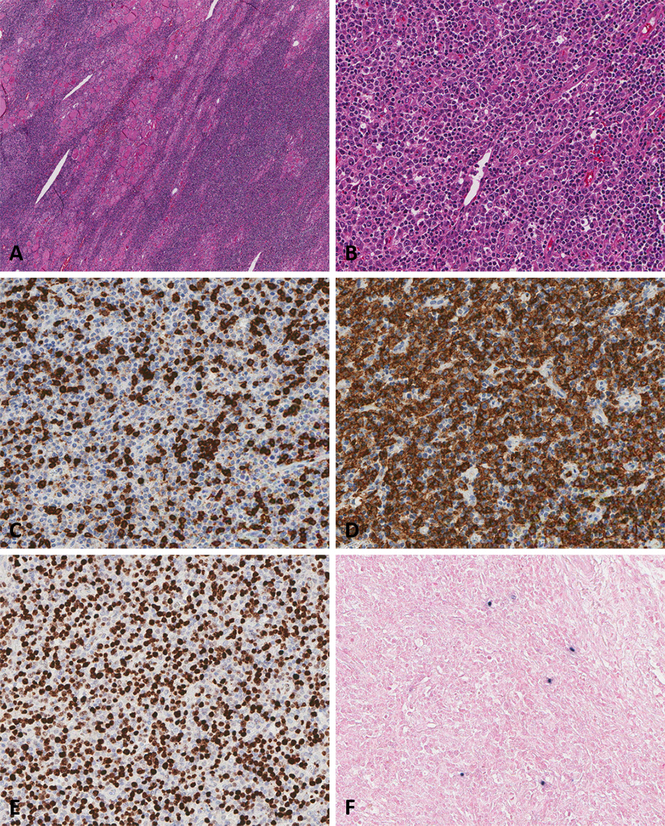
- Primary thyroid diffuse large B-cell lymphoma: fine needle aspiration and histological correlation
- Woo Sung Moon, Yong Tae Hong, Ae Ri Ahn
- J Pathol Transl Med. 2025;59(6):467-471. Published online November 3, 2025
- DOI: https://doi.org/10.4132/jptm.2025.08.28
- 4,307 View
- 120 Download
-
 Abstract
 PDF
PDF - Primary thyroid lymphoma (PTL) is a rare type of cancer that arises within the thyroid gland, representing about 2%–8% of all thyroid malignancies. Fine-needle aspiration cytology is commonly used as the first-line diagnostic approach for thyroid nodules and can assist in identifying PTL when suggestive features are present. Herein, we report the case of a 59-year-old female patient who presented with a rapidly enlarging anterior neck mass over 20 days. Clinically, the case was challenging to distinguish from anaplastic thyroid carcinoma because of the sudden enlargement of the neck mass. However, pathological examination confirmed the diagnosis of primary thyroid diffuse large B-cell lymphoma. Fine-needle aspiration cytology proved valuable in avoiding unnecessary surgical resection and guiding appropriate treatment. Additionally, we provide a brief review of the clinical and cytopathological features of primary thyroid lymphomas.
- A scoring system for the diagnosis of non-alcoholic steatohepatitis from liver biopsy
- Kyoungbun Lee, Eun Sun Jung, Eunsil Yu, Yun Kyung Kang, Mee-Yon Cho, Joon Mee Kim, Woo Sung Moon, Jin Sook Jeong, Cheol Keun Park, Jae-Bok Park, Dae Young Kang, Jin Hee Sohn, So-Young Jin
- J Pathol Transl Med. 2020;54(3):228-236. Published online April 15, 2020
- DOI: https://doi.org/10.4132/jptm.2020.03.07

- 16,205 View
- 284 Download
- 11 Web of Science
- 11 Crossref
-
Abstract
PDF
- Background
Liver biopsy is the essential method to diagnose non-alcoholic steatohepatitis (NASH), but histological features of NASH are too subjective to achieve reproducible diagnoses in early stages of disease. We aimed to identify the key histological features of NASH and devise a scoring model for diagnosis.
Methods
Thirteen pathologists blindly assessed 12 histological factors and final histological diagnoses (‘not-NASH,’ ‘borderline,’ and ‘NASH’) of 31 liver biopsies that were diagnosed as non-alcoholic fatty liver disease (NAFLD) or NASH before and after consensus. The main histological parameters to diagnose NASH were selected based on histological diagnoses and the diagnostic accuracy and agreement of 12 scoring models were compared for final diagnosis and the NAFLD Activity Score (NAS) system.
Results
Inter-observer agreement of final diagnosis was fair (κ = 0.25) before consensus and slightly improved after consensus (κ = 0.33). Steatosis at more than 5% was the essential parameter for diagnosis. Major diagnostic factors for diagnosis were fibrosis except 1C grade and presence of ballooned cells. Minor diagnostic factors were lobular inflammation ( ≥ 2 foci/ × 200 field), microgranuloma, and glycogenated nuclei. All 12 models showed higher inter-observer agreement rates than NAS and post-consensus diagnosis (κ = 0.52–0.69 vs. 0.33). Considering the reproducibility of factors and practicability of the model, summation of the scores of major (× 2) and minor factors may be used for the practical diagnosis of NASH.
Conclusions
A scoring system for the diagnosis of NAFLD would be helpful as guidelines for pathologists and clinicians by improving the reproducibility of histological diagnosis of NAFLD. -
Citations
Citations to this article as recorded by
- Preclinical liver toxicity models: Advantages, limitations and recommendations
Devaraj Ezhilarasan, Sivanesan Karthikeyan, Mustapha Najimi, Paramasivan Vijayalakshmi, Ganapathy Bhavani, Muthukrishnan Jansi Rani
Toxicology.2025; 511: 154020. CrossRef - Hepatic miR-93 promotes the pathogenesis of metabolic dysfunction-associated steatotic liver disease by suppressing SIRT1
Yo Han Lee, Jinyoung Lee, Joonho Jeong, Kieun Park, Bukyung Baik, Yuseong Kwon, Kimyeong Kim, Keon Woo Khim, Haneul Ji, Ji Young Lee, Kwangho Kim, Ji Won Kim, Tam Dao, Misung Kim, Tae Young Lee, Yong Ryoul Yang, Haejin Yoon, Dongryeol Ryu, Seonghwan Hwang
Metabolism.2025; 169: 156266. CrossRef - Deep learning-based method for grading histopathological liver fibrosis in rodent models of metabolic dysfunction-associated steatohepatitis
Soo Min Ko, Jae-ik Shin, Yiyu Hong, Hyunji Kim, Insuk Sohn, Ji-Young Lee, Hyo-Jeong Han, Da Som Jeong, Yerin Lee, Woo-Chan Son
Frontiers in Medicine.2025;[Epub] CrossRef - Liver biopsy in the modern era: from traditional techniques to artificial intelligence and multi-omics integration
Nasar Alwahaibi, Maryam Alwahaibi
Frontiers in Medicine.2025;[Epub] CrossRef - Presurgery health influences outcomes following vertical sleeve gastrectomy in adolescents
Debi Swertfeger, Ahlee Kim, Hannah Sexmith, Maria E. Moreno‐Fernandez, W. Sean Davidson, Michael Helmrath, Todd Jenkins, Tsuyoshi Okura, Esmond Geh, Stavra A. Xanthakos, Sara Szabo, Takahisa Nakamura, Senad Divanovic, Amy Sanghavi Shah
Obesity.2024; 32(6): 1187. CrossRef - Immunobiotic Bacteria Attenuate Hepatic Fibrosis through the Modulation of Gut Microbiota and the Activation of Aryl‐Hydrocarbon Receptors Pathway in Non‐Alcoholic Steatohepatitis Mice
Paulraj Kanmani, Julio Villena, Soo‐kyoung Lim, Eun‐Ji Song, Young‐Do Nam, Hojun Kim
Molecular Nutrition & Food Research.2024;[Epub] CrossRef - Lipid nanoparticle-mediated hepatocyte delivery of siRNA and silibinin in metabolic dysfunction-associated steatotic liver disease
Yifu Lyu, Xiuyi Yang, Lei Yang, Jinyu Dai, Huanyu Qin, Yunuo Zhou, Yunan Huang, Yanmei Wang, Di Wu, Qindai Shuai, Qilong Li, Xiaofei Xin, Lifang Yin
Journal of Controlled Release.2024; 373: 385. CrossRef - Enhanced hepatoprotective effects of empagliflozin and vitamin D dual therapy against metabolic dysfunction‐associated steatohepatitis in mice by boosted modulation of metabolic, oxidative stress, and inflammatory pathways
Wesam F. Farrash, Shakir Idris, Mohamed E. Elzubier, Elshiekh B. A. Khidir, Akhmed Aslam, Abdulrahman Mujalli, Riyad A. Almaimani, Ahmad A. Obaid, Mahmoud Z. El‐Readi, Mohammad A. Alobaidy, Afnan Salaka, Afnan M. Shakoori, Alaa M. Saleh, Faisal Minshawi,
International Journal of Experimental Pathology.2024; 105(6): 219. CrossRef - Bilirubin, a hepatoprotective agent that activates SIRT1, PGC-1α, and PPAR-α, while inhibiting NF-κB in rats with metabolic-associated fatty liver disease
Motahareh Taghizadeh, Mohammad Hasan Maleki, Omid Vakili, Ramin Tavakoli, Parvin Zarei, Amirreza Dehghanian, Hossein Bordbar, Sayed Mohammad Shafiee
Scientific Reports.2024;[Epub] CrossRef - Changes in indications for outpatient percutaneous liver biopsy over 5 years: from hepatitis C to fatty liver disease
Marlone Cunha-Silva, Luíza D. Torres, Mariana F. Fernandes, Tirzah de M. Lopes Secundo, Marina C.G. Moreira, Ademar Yamanaka, Leonardo T. Monici, Larissa B. Eloy da Costa, Daniel F. Mazo, Tiago Sevá-Pereira
Gastroenterología y Hepatología.2022; 45(8): 579. CrossRef - Changes in indications for outpatient percutaneous liver biopsy over 5 years: from hepatitis C to fatty liver disease
Marlone Cunha-Silva, Luíza D. Torres, Mariana F. Fernandes, Tirzah de M. Lopes Secundo, Marina C.G. Moreira, Ademar Yamanaka, Leonardo T. Monici, Larissa B. Eloy da Costa, Daniel F. Mazo, Tiago Sevá-Pereira
Gastroenterología y Hepatología (English Edition).2022; 45(8): 579. CrossRef
- Preclinical liver toxicity models: Advantages, limitations and recommendations
- Association between Expression of 8-OHdG and Cigarette Smoking in Non-small Cell Lung Cancer
- Ae Ri An, Kyoung Min Kim, Ho Sung Park, Kyu Yun Jang, Woo Sung Moon, Myoung Jae Kang, Yong Chul Lee, Jong Hun Kim, Han Jung Chae, Myoung Ja Chung
- J Pathol Transl Med. 2019;53(4):217-224. Published online March 11, 2019
- DOI: https://doi.org/10.4132/jptm.2019.02.20
- 11,013 View
- 251 Download
- 24 Web of Science
- 24 Crossref
-
Abstract
PDF
- Background
Exposure to cigarette smoking (CS) is a major risk factor for the development of lung cancer. CS is known to cause oxidative DNA damage and mutation of tumor-related genes, and these factors are involved in carcinogenesis. 8-Hydroxydeoxyguanosine (8-OHdG) is considered to be a reliable biomarker for oxidative DNA damage. Increased levels of 8-OHdG are associated with a number of pathological conditions, including cancer. There are no reports on the expression of 8-OHdG by immunohistochemistry in non-small cell lung cancer (NSCLC).
Methods
We investigated the expression of 8-OHdG and p53 in 203 NSCLC tissues using immunohistochemistry and correlated it with clinicopathological features including smoking.
Results
The expression of 8-OHdG was observed in 83.3% of NSCLC. It was significantly correlated with a low T category, negative lymph node status, never-smoker, and longer overall survival (p < .05) by univariate analysis. But multivariate analysis revealed that 8-OHdG was not an independent prognostic factor for overall survival in NSCLC patients. The aberrant expression of p53 significantly correlated with smoking, male, squamous cell carcinoma, and Ki-67 positivity (p < .05).
Conclusions
The expression of 8-OHdG was associated with good prognostic factors. It was positively correlated with never-smokers in NSCLC, suggesting that oxidative damage of DNA cannot be explained by smoking alone and may depend on complex control mechanisms. -
Citations
Citations to this article as recorded by- N-acetyl-cysteine alleviates nandrolone decanoate-induced hippocampal cell apoptosis in rats via reversing protein expressions of S1P1, Akt and FOXO3a signaling pathway
Alireza Shirpoor, Zahra Zarrini, Roya Naderi
Steroids.2026; 228: 109759. CrossRef - The distinct roles of ROS in tumor immunity: from mechanisms to immunotherapeutic applications
Jiayi Li, Chen Huang, Pan Tang, Ruiyan Wu, Quanyou Wu, Chenliang Zhang
Journal of Hematology & Oncology.2026;[Epub] CrossRef - Sustainable framework for automated segmentation and prediction of lung cancer in CT image using CapsNet with U-net segmentation
S.R. Vijayakumar, S. Aarthy, D. Deepa, P. Suresh
Biomedical Signal Processing and Control.2025; 99: 106873. CrossRef - Endolysosomal cation channel MCOLN as the novel regulator of redox homeostasis
Yahao Gao, Lei Xu, Ying Chen
Biochimica et Biophysica Acta (BBA) - Molecular Basis of Disease.2025; 1871(7): 167910. CrossRef - Catalase: The golden key to regulate oxidative stress in breast cancer
Jia-Wei Liu, Wen-Jia Chen, Yang-Zheng Lan, Jing Liu
World Journal of Clinical Oncology.2025;[Epub] CrossRef - Association of sirtuin 1 rs10997868 and rs730821 polymorphisms with sirtuin 1 and hydroxy-2′-deoxyguanosine levels in healthy smokers: A case–control study
Samar Sultan
Journal of International Medical Research.2025;[Epub] CrossRef - Increased pretreatment triglyceride glucose-body mass index associated with poor prognosis in patients with advanced non-small cell lung cancer
Shaoming Guo, Yi Zhao, Yue Jiang, Huaping Ye, Ying Wang
Clinical Nutrition ESPEN.2024; 59: 412. CrossRef - Oxidative Damage and Telomere Length as Markers of Lung Cancer Development among Chronic Obstructive Pulmonary Disease (COPD) Smokers
Elizabeth Córdoba-Lanús, Luis M. Montuenga, Angélica Domínguez-de-Barros, Alexis Oliva, Delia Mayato, Ana Remírez-Sanz, Francisca Gonzalvo, Bartolomé Celli, Javier J. Zulueta, Ciro Casanova
Antioxidants.2024; 13(2): 156. CrossRef - Automated determination of 8-OHdG in cells and tissue via immunofluorescence using a specially created antibody
Tobias Jung, Nicole Findik, Bianca Hartmann, Katja Hanack, Kai Grossmann, Dirk Roggenbuck, Marc Wegmann, René Mantke, Markus Deckert, Tilman Grune
Biotechnology Reports.2024; 42: e00833. CrossRef - Combination treatment of zinc and selenium intervention ameliorated BPA-exposed germ cell damage in SD rats: elucidation of molecular mechanisms
Chittaranjan Sahu, Gopabandhu Jena
Naunyn-Schmiedeberg's Archives of Pharmacology.2024; 397(9): 6685. CrossRef - Interplay of arsenic exposure and cigarette smoking on oxidative DNA damage in healthy males
Sepideh Nemati-Mansour, Mohammad Mosaferi, Javad Babaie, Asghar Mohammadpoorasl, Reza Dehghanzadeh, Leila Nikniaz, Mohammad Miri
Environmental Sciences Europe.2024;[Epub] CrossRef - The role of tissue persistent organic pollutants and genetic polymorphisms in patients with benign and malignant kidney tumors
Rasih Kocagöz, İlgen Onat, Merve Demirbügen Öz, Burak Turna, Banu Sarsık Kumbaracı, Mehmet Nurullah Orman, Halit Sinan Süzen, Hilmi Orhan
Environmental Toxicology and Pharmacology.2024; 110: 104495. CrossRef - Mitochondrial Plasticity and Glucose Metabolic Alterations in Human Cancer under Oxidative Stress—From Viewpoints of Chronic Inflammation and Neutrophil Extracellular Traps (NETs)
Hui-Ting Lee, Chen-Sung Lin, Chao-Yu Liu, Po Chen, Chang-Youh Tsai, Yau-Huei Wei
International Journal of Molecular Sciences.2024; 25(17): 9458. CrossRef - Oxidative DNA Damage and Arterial Hypertension in Light of Current ESC Guidelines
Radka Hazuková, Zdeněk Zadák, Miloslav Pleskot, Petr Zdráhal, Martin Pumprla, Miloš Táborský
International Journal of Molecular Sciences.2024; 25(23): 12557. CrossRef - Significance of 8-OHdG Expression as a Predictor of Survival in Colorectal Cancer
Myunghee Kang, Soyeon Jeong, Sungjin Park, Seungyoon Nam, Jun-Won Chung, Kyoung Oh Kim, Jungsuk An, Jung Ho Kim
Cancers.2023; 15(18): 4613. CrossRef - Serum 8-Hydroxy-2′-deoxyguanosine Predicts Severity and Prognosis of Patients with Acute Exacerbation of Chronic Obstructive Pulmonary Disease
Peng Cao, Chen Zhang, Dong-Xu Hua, Meng-Die Li, Bian-Bian Lv, Lin Fu, Hui Zhao
Lung.2022; 200(1): 31. CrossRef - Redox signaling at the crossroads of human health and disease
Jing Zuo, Zhe Zhang, Maochao Luo, Li Zhou, Edouard C. Nice, Wei Zhang, Chuang Wang, Canhua Huang
MedComm.2022;[Epub] CrossRef - Assessment of MDA and 8-OHdG expressions in ovine pulmonary adenocarcinomas by immunohistochemical and immunofluorescence methods
Emin Karakurt, Enver Beytut, Serpil Dağ, Hilmi Nuhoğlu, Ayfer Yıldız, Emre Kurtbaş
Acta Veterinaria Brno.2022; 91(3): 235. CrossRef - Dietary Antioxidants and Lung Cancer Risk in Smokers and Non-Smokers
Naser A. Alsharairi
Healthcare.2022; 10(12): 2501. CrossRef - Targeting oxidative stress in disease: promise and limitations of antioxidant therapy
Henry Jay Forman, Hongqiao Zhang
Nature Reviews Drug Discovery.2021; 20(9): 689. CrossRef - Association between tobacco substance usage and a missense mutation in the tumor suppressor gene P53 in the Saudi Arabian population
Mikhlid H. Almutairi, Bader O. Almutairi, Turki M. Alrubie, Sultan N. Alharbi, Narasimha R. Parine, Abdulwahed F. Alrefaei, Ibrahim Aldeailej, Abdullah Alamri, Abdelhabib Semlali, Alvaro Galli
PLOS ONE.2021; 16(1): e0245133. CrossRef - Measurement of uranium concentrations in urine samples of adult healthy groups in Najaf governorate with estimation of urine concentrations of 8-OHdG compound as biomarker for DNA damage
Samia K. Abbas, Dhuha S. Saleh, Hayder S. Hussain
Journal of Physics: Conference Series.2021; 1879(3): 032097. CrossRef - Common Data Model and Database System Development for the Korea Biobank Network
Soo-Jeong Ko, Wona Choi, Ki-Hoon Kim, Seo-Joon Lee, Haesook Min, Seol-Whan Oh, In Young Choi
Applied Sciences.2021; 11(24): 11825. CrossRef - EVALUATION OF OXIDATIVE STATUS IN PATIENTS WITH CHRONIC PERIODONTITIS AND ADDITIONAL TOBACCO ABUSE: A CROSS-SECTIONAL STUDY
Didem ÖZKAL EMİNOĞLU, Varol ÇANAKÇI
Atatürk Üniversitesi Diş Hekimliği Fakültesi Dergisi.2020; : 1. CrossRef
- N-acetyl-cysteine alleviates nandrolone decanoate-induced hippocampal cell apoptosis in rats via reversing protein expressions of S1P1, Akt and FOXO3a signaling pathway
- Iatrogenic Gastric Pseudolipomatosis during Endoscopic Submucosal Dissection
- Sang Wook Kim, Woo Sung Moon
- J Pathol Transl Med. 2017;51(5):513-515. Published online July 31, 2017
- DOI: https://doi.org/10.4132/jptm.2017.04.25
- 8,822 View
- 101 Download
- 3 Web of Science
- 3 Crossref
-
PDF
-
Citations
Citations to this article as recorded by- A Rare Case Report of Colonic Pseudolipomatosis with Distinct Endoscopic and Histological Features
Qi Zhao, Hai-Tao Yu, Qun-Ying Wang, Wei Zhang, Wen-Zhu Dong
Case Reports in Gastroenterology.2025; 19(1): 127. CrossRef - Endoscope disinfectant-induced colonic pseudolipomatosis: case series of a rare condition
Charfeddine Baccouche, Myriam Ayari, Imen Abdelaali, Amen Dhaoui, Taieb Jomni, Mohamed Hedi Douggui
Future Science OA.2024;[Epub] CrossRef - Unusual mucosal lesion: A case of rectal pseudolipomatosis in a 60-year-old patient
Boubacar Efared, Balandougou Sylla, Nawal Hammas, Hinde El Fatemi, Laila Chbani
SAGE Open Medical Case Reports.2019;[Epub] CrossRef
- A Rare Case Report of Colonic Pseudolipomatosis with Distinct Endoscopic and Histological Features
- Interobserver Agreement on Pathologic Features of Liver Biopsy Tissue in Patients with Nonalcoholic Fatty Liver Disease
- Eun Sun Jung, Kyoungbun Lee, Eunsil Yu, Yun Kyung Kang, Mee-Yon Cho, Joon Mee Kim, Woo Sung Moon, Jin Sook Jeong, Cheol Keun Park, Jae-Bok Park, Dae Young Kang, Jin Hee Sohn, So-Young Jin
- J Pathol Transl Med. 2016;50(3):190-196. Published online April 18, 2016
- DOI: https://doi.org/10.4132/jptm.2016.03.01
- 15,760 View
- 278 Download
- 30 Web of Science
- 30 Crossref
-
Abstract
PDF
- Background
The histomorphologic criteria for the pathological features of liver tissue from patients with non-alcoholic fatty liver disease (NAFLD) remain subjective, causing confusion among pathologists and clinicians. In this report, we studied interobserver agreement of NAFLD pathologic features and analyzed causes of disagreement.
Methods
Thirty-one cases of clinicopathologically diagnosed NAFLD from 10 hospitals were selected. One hematoxylin and eosin and one Masson’s trichrome-stained virtual slide from each case were blindly reviewed with regard to 12 histological parameters by 13 pathologists in a gastrointestinal study group of the Korean Society of Pathologists. After the first review, we analyzed the causes of disagreement and defined detailed morphological criteria. The glass slides from each case were reviewed a second time after a consensus meeting. The degree of interobserver agreement was determined by multi-rater kappa statistics.
Results
Kappa values of the first review ranged from 0.0091–0.7618. Acidophilic bodies (k = 0.7618) and portal inflammation (k = 0.5914) showed high levels of agreement, whereas microgranuloma (k = 0.0984) and microvesicular fatty change (k = 0.0091) showed low levels of agreement. After the second review, the kappa values of the four major pathological features increased from 0.3830 to 0.5638 for steatosis grade, from 0.1398 to 0.2815 for lobular inflammation, from 0.1923 to 0.3362 for ballooning degeneration, and from 0.3303 to 0.4664 for fibrosis.
Conclusions
More detailed histomorphological criteria must be defined for correct diagnosis and high interobserver agreement of NAFLD. -
Citations
Citations to this article as recorded by- Recent Advances in the Application of Machine Learning Models in Metabolic Dysfunction–Associated Steatotic Liver Disease
Fang Yang, Xueyue Sun, Kui Jiang, Mingxin Zhang, Chao Sun
Diabetes/Metabolism Research and Reviews.2026;[Epub] CrossRef - Double Graph Attention Network for predicting non-alcoholic fatty liver disease in patients with type 2 diabetes
Tianbin Chen, Yongbin Zeng, Jinlin Wang, Xiao Sun, Sihao Liu, Ya Fu, Qiang Yi, Qishui Ou, Kai Yan, Zhiheng Zhou
Artificial Intelligence in Medicine.2026; 174: 103369. CrossRef - Liver Biopsy in Metabolic-Associated Steatotic Liver Disease: Accuracy, Challenges, and Alternatives
Luca Borz-Baba, Adem Aydin, Russell Parvin
Cureus.2026;[Epub] CrossRef - Quantitative regression of qFibrosis with resmetirom: Exploratory histologic endpoints from the MAESTRO-NASH phase III clinical trial
Jörn M. Schattenberg, Pierre Bedossa, Cynthia D. Guy, Rebecca Taub, Dominic Labriola, Hang Zhang, James Hennan, Raul C. Camacho, Elaine Chng, Yayun Ren, Dean Tai, Stephen A. Harrison
Journal of Hepatology.2026; 85(2): 202. CrossRef - Chronic polypharmacy, monotherapy, and deprescribing: Understanding complex effects on the hepatic proteome of aging mice
Kevin Winardi, John Mach, Matthew J. McKay, Mark P. Molloy, Sarah J. Mitchell, Michael R. MacArthur, Catriona McKenzie, David G. Le Couteur, Sarah N. Hilmer
Aging Cell.2025;[Epub] CrossRef - Utility of AI digital pathology as an aid for pathologists scoring fibrosis in MASH
Desiree Abdurrachim, Serene Lek, Charlene Zhi Lin Ong, Chun Kit Wong, Yongqi Zhou, Aileen Wee, Gwyneth Soon, Timothy J. Kendall, Michael O. Idowu, Christopher Hendra, Ashmita Saigal, Radha Krishnan, Elaine Chng, Dean Tai, Gideon Ho, Thomas Forest, Annaswa
Journal of Hepatology.2025; 82(5): 898. CrossRef - Artificial intelligence scoring of liver biopsies in a phase II trial of semaglutide in nonalcoholic steatohepatitis
Vlad Ratziu, Sven Francque, Cynthia A. Behling, Vanja Cejvanovic, Helena Cortez-Pinto, Janani S. Iyer, Niels Krarup, Quang Le, Anne-Sophie Sejling, Dina Tiniakos, Stephen A. Harrison
Hepatology.2024; 80(1): 173. CrossRef - Classification of the Stages of Nonalcoholic Steatohepatitis via Federated General Visual Representation Learning
Mehmet Nergiz
International Journal of Imaging Systems and Technology.2024;[Epub] CrossRef - Outcome prediction in metabolic dysfunction‐associated steatotic liver disease using stain‐free digital pathological assessment
Timothy J. Kendall, Elaine Chng, Yayun Ren, Dean Tai, Gideon Ho, Jonathan A. Fallowfield
Liver International.2024; 44(10): 2511. CrossRef - Non-alcoholic fatty liver disease: the pathologist’s perspective
Wei-Qiang Leow, Anthony Wing-Hung Chan, Paulo Giovanni L. Mendoza, Regina Lo, Kihan Yap, Haeryoung Kim
Clinical and Molecular Hepatology.2023; 29(Suppl): S302. CrossRef - CT-based Hounsfield unit values reflect the degree of steatohepatitis in patients with low-grade fatty liver disease
Ha Neul Kim, Hong Jae Jeon, Hei Gwon Choi, In Sun Kwon, Woo Sun Rou, Jeong Eun Lee, Tae Hee Lee, Seok Hyun Kim, Byung Seok Lee, Kyung Sook Shin, Hyun Jung Lee, Hyuk Soo Eun
BMC Gastroenterology.2023;[Epub] CrossRef - Artificial intelligence and deep learning: New tools for histopathological diagnosis of nonalcoholic fatty liver disease/nonalcoholic steatohepatitis
Yoshihisa Takahashi, Erdenetsogt Dungubat, Hiroyuki Kusano, Toshio Fukusato
Computational and Structural Biotechnology Journal.2023; 21: 2495. CrossRef - An integrated gene-to-outcome multimodal database for metabolic dysfunction-associated steatotic liver disease
Timothy J. Kendall, Maria Jimenez-Ramos, Frances Turner, Prakash Ramachandran, Jessica Minnier, Michael D. McColgan, Masood Alam, Harriet Ellis, Donald R. Dunbar, Gabriele Kohnen, Prakash Konanahalli, Karin A. Oien, Lucia Bandiera, Filippo Menolascina, An
Nature Medicine.2023; 29(11): 2939. CrossRef - Improved pathology reporting in NAFLD/NASH for clinical trials
Caitlin Rose Langford, Marc H Goldinger, Darren Treanor, Clare McGenity, Jonathan R Dillman, Daniela S Allende, Robert Goldin, Elizabeth M Brunt, Kurt Zatloukal, Helmut Denk, Kenneth A Fleming
Journal of Clinical Pathology.2022; 75(2): 73. CrossRef - Standardizing the histological assessment of late posttransplantation biopsies from pediatric liver allograft recipients
Stefan G. Hübscher, Sandy Feng, Annette S. H. Gouw, Hironori Haga, Hyo Jeong Kang, Deirdre A. Kelly, Mina Komuta, Andrew Lesniak, Benjamin A. Popp, Henkjan J. Verkade, Eunsil Yu, Anthony J. Demetris
Liver Transplantation.2022; 28(9): 1475. CrossRef - Discordant pathological diagnosis of non‐alcoholic fatty liver disease: A prospective multicenter study
Takuya Kuwashiro, Hirokazu Takahashi, Hideyuki Hyogo, Yuji Ogawa, Kento Imajo, Masato Yoneda, Takashi Nakahara, Satoshi Oeda, Kenichi Tanaka, Yuichiro Amano, Shinji Ogawa, Atsushi Kawaguchi, Shinichi Aishima, Masayoshi Kage, Kazuaki Chayama, Atsushi Nakaj
JGH Open.2020; 4(3): 497. CrossRef - Obeticholic acid for the treatment of nonalcoholic steatohepatitis: Expectations and concerns
Stergios A. Polyzos, Jannis Kountouras, Christos S. Mantzoros
Metabolism.2020; 104: 154144. CrossRef - A scoring system for the diagnosis of non-alcoholic steatohepatitis from liver biopsy
Kyoungbun Lee, Eun Sun Jung, Eunsil Yu, Yun Kyung Kang, Mee-Yon Cho, Joon Mee Kim, Woo Sung Moon, Jin Sook Jeong, Cheol Keun Park, Jae-Bok Park, Dae Young Kang, Jin Hee Sohn, So-Young Jin
Journal of Pathology and Translational Medicine.2020; 54(3): 228. CrossRef - An Improved qFibrosis Algorithm for Precise Screening and Enrollment into Non-Alcoholic Steatohepatitis (NASH) Clinical Trials
Wei-Qiang Leow, Pierre Bedossa, Feng Liu, Lai Wei, Kiat-Hon Lim, Wei-Keat Wan, Yayun Ren, Jason Pik-Eu Chang, Chee-Kiat Tan, Aileen Wee, George Boon-Bee Goh
Diagnostics.2020; 10(9): 643. CrossRef - Deep learning quantification of percent steatosis in donor liver biopsy frozen sections
Lulu Sun, Jon N. Marsh, Matthew K. Matlock, Ling Chen, Joseph P. Gaut, Elizabeth M. Brunt, S. Joshua Swamidass, Ta-Chiang Liu
EBioMedicine.2020; 60: 103029. CrossRef - Magnetic resonance elastography SE-EPI vs GRE sequences at 3T in a pediatric population with liver disease
Juan S. Calle-Toro, Suraj D. Serai, Erum A. Hartung, David J. Goldberg, Bradley D. Bolster, Kassa Darge, Sudha A. Anupindi
Abdominal Radiology.2019; 44(3): 894. CrossRef - R2 relaxometry based MR imaging for estimation of liver iron content: A comparison between two methods
Juan S. Calle-Toro, Christian A. Barrera, Dmitry Khrichenko, Hansel J. Otero, Suraj D. Serai
Abdominal Radiology.2019; 44(9): 3058. CrossRef - Inhibition of mitochondrial fatty acid oxidation in drug-induced hepatic steatosis
Bernard Fromenty
Liver Research.2019; 3(3-4): 157. CrossRef - Standardising the interpretation of liver biopsies in non‐alcoholic fatty liver disease clinical trials
Rish K. Pai, David E. Kleiner, John Hart, Oyedele A. Adeyi, Andrew D. Clouston, Cynthia A. Behling, Dhanpat Jain, Sanjay Kakar, Mayur Brahmania, Lawrence Burgart, Kenneth P. Batts, Mark A. Valasek, Michael S. Torbenson, Maha Guindi, Hanlin L. Wang, Veeral
Alimentary Pharmacology & Therapeutics.2019; 50(10): 1100. CrossRef - NAFLD Histology: a Critical Review and Comparison of Scoring Systems
Rish K. Pai
Current Hepatology Reports.2019; 18(4): 473. CrossRef - Hepatic sonic hedgehog protein expression measured by computer assisted morphometry significantly correlates with features of non-alcoholic steatohepatitis
Michael Estep, Rohini Mehta, Gary Bratthauer, Lakshmi Alaparthi, Fanny Monge, Simon Ali, Dinan Abdelatif, Zahra Younoszai, Maria Stepanova, Zachary D. Goodman, Zobair M. Younossi
BMC Gastroenterology.2019;[Epub] CrossRef - Validation of intimate correlation between visceral fat and hepatic steatosis: Quantitative measurement techniques using CT for area of fat and MR for hepatic steatosis
Moon Hyung Choi, Joon-Il Choi, Michael Yong Park, Sung Eun Rha, Soon Nam Oh, Seung Eun Jung, Jae Young Byun, Stephan Kannengiesser, Yohan Son
Clinical Nutrition.2018; 37(1): 214. CrossRef - Ultrasound or MR elastography of liver: which one shall I use?
Meng Yin, Sudhakar K. Venkatesh
Abdominal Radiology.2018; 43(7): 1546. CrossRef - Feasibility and agreement of stiffness measurements using gradient-echo and spin-echo MR elastography sequences in unselected patients undergoing liver MRI
Guilherme Moura Cunha, Kevin J Glaser, Anke Bergman, Rodrigo P Luz, Eduardo H de Figueiredo, Flavia Paiva Proença Lobo Lopes
The British Journal of Radiology.2018; : 20180126. CrossRef - Second harmonic generation microscopy provides accurate automated staging of liver fibrosis in patients with non-alcoholic fatty liver disease
Pik Eu Chang, George Boon Bee Goh, Wei Qiang Leow, Liang Shen, Kiat Hon Lim, Chee Kiat Tan, Manlio Vinciguerra
PLOS ONE.2018; 13(6): e0199166. CrossRef
- Recent Advances in the Application of Machine Learning Models in Metabolic Dysfunction–Associated Steatotic Liver Disease
- Immunohistochemical Expression and Clinical Significance of Suggested Stem Cell Markers in Hepatocellular Carcinoma
- Jong Jin Sung, Sang Jae Noh, Jun Sang Bae, Ho Sung Park, Kyu Yun Jang, Myoung Ja Chung, Woo Sung Moon
- J Pathol Transl Med. 2016;50(1):52-57. Published online November 18, 2015
- DOI: https://doi.org/10.4132/jptm.2015.10.09
- 12,962 View
- 78 Download
- 21 Web of Science
- 19 Crossref
-
Abstract
PDF
- Background
Increasing evidence has shown that tumor initiation and growth are nourished by a small subpopulation of cancer stem cells (CSCs) within the tumor mass. CSCs are posited to be responsible for tumor maintenance, growth, distant metastasis, and relapse after curative operation. We examined the expression of CSC markers in paraffin-embedded tissue sections of hepatocellular carcinoma (HCC) and correlated the results with clinicopathologic characteristics. Methods: Immunohistochemical staining for the markers believed to be expressed in the CSCs, including epithelial cell adhesion molecule (EpCAM), keratin 19 (K19), CD133, and CD56, was performed in 82 HCC specimens. Results: EpCAM expression was observed in 56% of the HCCs (46/82) and K19 in 6% (5/82). EpCAM expression in HCC significantly correlated with elevated α-fetoprotein level, microvessel invasion of tumor cells, and high histologic grade. In addition, Ep- CAM expression significantly correlated with K19 expression. The overall survival and relapsefree survival rates in patients with EpCAM-expressing HCC were relatively lower than those in patients with EpCAM-negative HCC. All but two of the 82 HCCs were negative for CD133 and CD56, respectively. Conclusions: Our results suggest that HCCs expressing EpCAM are associated with unfavorable prognostic factors and have a more aggressive clinical course than those not expressing EpCAM. Further, the expression of either CD133 or CD56 in paraffin-embedded HCC tissues appears to be rare. -
Citations
Citations to this article as recorded by- Spatial immune scoring system predicts hepatocellular carcinoma recurrence
Gengjie Jia, Peiqi He, Tianli Dai, Denise Goh, Jiabei Wang, Mengyuan Sun, Felicia Wee, Fuling Li, Jeffrey Chun Tatt Lim, Shuxia Hao, Yao Liu, Tony Kiat Hon Lim, Nye-Thane Ngo, Qingping Tao, Wei Wang, Ahitsham Umar, Björn Nashan, Yongchang Zhang, Chen Ding
Nature.2025; 640(8060): 1031. CrossRef - Evolving Landscape of Systemic Therapy for Hepatocellular Carcinoma in 2025
Karan Kumar, Vivek A. Saraswat
Journal of Clinical and Experimental Hepatology.2025; 15(5): 102547. CrossRef - Recent Progress in Systemic Therapy for Advanced Hepatocellular Carcinoma
Narayanan Sadagopan, Aiwu Ruth He
International Journal of Molecular Sciences.2024; 25(2): 1259. CrossRef - Diagnostic value of expressions of cancer stem cell markers for adverse outcomes of hepatocellular carcinoma and their associations with prognosis: A Bayesian network meta‑analysis
Zhengrong Ou, Shoushuo Fu, Jian Yi, Jingxuan Huang, Weidong Zhu
Oncology Letters.2024;[Epub] CrossRef - Clinicopathological and prognostic value of epithelial cell adhesion molecule in solid tumours: a meta-analysis
Peiwen Ding, Panyu Chen, Jiqi Ouyang, Qiang Li, Shijie Li
Frontiers in Oncology.2023;[Epub] CrossRef - PD-L1 Downregulation and DNA Methylation Inhibition for Molecular Therapy against Cancer Stem Cells in Hepatocellular Carcinoma
Caecilia Sukowati, Loraine Kay D. Cabral, Beatrice Anfuso, Francesco Dituri, Roberto Negro, Gianluigi Giannelli, Claudio Tiribelli
International Journal of Molecular Sciences.2023; 24(17): 13357. CrossRef - EpCAM, Ki67, and ESM1 Predict Hepatocellular Carcinoma Recurrence After Liver Transplantation
Aiat Shaban Hemida, Doha Maher Taie, Moshira Mohamed Abd El-Wahed, Mohammed Ibrahim Shabaan, Mona Saeed Tantawy, Nermine Ahmed Ehsan
Applied Immunohistochemistry & Molecular Morphology.2023; 31(9): 596. CrossRef - The clinical, prognostic and therapeutic significance of liver cancer stem cells and their markers
Izabela Zarębska, Arkadiusz Gzil, Justyna Durślewicz, Damian Jaworski, Paulina Antosik, Navid Ahmadi, Marta Smolińska-Świtała, Dariusz Grzanka, Łukasz Szylberg
Clinics and Research in Hepatology and Gastroenterology.2021; 45(3): 101664. CrossRef - Detection of oncogenic mutations in paired circulating tumor DNA and circulating tumor cells in patients with hepatocellular carcinoma
Zhouhong Ge, Jean C.A. Helmijr, Maurice P.H.M. Jansen, Patrick P.C. Boor, Lisanne Noordam, Maikel Peppelenbosch, Jaap Kwekkeboom, Jaco Kraan, Dave Sprengers
Translational Oncology.2021; 14(7): 101073. CrossRef - Hepatocellular Carcinoma Score and Subclassification Into Aggressive Subtypes Using Immunohistochemical Expression of p53, β-Catenin, CD133, and Ki-67
Asmaa G. Abdou, Nanis S. Holah, Dina S. Elazab, Walaa G. El-Gendy, Mohammed T. Badr, Dalia R. Al-Sharaky
Applied Immunohistochemistry & Molecular Morphology.2021; 29(1): 20. CrossRef - The prognostic significance of neuroendocrine markers and somatostatin receptor 2 in hepatocellular carcinoma
Keigo Murakami, Hiroyuki Kumata, Shigehito Miyagi, Takashi Kamei, Hironobu Sasano
Pathology International.2021; 71(10): 682. CrossRef - Predictors of recurrence and survival of hepatocellular carcinoma: A prospective study including transient elastography and cancer stem cell markers
Hend Ibrahim Shousha, Rabab Fouad, Tamer Mahmoud Elbaz, Dina Sabry, Mohamed Mahmoud Nabeel, Ahmed Hosni Abdelmaksoud, Aisha Mahmoud Elsharkawy, Zeinab Abdellatif Soliman, Ghada Habib, Ashraf Omar Abdelaziz
Arab Journal of Gastroenterology.2020; 21(2): 95. CrossRef - Napabucasin Reduces Cancer Stem Cell Characteristics in Hepatocellular Carcinoma
Ya Li, Qiuju Han, Huajun Zhao, Quanjuan Guo, Jian Zhang
Frontiers in Pharmacology.2020;[Epub] CrossRef - The mRNA Distribution of Cancer Stem Cell Marker CD90/Thy-1 Is Comparable in Hepatocellular Carcinoma of Eastern and Western Populations
An B. Luong, Huy Q. Do, Paola Tarchi, Deborah Bonazza, Cristina Bottin, Loraine Kay D. Cabral, Long D. C. Tran, Thao P. T. Doan, Lory S. Crocè, Hoa L. T. Pham, Claudio Tiribelli, Caecilia H. C. Sukowati
Cells.2020; 9(12): 2672. CrossRef - The prognostic relationship between histopathological and immunohistochemical features of hepatocellular carcinoma, intrahepatic cholangiocarcinoma and mixed type
Fatmagül Kuşku Çabuk, Nuray Başsüllü, İlknur Türkmen, Murat Dayangaç, Murat Akyıldız, Yıldıray Yüzer, Yaman Tokat, Gülen Bülbül Doğusoy
Polish Journal of Pathology.2020; 71(2): 79. CrossRef - Histological architectural classification determines recurrence pattern and prognosis after curative hepatectomy in patients with hepatocellular carcinoma
Hirohisa Okabe, Tomoharu Yoshizumi, Yo-ichi Yamashita, Katsunori Imai, Hiromitsu Hayashi, Shigeki Nakagawa, Shinji Itoh, Norifumi Harimoto, Toru Ikegami, Hideaki Uchiyama, Toru Beppu, Shinichi Aishima, Ken Shirabe, Hideo Baba, Yoshihiko Maehara, Motoyuki
PLOS ONE.2018; 13(9): e0203856. CrossRef - Overexpression of epithelial cell adhesion molecule as a predictor of poor outcome in patients with hepatocellular carcinoma
Chih‑Jan Ko, Chia‑Jung Li, Meng‑Yu Wu, Pei‑Yi Chu
Experimental and Therapeutic Medicine.2018;[Epub] CrossRef - Clinicopathologic Significance of Survivin Expression in Relation to CD133 Expression in Surgically Resected Stage II or III Colorectal Cancer
Wanlu Li, Mi-Ra Lee, EunHee Choi, Mee-Yon Cho
Journal of Pathology and Translational Medicine.2017; 51(1): 17. CrossRef - PIN1 in hepatocellular carcinoma is associated with TP53 gene status
Jun Sang Bae, Sang Jae Noh, Kyoung Min Kim, Kyu Yun Jang, Ho Sung Park, Myoung Ja Chung, Byung-Hyun Park, Woo Sung Moon
Oncology Reports.2016; 36(4): 2405. CrossRef
- Spatial immune scoring system predicts hepatocellular carcinoma recurrence
- Nesidioblastosis and Pancreatic Non-functioning Islet Cell Tumor in an Adult with Type 2 Diabetes Mellitus
- Ji Eun Choi, Sang Jae Noh, Jong Jin Sung, Woo Sung Moon
- Korean J Pathol. 2013;47(5):489-491. Published online October 25, 2013
- DOI: https://doi.org/10.4132/KoreanJPathol.2013.47.5.489
- 10,756 View
- 72 Download
- 9 Crossref
-
PDF
-
Citations
Citations to this article as recorded by- Diffuse, Adult-Onset Nesidioblastosis/Non-Insulinoma Pancreatogenous Hypoglycemia Syndrome (NIPHS): Review of the Literature of a Rare Cause of Hyperinsulinemic Hypoglycemia
Martin Philipp Dieterle, Ayman Husari, Sophie Nicole Prozmann, Hendrik Wiethoff, Albrecht Stenzinger, Manuel Röhrich, Uwe Pfeiffer, Wolfgang Rüdiger Kießling, Helena Engel, Harald Sourij, Thorsten Steinberg, Pascal Tomakidi, Stefan Kopf, Julia Szendroedi
Biomedicines.2023; 11(6): 1732. CrossRef - Postprandial hypoglycemia after upper gastrointestinal tract surgery: prevalence and pathophysiology (part 1)
M. Yu. Yukina, M. O. Chernova, E. A. Troshina, V. V. Evdoshenko, N. M. Platonova
Almanac of Clinical Medicine.2021; 49(4): 285. CrossRef - Nésidioblastose traitée par pasiréotide LAR : à propos d’un cas
Alexia Rouland, Benjamin Bouillet, Pauline Legris, Isabelle Simoneau, Jean-Michel Petit, Bruno Vergès
Médecine des Maladies Métaboliques.2021; 15(6): 619. CrossRef - Concurrent Adult-Onset Diffuse β-Cell Nesidioblastosis and Pancreatic Neuroendocrine Tumor: A Case Report and Review of the Literature
Mushfig Orujov, Keith K. Lai, Catherine L. Forse
International Journal of Surgical Pathology.2019; 27(8): 912. CrossRef - Nesidioblastosis in an Adult with Short Gut Syndrome and Type 2 Diabetes
Mimi Wong, Luke Conway, Caroline Cooper, Ashim Sinha, Nirjhar Nandi
AACE Clinical Case Reports.2019; 5(6): e375. CrossRef - Superimposed effect of ovariectomy on type 2 diabetes mellitus in Wistar rats
Minerva K. Fahmy, Hayam G. Sayyed, Eman A. Abd Elrahim, Rana T.A. Farag
Alexandria Journal of Medicine.2018; 54(2): 129. CrossRef - Diagnostic Performance of 48-Hour Fasting Test and Insulin Surrogates in Patients With Suspected Insulinoma
Keijiro Ueda, Ken Kawabe, Lingaku Lee, Yuichi Tachibana, Nao Fujimori, Hisato Igarashi, Yoshinao Oda, Robert T. Jensen, Ryoichi Takayanagi, Tetsuhide Ito
Pancreas.2017; 46(4): 476. CrossRef - Nesidioblastosis (diagnosis, surgical treatment)
A. G. Kriger, A. V. Smirnov, D. V. Kalinin, A. V. Glotov, S. V. Berelavichus, G. I. Konyaeva, A. N. Lebedeva, N. A. Karel’skaya, V. N. Tsygankov
Khirurgiya. Zhurnal im. N.I. Pirogova.2015; (10): 16. CrossRef - A Rare Complication of Gastric Bypass (Weight Loss) Surgery
Betül Ünal, Özlem Ceren Uzun, Cumhur İbrahim Başsorgun, Okan Erdoğan, Gülsüm Özlem Elpek
International Journal of Surgical Pathology.2015; 23(1): 68. CrossRef
- Diffuse, Adult-Onset Nesidioblastosis/Non-Insulinoma Pancreatogenous Hypoglycemia Syndrome (NIPHS): Review of the Literature of a Rare Cause of Hyperinsulinemic Hypoglycemia
- Multicystic Biliary Hamartoma of the Liver
- Ji Soo Song, Sang Jae Noh, Baik Hwan Cho, Woo Sung Moon
- Korean J Pathol. 2013;47(3):275-278. Published online June 25, 2013
- DOI: https://doi.org/10.4132/KoreanJPathol.2013.47.3.275
- 10,526 View
- 92 Download
- 17 Crossref
-
Abstract
PDF
Multicystic biliary hamartoma (MCBH) is a very rare hamartomatous cystic nodule of the liver, which has recently been described as a new entity of a hepatic nodular lesion. We report a unique case of MCBH with a review of the literatures. A hepatic multicystic mass of segment 3 was detected in a 52-year-old male by abdominal computed tomography, and resection of this lesion was performed. Macroscopic examination revealed a 2.7×2.0 cm nodular mass with a multicystic honeycomb cut surface. Histologically, this lesion consisted of multiple dilated cystic ducts lined by biliary type epithelial cells, periductal glands and connective tissue, which included small amounts of hepatic parenchyma and blood vessels. Recognition of this unusual lesion is essential to avoid confusion with other cystic tumors of the liver, and to learn more about its natural history and response to treatment.
-
Citations
Citations to this article as recorded by- Multicystic biliary hamartoma with long-term gradual enlargement treated by laparoscopic partial hepatectomy
Satoshi Nishiwada, Tetsuya Tanaka, Yuki Kirihataya, Takeshi Takei, Tomomi Sadamitsu, Masato Takano, Masayoshi Sawai, Atsushi Yoshimura
Clinical Journal of Gastroenterology.2025; 18(3): 527. CrossRef - Technical Considerations in EEG Source Imaging
Benjamin H. Brinkmann
Journal of Clinical Neurophysiology.2024; 41(1): 2. CrossRef - A Case of Multicystic Biliary Hamartoma with a Marked Peribiliary Gland Component Successfully Treated by Purely Laparoscopic Anatomical Liver Resection
Keita Kai, Takao Ide, Tomokazu Tanaka, Kumpei Yukimoto, Hiroyuki Irie, Hirokazu Noshiro, Shinichi Aishima
Journal of Gastrointestinal Cancer.2023; 54(3): 996. CrossRef - Characteristics of multicystic biliary hamartoma: A case report
Jia Lian, Lixia Sun, Yankai Yang, Jun Li, Ye Zhang, Guiqiu Liu, Weijuan Hu
Frontiers in Surgery.2023;[Epub] CrossRef - Recurrent sepsis in a patient with biliary hamartomas
Maria Beatriz Santos, Magda Ponta Garça, Bárbara Vieira, Paulo Ávila, Alexandra Freitas
European Journal of Case Reports in Internal Medicine.2023;[Epub] CrossRef - Hamartoma multiquístico de vías biliares
Victoria Carmona, Iago Justo, Yolanda Rodríguez-Gil, Alberto Marcacuzco, Carmelo Loinaz, Carlos Jiménez
Cirugía Española.2022; 100(12): 800. CrossRef - Multicystic Biliary Hamartoma With Xanthogranulomatous Inflammation on 18F-FDG PET/CT
Nahomi Shono, Yoichi Otomi, Hideki Otsuka, Takayoshi Shinya, Masafumi Harada
Clinical Nuclear Medicine.2022; 47(10): 882. CrossRef - Intrahepatic multicystic biliary hamartoma: A case report
Chen-Yu Wang, Fu-Yang Shi, Wei-Feng Huang, Yan Tang, Ting Li, Guo-Lin He
World Journal of Clinical Cases.2022; 10(26): 9361. CrossRef - A Case of Multicystic Biliary Hamartoma Treated with Left Medial Sectionectomy
Naomi KUROKI, Tomoaki TANAKA, Takanobu SUGASE, Syoji TANIGUCHI, Takashi GOTO, Rintaro KOGA, Takumi KIWAKI, Hiroyuki TANAKA
Nihon Rinsho Geka Gakkai Zasshi (Journal of Japan Surgical Association).2022; 83(2): 395. CrossRef - Multicystic biliary hamartoma
Victoria Carmona, Iago Justo, Yolanda Rodríguez-Gil, Alberto Marcacuzco, Carmelo Loinaz, Carlos Jiménez
Cirugía Española (English Edition).2022; 100(12): 800. CrossRef - Case Report: Incidentally Discovered a Rare Cystic Lesion of Liver: Multicystic Biliary Hamartoma
Wentao Mu, Peng Su, Shanglei Ning
Pathology and Oncology Research.2021;[Epub] CrossRef - Bile Duct Hamartoma Mimicking Liver Metastasis in Suspected Porcelain Gallbladder: a Case Report
Gautham Krishnamurthy, Harjeet Singh, Sravya Deepika Ganti, Ganga Ram Verma
Journal of Gastrointestinal Cancer.2019; 50(4): 1022. CrossRef - A variant of multicystic biliary hamartoma presenting as an intrahepatic cystic neoplasm
Tetsuro Tominaga, Takafumi Abo, Naoe Kinoshita, Tomonori Murakami, Yasunori Sato, Yasuni Nakanuma, Kenich Harada, Junichi Masuda, Takeshi Nagayasu, Atsushi Nanashima
Clinical Journal of Gastroenterology.2015; 8(3): 162. CrossRef - Hamartoma biliar multiquístico intrahepático: presentación de un caso clínico
María Jezabel Fernández-Carrión, Ricardo Robles Campos, Asunción López Conesa, Roberto Brusadín, Pascual Parrilla Paricio
Cirugía Española.2015; 93(9): e103. CrossRef - Intrahepatic Multicystic Biliary Hamartoma: Presentation of a Case Report
María Jezabel Fernández-Carrión, Ricardo Robles Campos, Asunción López Conesa, Roberto Brusadín, Pascual Parrilla Paricio
Cirugía Española (English Edition).2015; 93(9): e103. CrossRef - Multicystic biliary hamartoma: A report of a rare entity and a review of the literature
Rachel E. Beard, Eric U. Yee, Koenraad J. Mortele, Khalid Khwaja
International Journal of Surgery Case Reports.2014; 5(12): 919. CrossRef - Multicystic biliary hamartoma mimicking intrahepatic cholangiocarcinoma: report of a case
Tomoaki Yoh, Ryuji Okamura, Hiroyuki Nakayama, Xue Lin, Yuya Nakamura, Tatsushi Kato
Clinical Journal of Gastroenterology.2014; 7(5): 418. CrossRef
- Multicystic biliary hamartoma with long-term gradual enlargement treated by laparoscopic partial hepatectomy
- Mucinous Non-neoplastic Cyst of the Pancreas
- Jae Do Yang, Ji Soo Song, Sang Jae Noh, Woo Sung Moon
- Korean J Pathol. 2013;47(2):188-190. Published online April 24, 2013
- DOI: https://doi.org/10.4132/KoreanJPathol.2013.47.2.188
- 9,152 View
- 48 Download
- 6 Crossref
-
PDF
-
Citations
Citations to this article as recorded by- Molecular Characterization of Pancreatic Simple Mucinous Cysts With GNAS Mutation: A Case Report and Literature Review
Shoichiro Mizukami, Koji Imai, Hiroyuki Takahashi, Shingo Shimada, Nobue Tamamura, Miyuki Mori, Koji Nishikawa, Yusuke Ono, Mishie Tanino, Yusuke Mizukami, Hideki Yokoo
Pancreas.2026; 55(1): e75. CrossRef - Simple mucinous cyst: another potential cancer precursor in the pancreas? Case report with molecular characterization and systematic review of the literature
Anna Caterina Milanetto, Alice Sabrina Tonello, Giovanni Valotto, Giada Munari, Claudio Luchini, Matteo Fassan, Claudio Pasquali
Virchows Archiv.2021; 479(1): 179. CrossRef - Mucinous Non-neoplastic Cyst of the Pancreas
Jun Hyung Kim, Dong Eun Park, Keum Ha Choi
The Korean Journal of Gastroenterology.2019; 73(4): 235. CrossRef - Mucinous nonneoplastic cyst of the pancreas: CT and MRI appearances
Kousei Ishigami, Akihiro Nishie, Naoki Mochidome, Yoshiki Asayama, Yasuhiro Ushijima, Daisuke Kakihara, Daisuke Okamoto, Nobuhiro Fujita, Takao Ohtsuka, Yoshihiro Miyasaka, Tomoyuki Hida, Tomoharu Yoshizumi, Hiroshi Honda
Abdominal Radiology.2017; 42(12): 2827. CrossRef - Pathologic Evaluation and Reporting of Intraductal Papillary Mucinous Neoplasms of the Pancreas and Other Tumoral Intraepithelial Neoplasms of Pancreatobiliary Tract
Volkan Adsay, Mari Mino-Kenudson, Toru Furukawa, Olca Basturk, Giuseppe Zamboni, Giovanni Marchegiani, Claudio Bassi, Roberto Salvia, Giuseppe Malleo, Salvatore Paiella, Christopher L. Wolfgang, Hanno Matthaei, G. Johan Offerhaus, Mustapha Adham, Marco J.
Annals of Surgery.2016; 263(1): 162. CrossRef - Rare Nonneoplastic Cysts of Pancreas
Yeon Suk Kim, Jae Hee Cho
Clinical Endoscopy.2015; 48(1): 31. CrossRef
- Molecular Characterization of Pancreatic Simple Mucinous Cysts With GNAS Mutation: A Case Report and Literature Review
- Expression of CHOP in Squamous Tumor of the Uterine Cervix
- Hyun Hee Chu, Jun Sang Bae, Kyoung Min Kim, Ho Sung Park, Dong Hyu Cho, Kyu Yun Jang, Woo Sung Moon, Myoung Jae Kang, Dong Geun Lee, Myoung Ja Chung
- Korean J Pathol. 2012;46(5):463-469. Published online October 25, 2012
- DOI: https://doi.org/10.4132/KoreanJPathol.2012.46.5.463
- 10,410 View
- 40 Download
- 8 Crossref
-
Abstract
PDF
Background High-risk human papillomavirus (HR-HPV) infection and abnormal p53 expression are closely involved in carcinogenesis of squamous cell carcinoma (SqCC) of uterine cervix. Recent studies have suggested that virus-induced endoplasmic reticulum (ER) stress modulates various cell survival and cell death signaling pathways. The C/EBP homologous protein (CHOP) is associated with ER stress-mediated apoptosis and is also involved in carcinogenesis of several human cancers. We hypothesized that CHOP is involved in the carcinogenesis of uterine cervical cancer in association with HR-HPV and/or p53.
Methods Immunohistochemistry was used to analyze CHOP and p53 protein expression of tissue sections from 191 patients with invasive cancer or preinvasive lesions of the uterine cervix (61 cases of SqCC, 66 cases of cervical intraepithelial neoplasia [CIN] III, and 64 cases of CIN I).
Results CHOP was expressed in 59.4% of CIN I, 48.5% of CIN III, and 70.5% of SqCC cases. It was also significantly more frequent in invasive SqCC than in preinvasive lesions (p=0.042). Moreover, CHOP expression significantly correlated with HR-HPV infection and p53 expression (p=0.009 and p=0.038, respectively).
Conclusions Our results suggest that CHOP is involved in the carcinogenesis of the uterine cervix SqCC via association with HR-HPV and p53.
-
Citations
Citations to this article as recorded by- Interplay between the cellular stress pathway, stemness markers, and Helicobacter pylori infection in gastric cancer
Mehran Gholamin, Atena Mansouri, Mohammad Reza Abbaszadegan, Mohammad Ali Karimi, Hossein Barzegar, Fatemeh Fardi Golyan, Hanie Mahaki, Hamid Tanzadehpanah, Reihaneh Alsadat Mahmoudian
Gene Reports.2025; 40: 102263. CrossRef - Role of C-reactive protein in cervical intraepithelial neoplasia/cancer
Adriana Pedreañez, Yenddy Carrero, Renata Vargas, Juan P.Hernández Fonseca, Jesús Mosquera
Pathology - Research and Practice.2025; 276: 156274. CrossRef - Expression of GRP78 and its copartners in HEK293 and pancreatic cancer cell lines (BxPC-3/PANC-1) exposed to MRI and CT contrast agents
Ali Ahmed Azzawri, Ibrahim Halil Yildirim, Zeynep Yegin, Abdurrahim Dusak
Nucleosides, Nucleotides & Nucleic Acids.2024; 43(5): 391. CrossRef - Endoplasmic Reticulum Stress and Homeostasis in Reproductive Physiology and Pathology
Elif Guzel, Sefa Arlier, Ozlem Guzeloglu-Kayisli, Mehmet Tabak, Tugba Ekiz, Nihan Semerci, Kellie Larsen, Frederick Schatz, Charles Lockwood, Umit Kayisli
International Journal of Molecular Sciences.2017; 18(4): 792. CrossRef - Endoplasmic reticulum stress pathway PERK‐eIF2α confers radioresistance in oropharyngeal carcinoma by activating NF‐κB
Qiao Qiao, Chaonan Sun, Chuyang Han, Ning Han, Miao Zhang, Guang Li
Cancer Science.2017; 108(7): 1421. CrossRef - Curcumin induces ER stress-mediated apoptosis through selective generation of reactive oxygen species in cervical cancer cells
Boyun Kim, Hee Seung Kim, Eun-Ji Jung, Jung Yun Lee, Benjamin K. Tsang, Jeong Mook Lim, Yong Sang Song
Molecular Carcinogenesis.2016; 55(5): 918. CrossRef - Down-regulation of C/EBP homologous protein (CHOP) expression in gastric cardia adenocarcinoma: Their relationship with clinicopathological parameters and prognostic significance
Xiao-Juan Zhu, She-Gan Gao, San-Qiang Li, Zhen-Guo Shi, Zhi-Kun Ma, Shan-Shan Zhu, Xiao-Shan Feng
Clinics and Research in Hepatology and Gastroenterology.2015; 39(3): 391. CrossRef - MG289 in <i>Mycoplasma genitalium</i> Enhances Microbial Invasion and Bacterial Persistence in Benign Human Prostate Cells
Wasia Rizwani, Leticia Reyes, Jeongsoon Kim, Steve Goodison, Charles J. Rosser
Open Journal of Urology.2013; 03(06): 232. CrossRef
- Interplay between the cellular stress pathway, stemness markers, and Helicobacter pylori infection in gastric cancer
- Expression of Cortactin and Focal Adhesion Kinase in Colorectal Adenocarcinoma: Correlation with Clinicopathologic Parameters and Their Prognostic Implication
- Yo Na Kim, Ji Eun Choi, Jun Sang Bae, Kyu Yun Jang, Myoung Ja Chung, Woo Sung Moon, Myoung Jae Kang, Dong Geun Lee, Ho Sung Park
- Korean J Pathol. 2012;46(5):454-462. Published online October 25, 2012
- DOI: https://doi.org/10.4132/KoreanJPathol.2012.46.5.454
- 10,465 View
- 47 Download
- 7 Crossref
-
Abstract
PDF
Background Cortactin and focal adhesion kinase (FAK) are two important components among actin cross-linking proteins that play a central role in cell migration.
Methods The aims of this study were to evaluate the expression of cortactin and FAK in normal colorectal mucosa and colorectal adenocarcinoma (CRC) using tissue microarray of 2 mm cores to correlate their expression with other clinicopathological factors and, investigate their prognostic significance.
Results Twenty (9%) and 24 cases (11%) of normal colorectal mucosa were immunoreactive for cortactin and FAK. In addition, 184 (84%) and 133 cases (61%) of CRCs were immunoreactive for cortactin and FAK, respectively. Cortactin expression was associated with histologic differentiation and FAK expression. Cortactin, but not FAK expression was also correlated with poor overall and relapse-free survival and served well as an independent prognostic factor for poor survival.
Conclusions Cortactin expression, in association with FAK expression, may plays an important role in tumor progression. Furthermore, it may also be a satisfactory biomarker to predict tumor progression and survival in CRC patients.
-
Citations
Citations to this article as recorded by- Identification of a Subset of Stage I Colorectal Cancer Patients With High Recurrence Risk
Lik Hang Lee, Lindy Davis, Lourdes Ylagan, Angela R Omilian, Kristopher Attwood, Canan Firat, Jinru Shia, Philip B Paty, William G Cance
JNCI: Journal of the National Cancer Institute.2022; 114(5): 732. CrossRef - Profiling the expression of pro-metastatic genes in association with the clinicopathological features of primary breast cancer
Seyed-Mohammad Mazloomi, Mitra Foroutan-Ghaznavi, Vahid Montazeri, Gholamreza Tavoosidana, Ashraf Fakhrjou, Hojjatollah Nozad-Charoudeh, Saeed Pirouzpanah
Cancer Cell International.2021;[Epub] CrossRef - PZR promotes metastasis of colorectal cancer through increasing FAK and Src phosphorylation
Dan Tan, Wenpeng Zhang, Yu Tao, Yesseyeva Galiya, Mingliang Wang
Acta Biochimica et Biophysica Sinica.2019; 51(4): 356. CrossRef - Overexpression and Tyr421-phosphorylation of cortactin is induced by three-dimensional spheroid culturing and contributes to migration and invasion of pancreatic ductal adenocarcinoma (PDAC) cells
Katharina Stock, Rebekka Borrink, Jan-Henrik Mikesch, Anna Hansmeier, Jan Rehkämper, Marcel Trautmann, Eva Wardelmann, Wolfgang Hartmann, Jan Sperveslage, Konrad Steinestel
Cancer Cell International.2019;[Epub] CrossRef - Cortactin promotes colorectal cancer cell proliferation by activating the EGFR-MAPK pathway
Xiaojian Zhang, Kun Liu, Tao Zhang, Zhenlei Wang, Xuan Qin, Xiaoqian Jing, Haoxuan Wu, Xiaopin Ji, Yonggang He, Ren Zhao
Oncotarget.2017; 8(1): 1541. CrossRef - Prognostic Value of Focal Adhesion Kinase (FAK) in Human Solid Carcinomas: A Meta-Analysis
Xiao-Qing Zeng, Na Li, Li-Li Ma, Yu-Jen Tseng, Nai-Qing Zhao, Shi-Yao Chen, Han-Chung Wu
PLOS ONE.2016; 11(9): e0162666. CrossRef - Regulators of Actin Dynamics in Gastrointestinal Tract Tumors
Konrad Steinestel, Eva Wardelmann, Wolfgang Hartmann, Inga Grünewald
Gastroenterology Research and Practice.2015; 2015: 1. CrossRef
- Identification of a Subset of Stage I Colorectal Cancer Patients With High Recurrence Risk
- Expressions of E-cadherin, Cortactin and MMP-9 in Pseudoepitheliomatous Hyperplasia and Squamous Cell Carcinoma of the Head and Neck: Their Relationships with Clinicopathologic Factors and Prognostic Implication
- Tack Kune You, Kyoung Min Kim, Sang Jae Noh, Jun Sang Bae, Kyu Yun Jang, Myoung Ja Chung, Woo Sung Moon, Myoung Jae Kang, Dong Geun Lee, Ho Sung Park
- Korean J Pathol. 2012;46(4):331-340. Published online August 23, 2012
- DOI: https://doi.org/10.4132/KoreanJPathol.2012.46.4.331
- 11,237 View
- 86 Download
- 15 Crossref
-
Abstract
PDF
Background E-cadherin, cortactin, and matrix metalloproteinase (MMP)-9 have roles in tumor development or progression, but their expression has not been fully investigated in pseudoepitheliomatous hyperplasia (PEH) and squamous cell carcinoma (SCC) of the head and neck.
Methods We evaluated the immunohistochemical expression of E-cadherin, cortactin, and MMP-9 in 29 cases of PEH and 97 cases of SCC. Additionally, we evaluated their relationship with clinicopathologic factors and prognostic implications in SCC.
Results Thirty-five cases of SCC showed reduced expression of E-cadherin, whereas none of the PEH did. A total of 20 cases and 11 cases of SCC were immunoreactive for cortactin and MMP-9, respectively, whereas none of the PEH did. In SCC, reduced expression of E-cadherin was correlated with cortactin expression and invasion depth. Cortactin expression was correlated with differentiation, T classification, and recurrence and/or metastasis. MMP-9 expression was correlated with invasion depth. Cortactin expression was correlated with poor overall survival and relapse-free survival and it was an independent prognostic factor.
Conclusions The reduced expression of E-cadherin and the expression of cortactin may be helpful for the differential diagnosis of PEH and SCC. Furthermore, cortactin expression in association with reduced E-cadherin expression is correlated with poor prognosis in SCC.
-
Citations
Citations to this article as recorded by- HIV-1 Tat-induced disruption of epithelial junctions and epithelial-mesenchymal transition of oral and genital epithelial cells lead to increased invasiveness of neoplastic cells and the spread of herpes simplex virus and cytomegalovirus
Sharof Tugizov
Frontiers in Immunology.2025;[Epub] CrossRef - Ultrastructural and immunohistochemical evaluation of hyperplastic soft tissues surrounding dental implants in fibular jaws
Kezia Rachellea Mustakim, Mi Young Eo, Mi Hyun Seo, Hyeong-Cheol Yang, Min-Keun Kim, Hoon Myoung, Soung Min Kim
Scientific Reports.2024;[Epub] CrossRef - Virus-associated disruption of mucosal epithelial tight junctions and its role in viral transmission and spread
Sharof Tugizov
Tissue Barriers.2021;[Epub] CrossRef - Leishmaniasis: still a diagnostic challenge?
Ricardo Tadeu Villa
Journal of Dermatology & Cosmetology.2021; 5(2): 23. CrossRef - COMPARISON OF EXPRESSION OF E-CADHERIN IN ORAL PSEUDOEPITHELIOMATOUS HYPERPLASIA AND ORAL SQUAMOUS CELL CARCINOMA
Ayesha Mukhtar Awan, Iram Naz, Muhammad Khurram Mahmood, Hafeez Uddin
Gomal Journal of Medical Sciences.2020; 17(3): 70. CrossRef - EXPRESSION OF MATRIX METALLOPROTEINASE-9 IN ORAL SQUAMOUS CELL CARCINOMA AND ORAL PSEUDOEPITHELIOMATOUS HYPERPLASIA
Ayesha Mukhtar Awan, Iram Naz, Muhammad Khurram Mahmood, Hafeez Uddin
Gomal Journal of Medical Sciences.2020; 18(01): 24. CrossRef - An update of knowledge on cortactin as a metastatic driver and potential therapeutic target in oral squamous cell carcinoma
Pablo Ramos‐García, Miguel Ángel González‐Moles, Lucía González‐Ruiz, Ángela Ayén, Isabel Ruiz‐Ávila, Francisco José Navarro‐Triviño, José Antonio Gil‐Montoya
Oral Diseases.2019; 25(4): 949. CrossRef - Prognostic and clinicopathological significance of CTTN/cortactin alterations in head and neck squamous cell carcinoma: Systematic review and meta‐analysis
Pablo Ramos‐García, Miguel Ángel González‐Moles, Ángela Ayén, Lucía González‐Ruiz, Isabel Ruiz‐Ávila, José Antonio Gil‐Montoya
Head & Neck.2019; 41(6): 1963. CrossRef - The effect of centromere protein U silencing by lentiviral mediated RNA interference on the proliferation and apoptosis of breast cancer
Shuang‑Yan Lin, Yan‑Bo Lv, Gen‑Xiang Mao, Xu‑Jiao Chen, Fang Peng
Oncology Letters.2018;[Epub] CrossRef - Glycosylation: a hallmark of cancer?
Bhairavi N. Vajaria, Prabhudas S. Patel
Glycoconjugate Journal.2017; 34(2): 147. CrossRef - Differential expression of the sirtuin family in renal cell carcinoma: Aspects of carcinogenesis and prognostic significance
Seong Uk Jeh, Jung Je Park, Jong Sil Lee, Dong Chul Kim, Jungmo Do, Sin Woo Lee, See Min Choi, Jae Seog Hyun, Deok Ha Seo, Chunwoo Lee, Sung Chul Kam, Ky Hyun Chung, Jeong Seok Hwa
Urologic Oncology: Seminars and Original Investigations.2017; 35(12): 675.e9. CrossRef - Cortactin promotes colorectal cancer cell proliferation by activating the EGFR-MAPK pathway
Xiaojian Zhang, Kun Liu, Tao Zhang, Zhenlei Wang, Xuan Qin, Xiaoqian Jing, Haoxuan Wu, Xiaopin Ji, Yonggang He, Ren Zhao
Oncotarget.2017; 8(1): 1541. CrossRef - Cortactin in cancer cell migration and invasion
Miao Yin, Wenqing Ma, Liguo An
Oncotarget.2017; 8(50): 88232. CrossRef - Association of SIRT1 and HMGA1 expression in non-small cell lung cancer
SHUANG-YAN LIN, FANG PENG
Oncology Letters.2016; 11(1): 782. CrossRef - Expression of SIRT1 and cortactin is associated with progression of non-small cell lung cancer
Sang Jae Noh, Hyun Ah Baek, Ho Sung Park, Kyu Yun Jang, Woo Sung Moon, Myoung Jae Kang, Dong Geun Lee, Min Ho Kim, Ju Hyung Lee, Myoung Ja Chung
Pathology - Research and Practice.2013; 209(6): 365. CrossRef
- HIV-1 Tat-induced disruption of epithelial junctions and epithelial-mesenchymal transition of oral and genital epithelial cells lead to increased invasiveness of neoplastic cells and the spread of herpes simplex virus and cytomegalovirus
- Fine Needle Aspiration Cytology of Metastatic Adenocarcinoma of the Gingiva from the Lung: A Case Report
- Tack Kune You, So Ri Kim, Ho Sung Park, Kyu Yun Jang, Woo Sung Moon, Myoung Ja Chung, Dong Geun Lee, Myoung Jae Kang
- Korean J Pathol. 2012;46(1):101-104. Published online February 23, 2012
- DOI: https://doi.org/10.4132/KoreanJPathol.2012.46.1.101
- 9,239 View
- 44 Download
-
Abstract
PDF
Metastases of malignant tumors to the oral region from distant sites are uncommon. A 45-year-old man with painless gingival swelling was diagnosed with adenocarcinoma of the lung. On cytology, clusters of tumor cells on mucous background revealed enlarged nuclei, indistinct cell borders, and irregular nuclear membranes. Some cells showed nuclear inclusions, nuclear grooves and small nucleoli. These findings are indicative of metastatic adenocarcinoma. We present a case of gingival metastasis from a lung adenocarcinoma.
- In Situ Follicular Lymphoma Developed after Hodgkin Lymphoma.
- Ho Sung Park, Sang Jae Noh, Jae Yong Kwak, Eun Kee Song, Myung Hee Sohn, Ho Lee, Woo Sung Moon, Kyu Yun Jang
- Korean J Pathol. 2011;45:S53-S57.
- DOI: https://doi.org/10.4132/KoreanJPathol.2011.45.S1.S53
- 4,275 View
- 23 Download
-
Abstract
PDF
- In situ follicular lymphoma is a newly defined entity among the lymphoid neoplasms and is defined as architecturally normal-appearing lymph nodes and other lymphoid tissues that have one or more follicles that demonstrate bcl-2 overexpressing centrocytes and centroblasts, with or without a monomorphic cytologic appearance suggestive of follicular lymphoma. Here we present a case of in situ follicular lymphoma diagnosed during the follow-up after a complete response to the treatment of lymphocyte-rich classical Hodgkin's lymphoma. In our case, because only a few germinal centers contained bcl-2 overexpressing cells, we missed the diagnosis of in situ follicular lymphoma in the initial histological examination. We could establish the diagnosis only after performing bcl-2 immunostaining in the sequential biopsy. Therefore, we recommend that careful histological examination along with bcl-2 immunostaining is needed in patients with suspicious clinical findings.
- Simultaneous Pancreatic Serous Microcystic Adenoma and Intraductal Papillary Mucinous Tumor of the Pancreas: A Case Report.
- Hyoung Jong Kwak, Young Kon Kim, Baik Hwan Cho, Woo Sung Moon
- Korean J Pathol. 2011;45:S29-S31.
- DOI: https://doi.org/10.4132/KoreanJPathol.2011.45.S1.S29
- 3,927 View
- 24 Download
-
Abstract
PDF
- Serous cystadenomas of the pancreas account for approximately a third of pancreatic cystic neoplasms. Their coexistence with a second tumor is extremely rare. We now report a case of a serous microcystic adenoma combined with an intraductal papillary mucinous tumor of the pancreas in a 69-year-old man. Abdominal computed tomography scans demonstrated an incidental cystic mass in the body with cystic dilatation of the duct in the head of the pancreas. Central pancreatectomy with pancreatico-jejunostomy, and cyst excision of the pancreatic head were performed. Histologic examination demonstrated a serous microcystic cystadenoma in the body coexisting with an intraductal papillary mucinous adenoma in the head of the pancreas. This case study highlights the importance of careful intra-operative and pathologic examination for synchronous pancreatic tumors.
- Intrathyroidal Thymic Tissue in an Adult.
- Woo Sung Moon, Seung Bae Hwang, Ki Hwan Hong
- Korean J Pathol. 2011;45(5):547-548.
- DOI: https://doi.org/10.4132/KoreanJPathol.2011.45.5.547
- 5,318 View
- 53 Download
- 2 Crossref
-
Abstract
PDF
- No abstract available.
-
Citations
Citations to this article as recorded by- Ectopic intrathyroidal thymic tissue in an adult patient: a case report and review of the literature
Sadegh Moradian, Sina Delazar, Farzad Yazdani, Maryam Mohammadzadeh
Journal of Medical Case Reports.2022;[Epub] CrossRef - Study of intrathyroid fat-containing lesions using CT imaging with literature review
Ki Hwan Kim, Hyung Suk Seo, Young Hen Lee, Ki Yeol Lee, Young-Sik Kim, Gil Soo Son, Sang-il Suh
Neuroradiology.2013; 55(11): 1405. CrossRef
- Ectopic intrathyroidal thymic tissue in an adult patient: a case report and review of the literature
- Solitary Fibrous Tumor of the Liver: A Case Report.
- Hee Chul Yu, Baik Hwan Cho, Young Kon Kim, Sang Jae Noh, Woo Sung Moon
- Korean J Pathol. 2010;44(5):536-539.
- DOI: https://doi.org/10.4132/KoreanJPathol.2010.44.5.536
- 4,801 View
- 27 Download
- 1 Crossref
-
Abstract
PDF
- Solitary fibrous tumor is an uncommon neoplasm of mesenchymal origin that primarily affects the pleura. This tumor has been rarely found in liver parenchyma. We present an additional case of a solitary fibrous tumor in the liver of a 46-year-old woman. A contrast-enhanced magnetic resonance image revealed a well-defined round hepatic mass with strong homogeneous enhancement on arterial phase imaging. The tumor was composed of cytologically bland spindle cells with alternating hypercellular and hypocellular sclerotic areas. Immunohistochemistry indicated that the tumor cells were positive for vimentin, CD34, CD99 and smooth muscle actin, but negative for cytokeratin, human melanoma black 45, CD117, bcl-2, and S-100 protein.
-
Citations
Citations to this article as recorded by- Meningeal Solitary Fibrous Tumors with Delayed Extracranial Metastasis
Nayoung Han, Hannah Kim, Soo Kee Min, Sun-Ha Paek, Chul-Kee Park, Seung-Hong Choi, U-Ri Chae, Sung-Hye Park
Journal of Pathology and Translational Medicine.2016; 50(2): 113. CrossRef
- Meningeal Solitary Fibrous Tumors with Delayed Extracranial Metastasis
- Fine Needle Aspiration Cytology of Gastric Glomus Tumor: A Case Report.
- Dong Geun Lee, Kyu Yun Jang, Myoung Ja Chung, Woo Sung Moon, Myoung Jae Kang, Ho Sung Park
- Korean J Pathol. 2010;44(4):448-452.
- DOI: https://doi.org/10.4132/KoreanJPathol.2010.44.4.448
- 4,660 View
- 49 Download
- 2 Crossref
-
Abstract
PDF
- Glomus tumors of the stomach are rare and are usually found as a solitary, intramural lesion. Here, we report a case of a gastric glomus tumor in a 60-year-old woman diagnosed by endoscopic ultrasound-guided fine-needle aspiration cytology. Endoscopic ultrasound revealed a 4 x 3 cm-sized, round, isoechoic mass at the fourth layer of the gastric wall. Smears revealed cohesive clusters of small, uniform, round to polygonal cells with scant cytoplasm and round, hyperchromatic nuclei with homogeneous chromatin. Immunocytochemistry by liquid-based cytology was positive for smooth muscle actin. The cytologic diagnosis of a glomus tumor was confirmed by a specimen from the laparoscopic resection. Although the cytologic features of glomus tumors are quite distinctive, an immunocytochemical stain from a liquid-based cytology preparation can further help to ascertain the diagnosis.
-
Citations
Citations to this article as recorded by- Glomus Tumor of the Stomach: A Systematic Review and Illustrative Case Report
Andrea Pansa, Laura Samà, Laura Ruspi, Federico Sicoli, Ferdinando Carlo Maria Cananzi, Vittorio Quagliuolo
Digestive Diseases.2023; 41(1): 17. CrossRef - Cytologic analysis of a glomus tumor in the left second toe: Case report
Jay Hwang, Susan McDowell, Bradley Cole, Aaron Huber, Maria Cecilia D. Reyes
Diagnostic Cytopathology.2022;[Epub] CrossRef
- Glomus Tumor of the Stomach: A Systematic Review and Illustrative Case Report
- Quality Control Program for Fresh Frozen Tissue and Its Results of Chonbuk National University Hospital National Biobank of Korea.
- Shin Young Park, Hyun Ah Baek, Hyoung Jong Kwak, Sang Hyun Hong, Ho Sung Park, Kyu Yun Jang, Woo Sung Moon, Myoung Jae Kang, Dong Geun Lee, Myoung Ja Chung
- Korean J Pathol. 2010;44(3):295-301.
- DOI: https://doi.org/10.4132/KoreanJPathol.2010.44.3.295
- 5,877 View
- 60 Download
- 1 Crossref
-
Abstract
PDF
- BACKGROUND
Molecular tools for tissue profiling generally require collection of fresh frozen tissues (FFT) as sources of high-quality DNA and RNA. Nowadays, researchers carry out large-scale, multi-center studies and they request inter-institutional minimal intrinsic bias, some fundamental similarities, and the same standardized and validated procedures.
METHODS
This study reports standardized quality control procedure for fresh frozen tissue of the National Biobank of Korea.
RESULTS
The main procedures for quality control for FFT are as follows: records related to sample collection such as labeling of samples, transport temperature, lag time from excision of tissue to freezing, and sample size were reviewed for all fresh frozen samples. The stability of RNA and DNA in fresh frozen tissue was evaluated for 3% of collected samples and purity was assessed (ratio of the absorbance at 260 and 280 nm) as was integrity (agarose gel electrophoresis). Stained hematoxylin and eosin sections were reviewed by a pathologist to confirm the diagnosis and to assess how representative the frozen sample was.
CONCLUSIONS
We introduced that the quality-control criteria for fresh frozen tissue of the NBK. We expect that this study contributes to standardization of collection, storage, and quality control of fresh frozen tissue. -
Citations
Citations to this article as recorded by- Influence of Cold Ischemia Time and Storage Period on DNA Quality and Biomarker Research in Biobanked Colorectal Cancer Tissues
Min Gyoung Pak, Mee Sook Roh
Kosin Medical Journal.2020; 35(1): 26. CrossRef
- Influence of Cold Ischemia Time and Storage Period on DNA Quality and Biomarker Research in Biobanked Colorectal Cancer Tissues
- The Prognostic Significance of the Tumor-Infiltrating FoxP3-Positive Regulatory T Cells in Gastric Carcinoma.
- Sang Jae Noh, Shin Young Park, Kyung Ryoul Kim, Chan Young Kim, Keun Sang Kwon, Ho Sung Park, Ho Lee, Myoung Ja Chung, Woo Sung Moon, Kyu Yun Jang
- Korean J Pathol. 2010;44(1):9-15.
- DOI: https://doi.org/10.4132/KoreanJPathol.2010.44.1.9
- 4,968 View
- 42 Download
- 2 Crossref
-
Abstract
PDF
- BACKGROUND
Regulatory T cells (Tregs) are known to be key regulators of immune responses in patients with autoimmune disease and infection and also for attenuating antitumor immunity by the host. It has been reported that high numbers of tumor-infiltrating Tregs might be associated with poor clinical outcomes for several malignant tumors. Therefore, this study aimed to examine the impact of tumor-infiltrating Tregs on the prognosis of gastric carcinoma patients.
METHODS
The immunohistochemical staining for anti-fork head Box P3 (FoxP3) antibody was performed by using a 3 mm core from the tumor specimens of each of the 173 gastric cancer patients for constructing a tissue microarray. FoxP3-positive Tregs were quantified by calculating the numbers of positive cells per 5 high-power fields on light microscopy. Thereafter, the 173 patients were subdivided into the low Tregs group (< or = 3/5 high power fields [HPF], n = 41) and the high Tregs group (> 3/5 HPF, n = 132).
RESULTS
The high Tregs group was significantly associated with a higher stage, more invasion depth and lymph node metastasis (p = 0.009, p = 0.036, p = 0.006, respectively). The high Tregs group showed significantly poorer overall survival and event-free survival (p = 0.004, p = 0.017, respectively) on the univariate analysis. The Tregs group and the tumor, node and metastasis stage were also independent prognostic factors that were significantly associated with overall survival (p = 0.025, p < 0.001, respectively) by multivariate analysis.
CONCLUSIONS
Our results indicated that a high number of tumor-infiltrating FoxP3-positive Tregs could be an indicator of poor long term survival for gastric carcinoma patients. -
Citations
Citations to this article as recorded by- Tumor-infiltrating PD1-Positive Lymphocytes and FoxP3-Positive Regulatory T Cells Predict Distant Metastatic Relapse and Survival of Clear Cell Renal Cell Carcinoma
Myoung Jae Kang, Kyoung Min Kim, Jun Sang Bae, Ho Sung Park, Ho Lee, Myoung Ja Chung, Woo Sung Moon, Dong Geun Lee, Kyu Yun Jang
Translational Oncology.2013; 6(3): 282. CrossRef - Significance of Foxp3 Positive Regulatory T Cell and Tumor Infiltrating T Lymphocyte in Triple Negative Breast Cancer
Hanna Kang, Harin Cheong, Min Sun Cho, Heasoo Koo, Woon Sup Han, Kyung Eun Lee, Byung In Moon, Sun Hee Sung
The Korean Journal of Pathology.2011; 45(1): 53. CrossRef
- Tumor-infiltrating PD1-Positive Lymphocytes and FoxP3-Positive Regulatory T Cells Predict Distant Metastatic Relapse and Survival of Clear Cell Renal Cell Carcinoma
- Expression and Prognostic Significance of Serum Response Factor in Cholangiocarcinoma.
- Shin Young Park, Kyu Yun Jang, Yo Na Kim, Hee Jin Kim, Ho Sung Park, Myoung Ja Chung, Hee Chul Yu, Baik Hwan Cho, Kyoung Ryul Kim, Woo Sung Moon
- Korean J Pathol. 2009;43(6):517-522.
- DOI: https://doi.org/10.4132/KoreanJPathol.2009.43.6.517
- 5,036 View
- 22 Download
- 2 Crossref
-
Abstract
PDF
- BACKGROUND
Serum response factor (SRF) is a transcriptional factor that plays an important role in cell growth and differentiation for several types of cells. The expression of SRF in cholangiocarcinoma (CC) and its potential role has not been examined. The aim of this study was to determine the relationship between the expression of SRF in CC and the clinicopathological parameters, as well as patient survival.
METHODS
We analyzed the expression of SRF in 84 surgically resected cases of CC (33 cases of intrahepatic CC [ICC] and 51 cases of extrahepatic CC [ECC]) by using immunohistochemistry. RESULTS: The positive expression of SRF was detected in 48.8% of the cases of CC (42.4% in ICC, 52.9% in ECC). SRF was predominantly expressed in the CC cells with intense labeling in the nucleus. A SRF expression was significantly associated with the cell proliferation rate (Ki-67 labeling index, p=0.046) and poor patient survival (p=0.002). The tumor differentiation (p=0.038), the T category (p<0.001), lymph node and distant metastasis (p<0.001, p=0.009) and nerve and vessel invasion (p=0.010, p=0.012) were also found to be significantly associated with a poor CC prognosis. CONCLUSIONS: These results suggest that the SRF may play a role in the tumor cell proliferation of CC, and its expression in tumor cells can provide additional prognostic information. -
Citations
Citations to this article as recorded by- Serum response factor induces epithelial to mesenchymal transition with resistance to sorafenib in hepatocellular carcinoma
JUN SANG BAE, SANG JAE NOH, KYOUNG MIN KIM, KYU YUN JANG, MYOUNG JA CHUNG, DAE GOHN KIM, WOO SUNG MOON
International Journal of Oncology.2014; 44(1): 129. CrossRef - Clinicopathologic significance of serum response factor expression in colorectal adenocarcinomas
Se Min Jang, Young Jin Jun, Hulin Han, Kang Hong Lee, Ki-Seok Jang, Seung Sam Paik
Basic and Applied Pathology.2011; 4(2): 46. CrossRef
- Serum response factor induces epithelial to mesenchymal transition with resistance to sorafenib in hepatocellular carcinoma
- The Expressions of Nerve Growth Factor and Its Receptor p75NGFR in Hepatocellular Carcinoma: Their Relation with the Clinicopathologic Factors.
- Woo Sung Moon, Kyu Yun Jang, Myoung Ja Chung, Myoung Jae Kang, Dong Geun Lee, Ho Lee, Ho Sung Park
- Korean J Pathol. 2009;43(2):145-151.
- DOI: https://doi.org/10.4132/KoreanJPathol.2009.43.2.145
- 5,303 View
- 24 Download
- 1 Crossref
-
Abstract
PDF
- BACKGROUND
Nerve growth factor (NGF) has been suggested to participate in tumor progression and it can interact with its receptor p75NGFR. In the present study, we investigated the expressions of NGF and p75NGFR in hepatocellular carcinoma (HCC).
METHODS
We performed immunohistochemistry for NGF, p75NGFR and PCNA in 45 cases of HCCs, and examined the relationships between the clinicopathologic factors and the immunohistochemical results.
RESULTS
NGF was detected in 84.4% (38/45) of the tumor cells and in 64.4% (29/45) of the non-tumorous hepatocytes. Furthermore, a NGF expression was present in 28.9% (13/45) of the endothelial cells in the HCCs, but in 80% (36/45) of the endothelial cells in the non-tumor liver tissue. The tumor cells were negative for p75NGFR in all the HCCs. Although a p75NGFR expression was present in all the nerve fibers in the non-tumor liver tissues, it was markedly reduced (42.2%; 19/45) in the HCCs and a p75NGFR expression was observed at the sinusoids or around the large vessels. The HCCs expressing NGF, either in the tumor cells or the endothelial cells, showed a larger size than those HCCs that didn't express NGF. The NGF positive tumors showed a tendency toward a higher PCNA-labeling index than did the negative tumors.
CONCLUSIONS
The changed localization of the NGF expression and the decreased expression of p75NGFR are associated with hepatic carcinogenesis. We suggest that a NGF expression may contribute to the progression of HCC. -
Citations
Citations to this article as recorded by- Expression of nerve growth factor and heme oxygenase-1 predict poor survival of breast carcinoma patients
Sang Jae Noh, Jun Sang Bae, Urangoo Jamiyandorj, Ho Sung Park, Keun Sang Kwon, Sung Hoo Jung, Hyun Jo Youn, Ho Lee, Byung-Hyun Park, Myoung Ja Chung, Woo Sung Moon, Myoung Jae Kang, Kyu Yun Jang
BMC Cancer.2013;[Epub] CrossRef
- Expression of nerve growth factor and heme oxygenase-1 predict poor survival of breast carcinoma patients
- Intraosseous Neurilemmoma of the Mandible: A Case Report.
- Kyu Yun Jang, Woo Sung Moon, Ho Sung Park
- Korean J Pathol. 2009;43(1):88-91.
- DOI: https://doi.org/10.4132/KoreanJPathol.2009.43.1.88
- 4,502 View
- 35 Download
- 6 Crossref
-
Abstract
PDF
- Neurilemmoma (Schwannoma) is a benign nerve sheath tumor that's composed entirely of well-differentiated Schwann cells. Intraosseous neurilemmomas are rare and they represent less than 1% of all benign primary bone tumors. We report here on an additional case of intraosseous neurilemmoma that was located in the mandible of a 77-year-old woman. CT revealed an expansile, well-defined lesion on the right side of the mandibular body with thinning of the cortex. The lesion was surgically removed and it was found to be a 2x1.7 cm-sized, bright yellowish, hard mass with hemorrhage and cyst formation. Histologically, the mass was a moderately cellular neoplasm and it showed distinct nuclear palisading, numerous Verocay bodies and tumor cells that were positively immunohistostained for S-100 protein. Two months after the operation, the patient has remained in a good condition with no signs or symptoms of tumor recurrence.
-
Citations
Citations to this article as recorded by- Mandibular Schwannoma: A Systematic Review of 33 Case Reports
Ahmad Al Malak, Yasmina El Masri, Jad El Masri, Farah Sarmout, Mohammad Hassoun, Georges Aoun
Oral Diseases.2026; 32(3): 657. CrossRef - Intraosseous benign peripheral nerve sheath tumor of the jaws: report of 4 new cases and a comprehensive literature review
Brendo Vinícius Rodrigues Louredo, Paulo Victor Mendes Penafort, Ana Luiza Oliveira Corrêa Roza, Maria Cecília Querido De Oliveira, Ricardo Pelletti Ocaña, Alexandre Machado Torres, Samuel de Barros Ferreira Júnior, André Caroli Rocha, Rafael Cabral da Co
Oral Surgery, Oral Medicine, Oral Pathology and Oral Radiology.2025; 139(4): e104. CrossRef - Imaging Features of Intraosseous Schwannoma: A Case Series and Review of the Literature
Firoozeh Shomal Zadeh, Arash Azhideh, Jose G. Mantilla, Vijaya Kosaraju, Nitin Venugopal, Cree M. Gaskin, Atefe Pooyan, Ehsan Alipour, Majid Chalian
Diagnostics.2023; 13(9): 1610. CrossRef - Gnathic Schwannomas: A Report of Two Cases and Systematic Review of the Literature
Alberto Jose Peraza Labrador, Luciano Hermios Matos Valdez, Nestor Ricardo Gonzalez Marin, Karem Annelise Rodriguez Ibazetta, Marcelo Villacis, Joan Lopez Chacon, Hebert Ochoa Huaman, Harold Cuzcano Pariahuamán, Hosting Barría Angulo, Victoria Woo
Head and Neck Pathology.2023; 17(4): 984. CrossRef - Intraosseous schwannoma of the humerus: a rarity yet warrants consideration
Jagannath Kamath, Harshit Bhaskar Shetty, Arkesh Madegowda, Anusha S Bhatt
BMJ Case Reports.2021; 14(9): e240007. CrossRef - Intraosseous Schwannoma of the Jaws: An Updated Review of the Literature and Report of 2 New Cases Affecting the Mandible
Dru Perkins, Tudor I. Stiharu, James Q. Swift, Tran Volong Dao, Gisele N. Mainville
Journal of Oral and Maxillofacial Surgery.2018; 76(6): 1226. CrossRef
- Mandibular Schwannoma: A Systematic Review of 33 Case Reports
- Meningeal Melanocytoma Associated with Ota's Nevus: Report of a case.
- Woo Sung Moon, Joo Heon Kim, Dong Geun Lee, Ho Yeul Choi, Sang Ho Kim
- Korean J Pathol. 1992;26(6):605-609.
- 2,550 View
- 19 Download
-
Abstract
PDF
- Primary meningeal melanocytoma of the central nervous system is extremely rare. We report a case of meningeal melanocytoma associated with Ota's nevus as a recurrent form in a 53-year old male. The meningeal melanocytoma was removed from right parietooccipital lobe 4 years ago and recurred in right parietal, occipital and left frontal lobes. Ultrastructurally, the tumor cells were characterized by the presence of numerous melanosomes and premelanosomes in their cytoplasm. Moreover, the tumor was lacking in histologic and ultrastructural features of pigmented meningioma, melanotic schwannoma and prolonged clinical course was different from primary meningeal melanoma or metastatic malignant melanoma.
- Gastric Collision Tumor (Adenocarcinoma and Neuro-endocrine Carcinoma): A Report of Two Cases.
- Ho Sung Park, Ja Myoung Chung, Woo Sung Moon, Myoung Jae Kang, Dong Geun Lee
- Korean J Pathol. 2001;35(1):76-79.
- 2,709 View
- 17 Download
-
Abstract
PDF
- Concurrence of adenocarcinoma and neuroendocrine carcinoma in the gastrointestinal tract has rarely been observed. We report two cases of gastric collision tumors (adenocarcinoma and neuroendocrine carcinoma) that developed in a 64-year-old man and a 71-year-old man. In both cases, there was a single ulcerative lesion in the stomach. Histologically, the gastric lesions were composed of two discrete lesions: tubular adenocarcinoma at the edge of an ulcer and neuroendocrine carcinoma in the ulcer base. We will discuss collision and composite tumors.
- Fine Needle Aspiration Cytology of Lipoblastoma: A Report of Two Cases.
- So Yeong Oh, Myoung Ja Chung, Woo Sung Moon, Myoung Jae Kang, Dong Geun Lee
- J Pathol Transl Med. 1998;9(2):241-244.
- 2,080 View
- 20 Download
-
Abstract
PDF
- Lipoblastoma is a rare benign neoplasm occurring exclusively in children below the age of three years. It affects chiefly the upper and lower extremities, and less commonly head and neck area, trunk, mediastinum, mesentery, and retroperito neum. We present two cases of lipoblastoma occurring in the mediastinum of a 21-month-old boy and in the back of a 15-month-old boy. The characteristic features of Fine needle aspiration cytology smears were the presence of immature fat cells in the form of spindle-shaped cells, stellate cells and vacuolated lipoblasts along with lipocytes against a myxomatous background. Two tumors were histologically confirmed to be lipoblastomas. Lipoblastoma can be cytologically diagnosed by considering the cytologic findings and the age of the patient.
- The Expression of Transforming Growth Factor-1 and Its Signaling Receptors in Human Colorectal Carcinoma.
- Gyeong Seon Kim, Joo Heon Kim, Woo Sung Moon, Myoung Ja Chung, Dong Geun Lee, Myoung Jae Kang
- Korean J Pathol. 2001;35(2):115-122.
- 2,239 View
- 14 Download
-
Abstract
PDF
- BACKGROUND
Resistance to the potent growth inhibitory effects of TGF- (transforming growth factor-) is a characteristic of many malignancies. TGF- insensitivity has been attributed to alterations in the number and function of the TGF- receptors as well as disturbances of downstream signal transduction. The aim of this study was to examine the expression of TGF-1 and its receptors in human colorectal cancer tissue and determine its relationship with cancer growth and with prognostic factors.
METHODS
Immunohistochemical staining of TGF-1, TGF-RI, and TGF-RII was performed on 20 human colorectal adenomas, 30 carcinomas and 10 normal mucosas as a control.
RESULTS
The staining indices of TGF-1, TGF-RI, and TGF-RII increased in adenomas and carcinomas compared with normal mucosas and adenomas, respectively. In adenomas the staining index of TGF-1 significantly increased with the severity of atypism. The staining index of TGF-RII increased in the carcinomas in the right colon and rectum, compared with those in the left colon.
CONCLUSION
The enhanced expression of TGF-1, TGF-RI and II in the colorectal carcinoma suggests an important role of colorectal carcinogenesis and tumor progression.
- Intraocular Ossification: A Case Report.
- Ho Sung Park, Tae Shik Kong, Kyu Yun Jang, Myoung Ja Chung, Woo Sung Moon, Dong Geun Lee, Myoung Jae Kang
- Korean J Pathol. 2004;38(3):188-190.
- 2,609 View
- 29 Download
-
Abstract
PDF
- Heterotopic bone formation in the eyeball is a rare finding. Some etiologic factors, such as trauma, chronic inflammation, and long-standing retinal detachment have been associated with the onset of intraocular ossification. We report here on a case of a 21-year-old woman with a history of blunt trauma fifteen years ago, who complained of right eye blindness. When the right eyeball eviceration was done, a hard, grayish mass was found. On histopathologic examination, the mass showed lamellar bone with fatty marrow and hyalinized tissue with dystrophic calcification. We diagnosed her case as intraocular ossification.
- Lymph Node Infarction After Fine-Needle Aspiration.
- Ho Sung Park, Kyu Yun Jang, Myoung Ja Chung, Woo Sung Moon, Dong Geun Lee, Myoung Jae Kang
- Korean J Pathol. 2004;38(3):204-207.
- 2,544 View
- 21 Download
-
Abstract
PDF
- Histologic alterations of lymph nodes following fine-needle aspiration have not been well described. Only two cases of lymph node infarction following fine-needle aspiration have currently been reported. We report here on a case of near total infarction of a lymph node that was detected 16 days after fine-needle aspiration in a 74-year old man. A fine-needle aspiration smear of the right inguinal lymph node showed scattered and clustered cells including lymphocytes, plasma cells, neutrophils that were seen as a reactive nodal hyperplasia in the clean background. There were no malignant cells, granulomas or necrotic debris. In the incisional biopsy of the same lymph node, the sections revealed a thin rim of viable lymphocytes, granular tissue was noted peripherally and extensive necrosis associated with vascular thrombi was noted centrally. There was no evidence of malignancy or granulomatous inflammation.
- Ovarian Mucinous Cystadenocarcinoma with Mural Nodules of Anaplastic Carcinoma: A case report.
- Kyu Yun Jang, Woo Sung Moon, Dong Geun Lee
- Korean J Pathol. 1999;33(3):217-220.
- 2,717 View
- 39 Download
-
Abstract
PDF
- A case of an ovarian mucinous cystadenocarcinoma with mural nodules of anaplastic carcinoma is reported. The patient was a 46-year-old female with a right ovarian cystic mass and underwent a total abdominal hysterectomy with bilateral salpingo-oophorectomy. The patient died of the disease in five months. Macroscopically, the right ovarian cyst wall contained numerous well-demarcated mural nodules. Histologically, the cysts were mucinous cystadenocarcinoma, and the nodules were composed of sarcoma-like spindle and polygonal cells with atypia and numerous mitoses. Spindle cells in the mural nodule showed epithelial characteristics by electron microscopy and immunohistochemistry. This case supports an epithelial origin of the mural nodule of anaplastic carcinoma in ovarian mucinous tumors.
- Carcinosarcoma in Recurrent Lesion of Serous Cystadenocarcinoma of the Ovary: A case report.
- Kyu Yun Jang, Woo Sung Moon, Dong Geun Lee
- Korean J Pathol. 1999;33(3):221-224.
- 2,182 View
- 11 Download
-
Abstract
PDF
- Neoplasms showing both carcinomatous and sarcomatous features are well established, and have been reported in practically every organ where carcinoma can occur. But the difference in terminology and difficulties in histopathologic interpretation have hampered adequate understanding of these neoplasms. We report a case of carcinosarcoma in the recurrent form of serous cystadenocarcinoma of the ovary. The patient was a 64-year-old female who underwent total hysterectomy and bilateral salpingoophorectomy three years ago, due to perforated papillary serous cysta denocarcinoma of the right ovary. Recurrent pelvic masses contained both carcinomatous and sarcomatous components. Morphological transition between carcinomatous and sarcomatous components, and epithelial characteristics in the sarcomatous component by immunohistochemistry were recognized. We postulate the histopathogenetic mechanism based on the phenotypic conversion of carcinoma into sarcoma in the carcinosarcoma.
- Fine Needle Aspiration Cytology of Peripheral T Cell Lymphoma, Lymphoepithelioid Cell Type: Report of A Case Mimicking Tuberculous Lymphadenitis .
- Ho Sung Park, Jong Myung Hong, Myoung Ja Chung, Woo Sung Moon
- J Pathol Transl Med. 1999;10(2):185-189.
- 2,044 View
- 13 Download
-
Abstract
PDF
- The diagnosis of peripheral T cell lymphoma is difficult due to the varying size and shape of the neoplastic lymphoid cells and the frequent admixture of nonneoplastic mature lymphyocytes, histiocytes, eosinophils, and plasma cells. We report a case of peripheral T cell lymphoma, lymphoepithelioid cell type, which was difficult to differentiate from tuberculous lymphadenitis due to the aggregates of epithelioid histiocytes mimicking granuloma and the past history of pulmonary tuberculosis. Fine needle aspiration cytology of the inguinal lymph node in a 63-year-old male was characterized by hypercellular aspirates composed of a mixture of small and intermediate-size lymphoid cells and large lymphoid cells with background of confluent epithelioid histiocytes. The neoplastic lymphocytes demonstrated significant nuclear irregularity with protrusion and indentations of the nuclear membrane, prominent nucleoli, and frequent mitotic figures. The diagnosis of peripheral T cell lymphoma was confirmed by histological and immunohistochemical studies.
- Rarity of EGFR and c-ErbB-2 Overexpressions in Hepatocellular Carcinoma: An Immunohistochemical Study.
- Woo Sung Moon, Hyun Jin Son, Ho Sung Park, Min Young Park
- Korean J Pathol. 2004;38(4):244-248.
- 2,516 View
- 13 Download
-
Abstract
PDF
- BACKGROUND
The overexpression of epidermal growth factor receptor (EGFR) and c-erbB-2 oncogenes has been implicated in the development of many types of cancer. However, the role of EGFR and c-erbB-2 overexpression in hepatocellular carcinoma (HCC) has not been fully elucidated.
METHODS
The aim of this study was to evaluate the immunohistochemical expression of EGFR and c-erbB-2 oncoprotein in a series of 52 HCCs.
RESULTS
All but one of the HCC tumor tissues were negative for EGFR monoclonal antibody, clone H11. All of the HCC tumor tissue samples were negative for EGFR monoclonal antibody, clone 29.1.1. However, strong EGFR immunoreactivity was detected in sinusoidal endothelial cells of HCC in 25 tumors (48%) using EGFR 29.1.1 antibody. The expression of c-erbB-2 was observed in 6% (3/52) of the HCCs. No significant correlation was found between p53 mutation and the expression of c-erbB-2.
CONCLUSION
Our results suggest that both EGFR and c-erbB-2 oncoprotein overexpressions in tumor cells are rare and do not seem to predominantly contribute to the malignant phenotype in HCC.
- Fine Needle Aspiration Cytology of Inflammatory Myofibroblastic Tumor in Mesentery: A Case Report.
- Hyun Jin Son, Joo Heon Kim, Woo Sung Moon, Myoung Jae Kang, Ho Yeul Choi
- J Pathol Transl Med. 2000;11(1):35-40.
- 2,363 View
- 19 Download
-
Abstract
PDF
- Since inflammatory myofibroblastic tumor was initially recognized in the lung, this tumor has been described in other extrapulmonary sites. In spite of relatively uniform histologic findings in various organs, a rarity in extrapulmonary sites and highly vascular characteristics frequently lead to a misdiagnosis in preoperative radiology and fine needle aspiration cytology. We present a case of inflammatory myofibro blastic tumor occurring in the mesentery of a 4-month-old girl. Fine needle aspira tion cytology smear disclosed characteristic spindle cells intermixed with prominent mature plasma cells and lymphocytes. According to the immunohistochemical staining, we recognized that the intervening spindle cells are myofibroblasts which have reactivity for the both actin and vimentin.
- Granulosa Cell Tumor of the Unilocular Cystic Type: A Case Report.
- Kyu Yun Jang, Myoung Ja Chung, Woo Sung Moon, Myoung Jae Kang, Dong Geun Lee
- Korean J Pathol. 2004;38(4):284-287.
- 2,781 View
- 65 Download
-
Abstract
PDF
- Unilocular cystic granulosa cell tumors (UCG) are extremely rare. Due to the relatively small mass of the tumor available for histologic examination, diagnosis of UCG is not easy. Here we present a case of UCG in a 54-year old female. A 12x10 cm unilocular cystic mass was identified in the right ovary. The tumor was thin-walled and consisted of a single large cavity with a smooth internal surface. In most areas there were no cells lining the cyst, however, in focal areas the unilocular cyst was lined by one or more layers of uniform granulosa cells, forming Call-Exner bodies. A diagnosis of UCG was made after multiple sections were examined. A careful histologic examination is required for the correct diagnosis of UCG.
- Pseudometastasis in Sentinel Lymph Nodes with Cytokeratin Debris-containing Histiocytes in Breast Cancer Patient: A Case Report.
- Keum Ha Choi, Eun Jung Cha, Ha Na Choi, Woo Sung Moon
- Korean J Pathol. 2007;41(6):427-429.
- 2,882 View
- 49 Download
-
Abstract
PDF
- Immunohistochemical staining for cytokeratins can detect false negative nodes in patients with breast carcinoma. We report on a patient with breast carcinoma and pseudometastasis detected by immunohistochemical staining within a negative sentinel lymph node. A 66-year-old woman underwent a simple mastectomy and sentinel lymph node biopsy. Immunohistochemical staining of the sentinel nodes for cytokeratin in permanent sections showed cells with intense cytoplasmic staining in the subcapsular sinus. The cells were negative for epithelial membrane antigen staining, but positive for CD68. In combination with morphologic findings and immunohistochemistry, cytokeratin-positive cells were confirmed as histiocytes with phagocytized cytokeratin debris. Careful correlation with histology and additional IHC could help avoid a misinterpretation of this type of pseudometastasis.
- Actinomycosis of the Penile Shaft Coexisting with Fibrous Pseudotumor of the Testis.
- Eun Jung Cha, Kyu Yun Jang, Ho Sung Park, Jong Kwan Park, Chang Seop Lee, Myoung Ja Chung, Woo Sung Moon, Dong Geun Lee, Myoung Jae Kang
- Korean J Pathol. 2008;42(1):50-53.
- 2,578 View
- 19 Download
-
Abstract
PDF
- Here, we present an uncommon case of the penile shaft actinomycosis with coexisting fibrous pseudotumors of the testis. A 37-year-old, circumcised man presented with one penile and eight scrotal masses. The penile mass having a healed surface ulceration was located at the right side of the penile shaft. It was relatively circumscribed without a fibrous capsule. The cut surface showed a yellow-brown color with central focal necrosis. The scrotal tumors were circumscribed, whorled, white masses 0.3-2.0 cm in diameters, and were attached to the tunica vaginalis and tunica albuginea. Microscopically, the penile mass showed active inflammatory changes containing actinomyces displaying characteristic sulfur granules. Testicular masses were fibrous pseudotumors composed of bland spindle and stellate cells lying in dense collagenous stroma. Actinomycosis of the penis has been reported to occur at the corona of the uncircumcised penis associated with pilonidal sinus. The present case was not associated with pilonidal sinus and, unusually, displayed co-existence with fibrous pseudotumors of the testis.
- Dedifferentiated Liposarcoma of the Retroperitoneum: A case report.
- Woo Sung Moon, Myoung Ja Jeong, Dong Geun Lee, Ho Yeoul Choi, Sang Ho Kim
- Korean J Pathol. 1993;27(3):296-298.
- 2,792 View
- 56 Download
-
Abstract
PDF
- We report a case of dedifferentiated liposarcoma of retroperitoneum as a recurrent form in a 41 year old male. The patient received a extirpation for retroperitoneal mass and diagnosed as myxoid liosarcoma 4 years ago. The patient experienced 3 recurrences over a period of 4 years and diagnosed as myxoid liposarcoma in the second, third recurrence also. Histologically, the mass was composed of several clearly distinct elements : well differentiated liposarcoma, myxoid liposarcoma, myxoid malignant fibrous histiocytoma, poorly differntiated sarcoma, and fibrosarcoma. Immunohistochemically, S-100 protein was expressed in the area of spindle cell sarcoma, well differentiated liposarcoma, and malignant fibrous histiocytoma but alpha-1-antichymotrypsin was only expressed in the area of myxoid malignant fibrous histiocytoma.
- Immunohistochemical and Ultrastructural Studies on the Histogenesis of Thyroid Undifferentiated Carcinoma.
- Myoung Ja Jeong, Woo Sung Moon, Young Hye Lee, Myoung Jae Kang, Ho Yeul Choi, Sang Ho Kim, Dong Geun Lee
- Korean J Pathol. 1995;29(6):756-765.
- 2,226 View
- 15 Download
-
Abstract
PDF
- Histologic, immunohistochemical and ultrastructural studies were performed on 6 cases of undifferentiated thyroid carcinoma to study the histogenesis of the undifferentiated thyroid carcinoma, to determine the most useful markers for diagnosing these tumors and to investigate the nature of osteoclast-like giant cells rarely observed in these tumors. For the immuno-histochemical study, a panel of antibodies to epithelial (cocktailed keratin, low molecular weight keratin, CEA), mesenchymal(vimentin, desmin, actin, FVIIIRAg) endocrine(calcitonin, chromogranin), lymphocytic(LCA), histiocytic(alpha-l-ACT, alpha-1-AT, lysozyme, CD68), and Schwann cell(S-100 protein) markers were used. The following results were obtained; 1) Well differentiated carcinoma was associated with 2 cases of spindle cell type and 1 case of giant cell type of undifferentiated thyroid carcinoma and a transitional zone between the well differentiated and undifferentiated lesions was observed. 2) All of the examined cases expressed keratin, and 3 tumors expressed CEA. 3) All the mesenchymal markers, LCA, S-100 protein, calcitonin, and chromogranin were not expressed. Vimentin was coexpressed with keratin in 4 cases. 4) Osteoclast-like giant cells were observed in 1 case of spindle cell type. They expressed CD68 but not keratin. 5) Ultrastructural study revealed the desmosomes between the tumor cells and non-neoplastic, follicular, thyroid epithelial cells. The above results indicate that undifferentiated thyroid carcinoma originates from follicular epithelial cell, keratin is the most useful marker for diagnosis of this tumor, and the osteoclast-like giant cells are histiocytic in nature and reactive, rather than neoplastic.
- Eccrine spiradenoma: A report of two cases.
- Woo Sung Moon, Dong Geun Lee, Myoung Ja Jeong, Myoung Jae Kang, Ho Yeul Choi, Sang Ho Kim
- Korean J Pathol. 1993;27(4):402-406.
- 2,276 View
- 17 Download
-
Abstract
PDF
- Eccrine spiradenomas are clinically characterized by a solitary, tender mass and they are situated on the upper parts of the body, predominantly on the ventral aspect. We herein report two cases of eccrine spiradenoma in 35-year-old man and 53-year-old woman, which located on upper extremity and inguinal region. The masses are round, well circumscribed and measuring 0.7x0.5x0.5 cm, 5x4.5x3 cm in size, respectively. Histopathologically, the tumor consists of masses of two types of cells, intensely staining cells and pale staining cells, usually arranged in chains, cords and pseudoglands. Immunostainings for low molecular cytokeratin, high molecular cytokeratin, carcinoembryonic antigen, and S-100 protein show positivity in neoplastic cells. Electron microscopically, the tumor was composed of three types of cells, i. e. 1) round or ovoid tumor cells in shape with scanty cytoplasm and poorly developed intracytoplasmic orgenelles, 2) spindle shaped dark cells interconnected by desmosomes, 3) large epithelial cells with abundant cytoplasm and cytoplasmic intermediate filaments which formed glandular structures. The large epithelial cells joint each other by desmosomal attachments and luminal cells featured small numbers of microvilli, but either secretory granules nor ductal type granules were noticed.
- Inflammatory Myofibroblastic Tumor of the Breast: A Case Report.
- Myoung Ja Chung, So Yeong Oh, Kyu Yun Jang, Woo Sung Moon, Myoung Jae Kang, Dong Geun Lee
- Korean J Pathol. 2005;39(1):54-58.
- 2,530 View
- 23 Download
-
Abstract
PDF
- Inflammatory myofibroblastic tumor (IMT) is characterized by a clonal proliferation of myofibroblasic spindle cells, and this is accompanied by a lymphoplasmacytic infiltration. In the majority of cases, this disease has occurred in the lungs and only 9 cases of IMT in the breast have been previously reported. We report here on an IMT in a 25-year-old-female who presented with a palpable mass in the right breast. Histologically, it was characterized by plump spindle cells admixed with prominent inflammation, that was composed of lymphocytes and plasma cells. Immunohistochemically, the spindle cells were positive for vimentin and -smooth muscle actin.
- Expression of Vascular Endothelial Growth Factors A,C and D in Gastric Adenocarcinoma.
- Myoung Ja Chung, Jin Wook Lee, Ki Hoon Yu, Doo Hyun Yang, Kyu Yun Jang, Woo Sung Moon, Myoung Jae Kang, Dong Geun Lee
- Korean J Pathol. 2005;39(2):99-105.
- 2,260 View
- 14 Download
-
Abstract
PDF
- BACKGROUND
Vascular endothelial growth factor (VEGF)-C and VEGF-D are novel growth factors that regulate lymphatic vessel growth. This study was designed to examine whether the expression of three VEGF family members, VEGF-A, VEGF-C and VEGF-D are associated with the clinicopathologic parameters, especially with lymph node metastasis, in advanced gastric carcinomas.
METHODS
Immunohistochemical staining was performed for VEGF-A, VEGF-C, and VEGF-D in the surgically resected specimens from 102 patients with advanced gastric carcinoma. The mRNA expressions of the three VEGF family members were assessed in 16 cases of tumor tissues and their corresponding non-neoplastic tissues.
RESULTS
Of the 102 gastric carcinomas, 74 (73%), 82 (80%), and 34 (33%) cases showed cytoplasmic immunoreactivity for VEGF-A, VEGF-C and VEGF-D, respectively. Both VEGF-A and VEGF-C expressions were associated with lymphatic invasion and lymph node metastasis (p<0.05), but the VEGF-D expression was not associated with them (p>0.05). In the tumor tissue, VEGF-C mRNA expression was greater, while VEGF-D mRNA expression was lower than in the nonneoplatic tissue adjacent to the tumor.
CONCLUSIONS
VEGF-A and VEGF-C may play important roles for the lymphatic spread of gastric carcinoma. We suggest that neutralizing both VEGF-A and VEGF-C may be reguired to block lymph node metastasis.
- Correlation of the Nuclear beta-catenin Expression with the Clinicopathological Parameters of Hepatocellular Carcinoma.
- Hyoung Jong Kwak, Ha Na Choi, Sung Ho Hwang, Keum Ha Choi, Ho Sung Park, Kyu Yun Jang, Myoung Ja Chung, Myoung Jae Kang, Dong Geun Lee, Woo Sung Moon
- Korean J Pathol. 2008;42(4):208-214.
- 2,657 View
- 24 Download
-
Abstract
PDF
- BACKGROUND
Hepatocellular carcinoma (HCC) is the most common primary malignant tumor of the human liver. However, the molecular changes and mechanisms that regulate the development and progression of HCC remain unclear. Beta-catenin is known as a multi-functional protein that acts as a regulator of the cadherin-mediated cell-cell adhesion system and also in the Wingless/Wnt signal transduction pathway. The aim of this study was to investigate the expression of beta-catenin and its possible role in HCC.
METHODS
We investigated the expression of beta-catenin, Ki-67, TP53, alpha-smooth muscle actin and CD34 by performing immunohistochemical staining for 61 specimens of HCC and their adjacent non-tumorous tissue. We also examined the relationship between the nuclear expression of beta-catenin and the clinicopathologic parameters.
RESULTS
The altered expression of beta-catenin was not detected in the nontumorous liver tissue. The nuclear expression of beta-catenin was observed in approximately 16% (10/61) of the HCC specimens. Double immunohistochemical staining for beta-catenin and E-cadherin showed a close relationship between nuclear translocation of beta-catenin and the loss of the membranous E-cadherin expression. Significant correlation was found between the nuclear translocation of beta-catenin and the tumor size, tumor necrosis and the presence of microvessel invasion and intrahepatic metastasis (p<0.05).
CONCLUSIONS
This data indicates that nuclear translocation of beta-catenin could play a role in the growth and progression of HCC.
- A Study of the Correlation between Prognostic Factors of Human Gastric Carcinomas and the Expression of CD44.
- Ho Lee, Hyung Chul Kim, Woo Sung Moon, Myoung Jae Kang
- Korean J Pathol. 1997;31(9):873-883.
- 2,133 View
- 11 Download
-
Abstract
PDF
- This study was performed to investigate the relationship between CD44 expression and depth of, tumor invasion histopathologic differentiation, tumor size, lymph node metastasis, and proliferating capacity of tumor cells in the gastric carcinoma. In 20 cases of early gastric carcinoma (EGC) and 40 cases of advanced gastric carcinoma (AGC), the immunohistochemical staining for CD44v3, CD44v5, and PCNA gave the following results. 1) In all 60 cases, the positive rates for CD44v3 and CD44v5 were 18.3% and 71.7%, respectively. 2) CD44v5 was expressed in 45% of EGC and 85% of AGC. 3) Larger tumors exhibited higher positive rates for CD44v5. 4) There were 28 cases of lymph node metastases out of 43 cases of CD44v5- positive primary gastric carcinomas (65.1%), and there were 4 cases of lymph node metastases out of 17 CD44v5-negative cases (23.5%). 5) There was no relationship between CD44v5 expression and PCNA index. Because the tumors that exhibit deep invasion, and large in size and have lymph node metastses tend to have more frequent expression of CD44v5, CD44v5 may be one of the useful prognostic markers for gastric carcinoma.
- Relationship between HPV Infection and bcl-2 Protein Expression and Apoptosis in Invasive and In Situ Squamous Cell Carcinoma of the Uterine Cervix.
- Myoung Ja Chung, Kyu Yun Jang, Woo Sung Moon, Myoung Jae Kang, Dong Geun Lee
- Korean J Pathol. 1999;33(9):702-708.
- 2,518 View
- 24 Download
-
Abstract
PDF
- Human papillomavirus (HPV) 16/18 is a causative agent of uterine cervical carcinoma. HPV 16/18 can alter cell cycle regulation through apoptosis. Bcl-2 is an important regulatory gene of apoptosis. A study was done to evaluate the relation between HPV 16/18 and bcl-2 and apoptosis in 21 cases of carcinoma in-situ (CIS), 5 cases of microinvasive carcinoma and 23 cases of invasive squamous cell carcinoma. HPV 16/18 was detected by hybrid capture system (HCS), bcl-2 protein by immunohistochemical method and apoptosis by using the hematoxylin-eosin stained slide. The results were as follows: Expression of the bcl-2 protein was 43% (9/21) in CIS and 26% (6/23) in invasive carcinoma. Expression of the bcl-2 protein was 42% (5/12) in CIS with HPV 16/18 infection, 44% in CIS without HPV 16/18 infection, 20% (2/10) in invasive carcinoma with HPV 16/18 infection and 31% (4/13) in invasive carcinoma without HPV 16/18 infection. Mean apoptotic index (mAI) was 3.36 in CIS, 5.23 in microinvasive and 6.25 in invasive carcinoma. mAI was 3.66 in CIS with HPV 16/18 infection, 2.86 in CIS without HPV 16/18 infection, 6.18 in invasive carcinoma with HPV 16/18 infection and 6.30 in invasive carcinoma without HPV 16/18 infection. Based on these results, we conclude that there are no correlation between HPV infection and bcl-2, and between HPV infection and apoptosis in invasive and in situ carcinoma of the uterine cervix, and apoptosis is increased according to tumor progression.
- Expression of Claudin-1, p53 and E-cadherin in Pseudoepitheliomatous Hyperplasia and Squamous Cell Carcinoma of the Head and Neck.
- Keum Ha Choi, Jae Hong Lim, Ju Hyung Lee, Keun Sang Kwon, Ho Lee, Ho Sung Park, Myoung Ja Chung, Woo Sung Moon, Jae Soon Eun, Dong Geun Lee, Kyu Yun Jang
- Korean J Pathol. 2008;42(5):287-293.
- 2,792 View
- 27 Download
-
Abstract
PDF
- BACKGROUND
Pseudoepitheliomatous hyperplasia (PEH) is a reactive proliferation of surface epithelium and can be confused with invasive squamous cell carcinoma (SCC) in head and neck biopsy specimens. To distinguish PEH from invasive SCC, immunohistochemical staining for claudin-1, E-cadherin and p53 was performed. METHODS: Eighteen cases of PEH and 29 invasive SCC from head and neck lesions were immunostained and examined. RESULTS: The invasive SCC showed increased staining of claudin-1 (p<0.001) and p53 (p<0.001) and decreased staining of E-cadherin (p=0.005) compared to the PEH specimens. The combined score calculated by adding the positive sum of claudin-1 and p53 and subtracting E-cadherin was useful for the differentiation of SCC from PEH (89.7% sensitivity and 88.9% specificity, p<0.001). CONCLUSION: The combined immunostaining for claudin-1, p53 and E-cadherin may help differentiate PEH from invasive SCC. The results of this study suggest that the increased expression of claudin-1 and p53 and the decreased expression of E-cadherin maybe markers for the aggressive growth of invasive SCC.
- Thrombospondin-1 and -2 Expressions in Hepatocellular Carcinomas: an Association with Tumor Angiogenesis and p53 Overexpression.
- Jae Sin Chung, Ho Sung Park, Hyun Jin Son, Myoung Jae Kang, Woo Sung Moon
- Korean J Pathol. 2005;39(4):215-221.
- 2,335 View
- 20 Download
-
Abstract
PDF
- Background
: It has been suggested that thrombospondin (TSP) is a p53-dependent negative regulator of tumor angiogenesis. TSP expression and localization in hepatocellular carcinomas (HCCs) and its association with overexpression of p53 protein were investigated. Methods : TSP-1 and -2 expressions were examined in 40 HCC specimens by immunohistochemical staining and in 4 HCC cell lines by Western blotting. In addition, p53 protein expression and microvessel density (MVD) were correlated with the TSP expression. Results : Strong immu- nopositivity for TSP-1 was observed in fibroblasts, vascular endothelial cells, and some vas- cular smooth muscle cells of the stroma in 18 cases (45%), and in tumor cells in 3 cases (7.5%) of 40 cases of HCC. Immunoreactivity for TSP-2 was observed in only the sinusoidal lining cells of the tumor in 15 cases (46%), and in tumor cells in 2 cases (6%) of 32 cases of HCC. TSP-1 expression was inversely correlated with MVD (p=0.028), but TSP-2 expression did not show any correlation with MVD. Although p53 was overexpressed in 17 cases, there was no significant correlation between TSP and p53 expressions. None of the HCC cell lines expressed TSP-1 or -2. Conclusions : These findings indicate that TSP-1 is mainly derived from nonparenchymal cells, and may decrease tumor angiogenesis in HCC.
- Expression of Epidermal Growth Factor Related Peptides, EGF-R, and c-erbB-2 and Their Relationship with the Prognostic Factors in Gastric Carcinoma.
- Joo Heon Kim, Jin Wook Lee, Woo Sung Moon, Myoung Jae Kang, Dong Geun Lee
- Korean J Pathol. 1999;33(11):1039-1046.
- 2,197 View
- 11 Download
-
Abstract
PDF
- Recent investigations have revealed that autocrine growth factors and their receptors are closely related and play an important role in controlling cancer cell growth. We performed an immunohistochemical study on the expression of epidermal growth factor (EGF), transforming growth factor-alpha (TGF-alpha), epidermal growth factor receptor (EGF-R), c-erbB-2, and PCNA labelling index in 60 cases of human gastric carcinomas. TGF-alpha was detected in 38 cases (63.3%), EGF in 26 cases (43.3%), EGF-R in 44 cases (73.3%), and c-erbB-2 in 18 cases (30%). These growth factors, EGF-R and c-erbB-2, were found more often in advanced gastric cancers. The PCNA labeling index was significantly higher in tumors with the expression of EGF-R or c-erbB-2. Tumors with simultaneous expression of EGF, TGF-alpha, EGF-R and c-erbB-2 was associated with a high PCNA labeling index. A correlation was observed between the synchronous expression of growth factors and its receptors and histological differentiation. The results suggest that the expression of EGF, TGF-alpha, EGF-R and c-erbB-2 are closely related and plays an important role in the growth and progression of human gastric carcinoma.
- Abdominal Endometriosis Diagnosed by Fine Needle Aspiration Cytology: A Report of Two Cases.
- Myoung Ja Chung, Yeon Jun Jeong, Ho Myong Hwang, Kyu Yoon Jang, Woo Sung Moon, Myoung Jae Kang, Dong Geun Lee
- J Pathol Transl Med. 2004;15(1):70-73.
- 2,430 View
- 17 Download
-
Abstract
PDF
- The incidence of endometriosis in post-operative abdominal scars is rare. We describe two cases of abdominal endometriosis diagnosed by fine needle aspiration (FNA). Both patients presented with subcutaneous masses at previous cesarean section scars with cyclic symptoms of pain. The cytologic smears were cellular and comprised two distinct cell populations consisting of epithelial and stromal components. An epithelial component consisted of flat sheets of polygonal cells and the second stromal component showed crowded clusters of spindle cells or isolated single cells. Hemosiderin-laden macrophages were found in the background. FNA offers a safe and effective tool for diagnosis of abdominal wall endometriosis.
- Polyvesicular Vitelline Tumor of the Ovary: A case report .
- Woo Sung Moon, Dong Geun Lee
- Korean J Pathol. 1999;33(11):1102-1106.
- 2,258 View
- 26 Download
-
Abstract
PDF
- Polyvesicular vitelline tumor of the ovary is an extremely rare variant of yolk sac tumor. We present a case of pure polyvesicular vitelline tumor in a 43-year-old woman. Light microscopy revealed a predominantly polyvesicular pattern embedded in mesoblastic stroma with the cysts showing two type of lining; tall columnar and cuboidal, or mesothelioid cells. The lining atypical cells showed occasional mitoses and intracytoplasmic PAS positive hyaline globules. In some areas, the cystic space contained a large amount of intraluminal hyaline material. Immunohistochemically, alpha- fetoprotein and alpha-1-antitrypsin were detected as a fine granular deposit in the cytoplasm of epithelial cells and hyaline globules. Electron microscopically, marked specialization of the vesicular lining cells suggested a differentiation toward gut structures and mature yolk sac.
 First
First Prev
Prev


