 E-submission
Search
- Page Path
- HOME > Search
- Recent topics on thyroid cytopathology: reporting systems and ancillary studies
- Mitsuyoshi Hirokawa, Ayana Suzuki
- J Pathol Transl Med. 2025;59(4):214-224. Published online June 30, 2025
- DOI: https://doi.org/10.4132/jptm.2025.04.18
- 6,447 View
- 351 Download
- 1 Web of Science
- 1 Crossref
-
 Abstract
 PDF
PDF - As fine-needle aspiration techniques and diagnostic methodologies for thyroid nodules have continued to evolve and reporting systems have been updated accordingly, we need to be up to date with the latest information to achieve accurate diagnoses. However, the diagnostic approaches and therapeutic strategies for thyroid nodules vary across laboratories and institutions. Several differences exist between Western and Eastern practices regarding thyroid fine-needle aspiration. This review describes the reporting systems for thyroid cytopathology and ancillary studies. Updated reporting systems enhance the accuracy, consistency, and clarity of cytology reporting, leading to improved patient outcomes and management strategies. Although a single global reporting system is optimal, reporting systems tailored to each country is acceptable. In such cases, compatibility must be ensured to facilitate data sharing. Ancillary methods include liquid-based cytology, immunocytochemistry, biochemical measurements, flow cytometry, molecular testing, and artificial intelligence, all of which improve diagnostic accuracy. These methods continue to evolve, and cytopathologists should actively adopt the latest methods and information to achieve more accurate diagnoses. We believe this review will be useful to practitioners of routine thyroid cytology.
-
Citations
Citations to this article as recorded by
- Importance of clinical, ultrasound and cytological characteristics in predicting malignancy in thyroid nodules with indeterminate cytology
Gabriel Gonçalves dos Santos, Wendy Muller Tirapani, Cínthia Minatel Riguetto, Icleia Siqueira Barreto, Denise Engelbrecht Zantut-Wittmann
Annals of Medicine.2026;[Epub] CrossRef
- Importance of clinical, ultrasound and cytological characteristics in predicting malignancy in thyroid nodules with indeterminate cytology
- Breast fine-needle aspiration cytology in the era of core-needle biopsy: what is its role?
- Ahrong Kim, Hyun Jung Lee, Jee Yeon Kim
- J Pathol Transl Med. 2025;59(1):26-38. Published online January 15, 2025
- DOI: https://doi.org/10.4132/jptm.2024.11.01
- Correction in: J Pathol Transl Med 2025;59(2):147
- 17,811 View
- 552 Download
- 6 Web of Science
- 7 Crossref
-
Abstract
PDF
- Fine-needle aspiration cytology (FNAC) has long been recognized as a minimally invasive, cost-effective, and reliable diagnostic tool for breast lesions. However, with the advent of core-needle biopsy (CNB), the role of FNAC has diminished in some clinical settings. This review aims to re-evaluate the diagnostic value of FNAC in the current era, focusing on its complementary use alongside CNB, the adoption of new approaches such as the International Academy of Cytology Yokohama System, and the implementation of rapid on-site evaluation to reduce inadequate sample rates. Advances in liquid-based cytology, receptor expression testing, molecular diagnostics, and artificial intelligence are discussed, highlighting their potential to enhance the diagnostic accuracy of FNAC. Despite challenges, FNAC remains a valuable diagnostic method, particularly in low-resource settings and specific clinical scenarios, and its role continues to evolve with technology.
-
Citations
Citations to this article as recorded by- Evaluation of Breast Lesions on Cytology Using International Academy of Cytology Yokohama Standardized Reporting System
Manish Jaiswal, Anurag Gupta, Tripti Verma, Pradyumn Singh, Rita Yadav, Akash Agarwal, Ashish Singhal, Nuzhat Husain, Shamrendra Narayan, Neha Singh
Diagnostic Cytopathology.2026; 54(3): 184. CrossRef - Personalizing therapies over the course of hormone receptor‐positive/HER2‐negative metastatic breast cancer
Akshara Singareeka Raghavendra, Senthil Damodaran, Carlos H. Barcenas, Suzanne A. Fuqua, Rachel M. Layman, Debu Tripathy
CA: A Cancer Journal for Clinicians.2026;[Epub] CrossRef - Transforming Breast Cancer Control in East Africa by Integrating Cytomorphology and Genetics Into National Policy
Josephine N Rioki, Mwangi Joseph, Rency Lel, Marshal Mweu, Lucy Muchiri
Cureus.2026;[Epub] CrossRef - Prélèvements mammaires percutanés
A. Ribrag, R. Foucher
EMC - Gynécologie.2026; 41(3): 1. CrossRef - Age and tumor size as independent predictors of malignancy in BI-RADS 4 and 5 breast lesions: A cross-sectional study in Vietnam
De Van Nguyen, Trung Van Pham, Tam Huu Dinh, Dung Ngoc Tran, Chung Thanh Dang, Dongling Wu
PLOS One.2026; 21(7): e0352690. CrossRef - Selective medical intelligence: Optimising AI-based breast cancer diagnosis classification through adaptive data filtering
Nicholas Christakis, Panagiotis Tirchas, Dimitris Drikakis
Neurocomputing.2026; 700: 134471. CrossRef - Bulk-lysis protocols as a sensitive method for investigation of circulating CK19 cells in the peripheral blood of patients with breast cancer by flow cytometry
Daniella Serafin Couto Vieira, Laura Otto Walter, Maria Eduarda Cunha da Silva, Lisandra de Oliveira Silva, Heloísa Zorzi Costa, Chandra Chiappin Cardoso, Fernando Carlos de Lander Schmitt, Maria Cláudia Santos-Silva
Analytical Methods.2025; 17(23): 4771. CrossRef
- Evaluation of Breast Lesions on Cytology Using International Academy of Cytology Yokohama Standardized Reporting System
- Noninvasive follicular thyroid neoplasm with papillary-like nuclear features: its updated diagnostic criteria, preoperative cytologic diagnoses and impact on the risk of malignancy
- Hee Young Na, So Yeon Park
- J Pathol Transl Med. 2022;56(6):319-325. Published online November 9, 2022
- DOI: https://doi.org/10.4132/jptm.2022.09.29
- 14,793 View
- 333 Download
- 9 Web of Science
- 9 Crossref
-
Abstract
PDF
- Due to the extremely indolent behavior, a subset of noninvasive encapsulated follicular variant papillary thyroid carcinomas has been classified as “noninvasive follicular thyroid neoplasm with papillary-like nuclear features (NIFTP)” since 2016 and is no longer considered carcinoma. Since the introduction of this new terminology, changes and refinements have been made in diagnostic criteria. Initially, the incidence of NIFTP was estimated substantial. However, the reported incidence of NIFTP varies greatly among studies and regions, with higher incidence in North American and European countries than in Asian countries. Thus, the changes in the risk of malignancy (ROM) in the Bethesda System for Reporting Thyroid Cytopathology (TBSRTC) differ inevitably among regions. Because more conservative surgery is recommended for NIFTPs, distinguishing NIFTPs from papillary thyroid carcinomas in preoperative fine-needle aspiration cytology became one of the major concerns. This review will provide comprehensive overview of updates on diagnostic criteria, actual incidence and preoperative cytologic diagnoses of NIFTP, and its impact on the ROM in TBSRTC.
-
Citations
Citations to this article as recorded by- Diagnosis of invasive encapsulated follicular variant papillary thyroid carcinoma by protein-based machine learning
Truong Phan-Xuan Nguyen, Minh-Khang Le, Sittiruk Roytrakul, Shanop Shuangshoti, Nakarin Kitkumthorn, Somboon Keelawat
Journal of Pathology and Translational Medicine.2025; 59(1): 39. CrossRef - Papillae, psammoma bodies, and/or many nuclear pseudoinclusions are helpful criteria but should not be required for a definitive cytologic diagnosis of papillary thyroid carcinoma: An institutional experience of 207 cases with surgical follow up
Tarik M. Elsheikh, Matthew Thomas, Jennifer Brainard, Jessica Di Marco, Erica Manosky, Bridgette Springer, Dawn Underwood, Deborah J. Chute
Cancer Cytopathology.2024; 132(6): 348. CrossRef - ThyroSeq overview on indeterminate thyroid nodules: An institutional experience
Sam Sirotnikov, Christopher C. Griffith, Daniel Lubin, Chao Zhang, Nabil F. Saba, Dehong Li, Amanda Kornfield, Amy Chen, Qiuying Shi
Diagnostic Cytopathology.2024; 52(7): 353. CrossRef - Oncocytic Noninvasive Follicular Thyroid Neoplasm with Papillary-Like Nuclear Features: A Case Report
Kaveripakam Ajay Joseph, Sana Ahuja, Sufian Zaheer
Indian Journal of Surgical Oncology.2024; 15(S4): 606. CrossRef - Cytologic hallmarks and differential diagnosis of papillary thyroid carcinoma subtypes
Agnes Stephanie Harahap, Chan Kwon Jung
Journal of Pathology and Translational Medicine.2024; 58(6): 265. CrossRef - Preoperative evaluation of thyroid nodules – Diagnosis and management strategies
Tapoi Dana Antonia, Lambrescu Ioana Maria, Gheorghisan-Galateanu Ancuta-Augustina
Pathology - Research and Practice.2023; 246: 154516. CrossRef - Reevaluating diagnostic categories and associated malignancy risks in thyroid core needle biopsy
Chan Kwon Jung
Journal of Pathology and Translational Medicine.2023; 57(4): 208. CrossRef - Strategies for Treatment of Thyroid Cancer
Deepika Yadav, Pramod Kumar Sharma, Rishabha Malviya, Prem Shankar Mishra
Current Drug Targets.2023; 24(5): 406. CrossRef - Identification of NIFTP-Specific mRNA Markers for Reliable Molecular Diagnosis of Thyroid Tumors
So-Yeon Lee, Jong-Lyul Park, Kwangsoon Kim, Ja Seong Bae, Jae-Yoon Kim, Seon-Young Kim, Chan Kwon Jung
Endocrine Pathology.2023; 34(3): 311. CrossRef
- Diagnosis of invasive encapsulated follicular variant papillary thyroid carcinoma by protein-based machine learning
- Papillary and medullary thyroid carcinomas coexisting in the same lobe, first suspected based on fine-needle aspiration cytology: a case report
- Hyun Hee Koh, Young Lyun Oh
- J Pathol Transl Med. 2022;56(5):301-308. Published online September 13, 2022
- DOI: https://doi.org/10.4132/jptm.2022.08.03

- 7,708 View
- 120 Download
- 6 Crossref
-
Abstract
PDF
- Because different types of thyroid malignancies have distinct embryological origins, coexisting tumors are rarely observed. We describe a coexisting papillary thyroid carcinoma (PTC) and medullary thyroid carcinoma (MTC) first suspected by fine-needle aspiration cytology (FNAC). A 57-year-old female presented with an irregular mass in the right thyroid lobe. The cytopathologic findings of fine-needle aspiration showed two components: a papillary-like arrangement consisting of cells with pale enlarged nuclei indicative of PTC and loose clusters comprised of oval cells with granular chromatin indicative of MTC. The diagnosis of a coexisting PTC and MTC was initially confirmed by calcitonin immunocytochemistry and later after total thyroidectomy. Although some surgical case reports of PTC and MTC coexisting in either the same or different lobes have been documented, a case suspected by FNAC before the surgery has rarely been reported. Because appropriate treatment and prognosis of PTC and MTC are different, cytopathologists should be aware of this rare entity.
-
Citations
Citations to this article as recorded by- Evaluation of Diagnostic Accuracy of Medullary Thyroid Carcinoma Using Fine‐Needle Aspiration Cytology—Based on a Single Tertiary Centre Experience
Si‐Yi Chen, Dong‐Mei Gu
Cytopathology.2026; 37(3): 255. CrossRef - Synchronous Presence of Papillary, Medullary, and Anaplastic Thyroid Tumors in a Single Patient: A Rare Case Report
Mohammed Al Essa, Reema Aldawish, Abdullah Alkhaldi, Ghaidaa Aljbli, Thamer Althunayan, Abdullah Alkarni, Abdullah Alsalamah
American Journal of Case Reports.2026;[Epub] CrossRef - Synchronous papillary and medullary thyroid carcinoma with distinct genetic mutations: A case report
Huanyu Jiang, Lijuan Zhou, Gang Zou, Haidong Zhang, Zhenkun Yu
Oral Oncology.2025; 161: 107191. CrossRef - Coexisting papillary and medullary thyroid carcinomas in a 60 year old male: a case report
Allahdad Khan, Anam Malik, Abdul Ahad Riaz, Muhammad Hussnain Sadiq, Muhammad Shahzaib Arshad, Alka Rani, Ibrahim Nagmeldin Hassan
Annals of Medicine & Surgery.2025; 87(10): 6740. CrossRef - Dedifferentiated Leiomyosarcoma of the Uterine Corpus with Heterologous Component: Clinicopathological Analysis of Five Consecutive Cases from a Single Institution and Comprehensive Literature Review
Suyeon Kim, Hyunsik Bae, Hyun-Soo Kim
Diagnostics.2024; 14(2): 160. CrossRef - Coexisting Medullary and Papillary Thyroid Carcinomas: A Case of Dual Neoplasia With a High Risk of Misdiagnosis
Santiago Sierra Castillo, Maria A Henao Rincón, David Aristizabal Colorado, David Alexander Vernaza Trujillo, Alin Abreu Lomba
Cureus.2024;[Epub] CrossRef
- Evaluation of Diagnostic Accuracy of Medullary Thyroid Carcinoma Using Fine‐Needle Aspiration Cytology—Based on a Single Tertiary Centre Experience
- Cytologic Diagnosis of Noninvasive Follicular Thyroid Neoplasm with Papillary-like Nuclear Features and Its Impact on the Risk of Malignancy in the Bethesda System for Reporting Thyroid Cytopathology: An Institutional Experience
- Milim Kim, Joung Eun Kim, Hyun Jeong Kim, Yul Ri Chung, Yoonjin Kwak, So Yeon Park
- J Pathol Transl Med. 2018;52(3):171-178. Published online April 3, 2018
- DOI: https://doi.org/10.4132/jptm.2018.04.03
- 12,901 View
- 210 Download
- 24 Web of Science
- 19 Crossref
-
Abstract
PDF
- Background
This study was performed to analyze cytologic diagnosis of noninvasive follicular thyroid neoplasm with papillary-like nuclear features (NIFTP) and its impact on the risk of malignancy (ROM) in the Bethesda System for Reporting Thyroid Cytopathology (TBSRTC).
Methods
Five thousand five hundred and forty-nine cases of thyroid fine-needle aspiration cytology (FNAC) diagnosed between 2012 and 2014 were included in this study. Diagnostic categories based on TBSRTC were compared with final surgical diagnoses, and the ROM in each category was calculated both when NIFTP was included in malignant lesions and when excluded from malignant lesions.
Results
Of the 5,549 thyroid FNAC cases, 1,891 cases underwent surgical resection. In final diagnosis, 1,700 cases were revealed as papillary thyroid carcinoma (PTC), and 25 cases were reclassified as NIFTP. The cytologic diagnoses of NIFTP were non-diagnostic in one, benign in five, atypia of undetermined significance (AUS) in 14, follicular neoplasm in two, and suspicious for malignancy in three cases. Collectively, NIFTP/encapsulated follicular variant of PTC (EFVPTC) were more frequently classified as benign, AUS, or follicular neoplasm and less frequently categorized as malignant compared to conventional PTCs. Exclusion of NIFTP from malignant diagnoses resulted in a slight decrease in malignancy rates in non-diagnostic, benign, AUS, follicular neoplasm, and suspicious for malignancy categories without any statistical significance.
Conclusions
The decrease in the ROM was not significant when NIFTP was excluded from malignant lesions. In thyroid FNACs, NIFTP/EFVPTCs were mostly classified into indeterminate categories. Therefore, it might be feasible to separate NIFTP/EFVPTC from conventional PTC on FNAC to guide clinicians to conservative management for patients with NIFTP/EFVPTC. -
Citations
Citations to this article as recorded by- High Rates of Unnecessary Surgery for Indeterminate Thyroid Nodules in the Absence of Molecular Test and the Cost-Effectiveness of Utilizing Molecular Test in an Asian Population: A Decision Analysis
Man Him Matrix Fung, Ching Tang, Gin Wai Kwok, Tin Ho Chan, Yan Luk, David Tak Wai Lui, Carlos King Ho Wong, Brian Hung Hin Lang
Thyroid®.2025; 35(2): 166. CrossRef - Spatial transcriptomics reveals prognosis‐associated cellular heterogeneity in the papillary thyroid carcinoma microenvironment
Kai Yan, Qing‐Zhi Liu, Rong‐Rong Huang, Yi‐Hua Jiang, Zhen‐Hua Bian, Si‐Jin Li, Liang Li, Fei Shen, Koichi Tsuneyama, Qing‐Ling Zhang, Zhe‐Xiong Lian, Haixia Guan, Bo Xu
Clinical and Translational Medicine.2024;[Epub] CrossRef - Cytological Features of “Non-invasive Follicular Tumour with Papillary Like Nuclear Features” – A Single Institutional Experience in India
K Amita, HB Rakshitha, M Sanjay, Prashantha Kalappa
Journal of Cytology.2023; 40(1): 28. CrossRef - Detailed fine needle aspiration cytopathology findings of noninvasive follicular thyroid neoplasm with papillary‐like nuclear features with nuclear grading correlated to that of biopsy and Bethesda category and systematic review
Sevgiye Kaçar Özkara, Gupse Turan
Diagnostic Cytopathology.2023; 51(12): 758. CrossRef - Non-Invasive Follicular Thyroid Neoplasm with Papillary-Like Nuclear Features Is Not a Cytological Diagnosis, but It Influences Cytological Diagnosis Outcomes: A Systematic Review and Meta-Analysis
Elina Haaga, David Kalfert, Marie Ludvíková, Ivana Kholová
Acta Cytologica.2022; 66(2): 85. CrossRef - Noninvasive follicular thyroid neoplasm with papillary-like nuclear features: its updated diagnostic criteria, preoperative cytologic diagnoses and impact on the risk of malignancy
Hee Young Na, So Yeon Park
Journal of Pathology and Translational Medicine.2022; 56(6): 319. CrossRef - Usage and Diagnostic Yield of Fine-Needle Aspiration Cytology and Core Needle Biopsy in Thyroid Nodules: A Systematic Review and Meta-Analysis of Literature Published by Korean Authors
Soon-Hyun Ahn
Clinical and Experimental Otorhinolaryngology.2021; 14(1): 116. CrossRef - Comprehensive DNA Methylation Profiling Identifies Novel Diagnostic Biomarkers for Thyroid Cancer
Jong-Lyul Park, Sora Jeon, Eun-Hye Seo, Dong Hyuck Bae, Young Mun Jeong, Yourha Kim, Ja Seong Bae, Seon-Kyu Kim, Chan Kwon Jung, Yong Sung Kim
Thyroid®.2020; 30(2): 192. CrossRef - Differences in surgical resection rate and risk of malignancy in thyroid cytopathology practice between Western and Asian countries: A systematic review and meta‐analysis
Huy Gia Vuong, Hanh Thi Tuyet Ngo, Andrey Bychkov, Chan Kwon Jung, Trang Huyen Vu, Kim Bach Lu, Kennichi Kakudo, Tetsuo Kondo
Cancer Cytopathology.2020; 128(4): 238. CrossRef - Noninvasive follicular neoplasm with papillary like nuclear features: A comprehensive analysis with a diagnostic algorithm
Chanchal Rana, Shreyamsa Manjunath, Pooja Ramakant, Kulranjan Singh, Suresh Babu, Anand Mishra
Diagnostic Cytopathology.2020; 48(4): 330. CrossRef - Noninvasive follicular thyroid neoplasm with papillary‐like nuclear features and the risk of malignancy in The Bethesda System for the Reporting of Thyroid Cytopathology
Danielle Elliott Range, Xiaoyin “Sara” Jiang
Diagnostic Cytopathology.2020; 48(6): 531. CrossRef - Did Introducing a New Category of Thyroid Tumors (Non-invasive Follicular Thyroid Neoplasm with Papillary-like Nuclear Features) Decrease the Risk of Malignancy for the Diagnostic Categories in the Bethesda System for Reporting Thyroid Cytopathology?
Janusz Kopczyński, Agnieszka Suligowska, Kornelia Niemyska, Iwona Pałyga, Agnieszka Walczyk, Danuta Gąsior-Perczak, Artur Kowalik, Kinga Hińcza, Ryszard Mężyk, Stanisław Góźdź, Aldona Kowalska
Endocrine Pathology.2020; 31(2): 143. CrossRef - High risk of malignancy in cases with atypia of undetermined significance on fine needle aspiration of thyroid nodules even after exclusion of NIFTP
Sevgiye Kaçar Özkara, Büşra Yaprak Bayrak, Gupse Turan
Diagnostic Cytopathology.2020; 48(11): 986. CrossRef - The importance of risk of neoplasm as an outcome in cytologic‐histologic correlation studies on thyroid fine needle aspiration
Yu‐Hsin Chen, Kristen L. Partyka, Rae Dougherty, Harvey M. Cramer, Howard H. Wu
Diagnostic Cytopathology.2020; 48(12): 1237. CrossRef - Preoperative diagnostic categories of fine needle aspiration cytology for histologically proven thyroid follicular adenoma and carcinoma, and Hurthle cell adenoma and carcinoma: Analysis of cause of under- or misdiagnoses
Hee Young Na, Jae Hoon Moon, June Young Choi, Hyeong Won Yu, Woo-Jin Jeong, Yeo Koon Kim, Ji-Young Choe, So Yeon Park, Paula Soares
PLOS ONE.2020; 15(11): e0241597. CrossRef - How is noninvasive follicular thyroid neoplasm with papillary-like nuclear features (NIFTP) shaping the way we interpret thyroid cytology?
Michiya Nishino
Journal of the American Society of Cytopathology.2019; 8(1): 1. CrossRef - Cytological Diagnoses Associated with Noninvasive Follicular Thyroid Neoplasms with Papillary-Like Nuclear Features According to the Bethesda System for Reporting Thyroid Cytopathology: A Systematic Review and Meta-Analysis
Massimo Bongiovanni, Luca Giovanella, Francesco Romanelli, Pierpaolo Trimboli
Thyroid.2019; 29(2): 222. CrossRef - Preoperative Diagnostic Categories of Noninvasive Follicular Thyroid Neoplasm with Papillary-Like Nuclear Features in Thyroid Core Needle Biopsy and Its Impact on Risk of Malignancy
Hee Young Na, Ji Won Woo, Jae Hoon Moon, June Young Choi, Woo-Jin Jeong, Yeo Koon Kim, Ji-Young Choe, So Yeon Park
Endocrine Pathology.2019; 30(4): 329. CrossRef - Papillary Thyroid Microcarcinoma: Reclassification to Non-Invasive Follicular Thyroid Neoplasm with Papillary-Like Nuclear Features (NIFTP): a Retrospective Clinicopathologic Study
Khurram Shafique, Virginia A. LiVolsi, Kathleen Montone, Zubair W. Baloch
Endocrine Pathology.2018; 29(4): 339. CrossRef
- High Rates of Unnecessary Surgery for Indeterminate Thyroid Nodules in the Absence of Molecular Test and the Cost-Effectiveness of Utilizing Molecular Test in an Asian Population: A Decision Analysis
- Cytological Features That Differentiate Follicular Neoplasm from Mimicking Lesions
- Kanghee Han, Hwa-Jeong Ha, Joon Seog Kong, Jung-Soon Kim, Jae Kyung Myung, Jae Soo Koh, Sunhoo Park, Myung-Soon Shin, Woo-Tack Song, Hye Sil Seol, Seung-Sook Lee
- J Pathol Transl Med. 2018;52(2):110-120. Published online January 29, 2018
- DOI: https://doi.org/10.4132/jptm.2018.01.17
- 18,260 View
- 234 Download
- 6 Web of Science
- 7 Crossref
-
Abstract
PDF
- Background
It is difficult to correctly diagnose follicular neoplasms (FNs) on fine-needle aspiration cytology (FNAC) because it shares many cytological features with other mimicking lesions. The aim of this study was to identify the cytological features that differentiate FNs from mimicking lesions.
Methods
We included the cytological slides from 116 cases of thyroid FN diagnosed on FNAC, and included their subsequent histological diagnoses. We evaluated the cytological architectural pattern and nuclear features of the lesions according to their histological groups.
Results
The final histological diagnoses of the 116 cases varied, and included 51 FNs (44%), 47 papillary thyroid carcinomas (40%) including follicular variant, and seventeen cellular nodular hyperplasias (15%). Regardless of the final histological diagnosis, microfollicular pattern was observed in most cases. On the other hand, trabecular pattern was identified in 34% of FNs, but not in any other lesions. Additionally, elongated nuclei and ground glass chromatin were found in only some papillary thyroid carcinomas.
Conclusions
This study shows that the trabecular pattern is a representative cytological feature of FNs that can be used to distinguish FNs from mimicking lesions. In addition, nuclear shape and chromatin pattern can be used to further confirm the diagnosis of FNs from mimicking lesions through FNAC. -
Citations
Citations to this article as recorded by- АКТУАЛЬНІ ТЕНДЕНЦІЇ ДІАГНОСТИКИ ТА ЛІКУВАННЯ ФОЛІКУЛЯРНИХ НЕОПЛАЗІЙ ЩИТОПОДІБНОЇ ЗАЛОЗИ
А. Я. Пасько, В. Д. Скрипко
Art of Medicine.2025; : 82. CrossRef - Diagnostic implication of thyroid spherules for cytological diagnosis of thyroid nodules
Heeseung Sohn, Kennichi Kakudo, Chan Kwon Jung
Cytopathology.2024; 35(3): 383. CrossRef - Fine needle aspiration cytology diagnoses of follicular thyroid carcinoma: results from a multicenter study in Asia
Hee Young Na, Miyoko Higuchi, Shinya Satoh, Kaori Kameyama, Chan Kwon Jung, Su-Jin Shin, Shipra Agarwal, Jen-Fan Hang, Yun Zhu, Zhiyan Liu, Andrey Bychkov, Kennichi Kakudo, So Yeon Park
Journal of Pathology and Translational Medicine.2024; 58(6): 331. CrossRef - Using Deep Convolutional Neural Networks for Enhanced Ultrasonographic Image Diagnosis of Differentiated Thyroid Cancer
Wai-Kin Chan, Jui-Hung Sun, Miaw-Jene Liou, Yan-Rong Li, Wei-Yu Chou, Feng-Hsuan Liu, Szu-Tah Chen, Syu-Jyun Peng
Biomedicines.2021; 9(12): 1771. CrossRef - The Role of Fine Needle Aspiration Biopsy with Bethesda System in the Evaluation of Thyroid Nodules
Gizem AKKAŞ AKGÜN, Figen ASLAN
Anadolu Kliniği Tıp Bilimleri Dergisi.2021; 26(1): 23. CrossRef - Comprehensive DNA Methylation Profiling Identifies Novel Diagnostic Biomarkers for Thyroid Cancer
Jong-Lyul Park, Sora Jeon, Eun-Hye Seo, Dong Hyuck Bae, Young Mun Jeong, Yourha Kim, Ja Seong Bae, Seon-Kyu Kim, Chan Kwon Jung, Yong Sung Kim
Thyroid®.2020; 30(2): 192. CrossRef - Preoperative diagnostic categories of fine needle aspiration cytology for histologically proven thyroid follicular adenoma and carcinoma, and Hurthle cell adenoma and carcinoma: Analysis of cause of under- or misdiagnoses
Hee Young Na, Jae Hoon Moon, June Young Choi, Hyeong Won Yu, Woo-Jin Jeong, Yeo Koon Kim, Ji-Young Choe, So Yeon Park, Paula Soares
PLOS ONE.2020; 15(11): e0241597. CrossRef
- АКТУАЛЬНІ ТЕНДЕНЦІЇ ДІАГНОСТИКИ ТА ЛІКУВАННЯ ФОЛІКУЛЯРНИХ НЕОПЛАЗІЙ ЩИТОПОДІБНОЇ ЗАЛОЗИ
- Importance of Individual Ghost Cells in Fine-Needle Aspiration Cytology Diagnosis of Pilomatricoma
- Kanghee Han, Hwa-Jeong Ha, Joon Seog Kong, Jae Kyung Myung, Sunhoo Park, Jung-Soon Kim, Myung-Soon Shin, Hye Sil Seol, Jae Soo Koh, Seung-Sook Lee
- J Pathol Transl Med. 2018;52(1):45-50. Published online January 15, 2018
- DOI: https://doi.org/10.4132/jptm.2017.10.18
- 11,621 View
- 163 Download
- 2 Web of Science
- 3 Crossref
-
Abstract
PDF
- Background
Although histological diagnosis of pilomatricoma is not difficult because of its unique histological features, cytological diagnosis through fine-needle aspiration cytology (FNAC) is often problematic due to misdiagnoses as malignancy.
Methods
We reviewed the cytological features of 14 cases of histologically-proven pilomatricoma from Korea Cancer Center Hospital, with a discussion on the diagnostic pitfalls of FNAC.
Results
Among 14 cases of pilomatricoma, 10 (71.4%) were correctly diagnosed through FNAC, and two (14.3%) were misdiagnosed as carcinoma. Cytologically, all cases had easily recognizable clusters of basaloid cells and foreign body-type multinucleated cells. Although ghost cells were also found in all cases, some were inconspicuous and hardly recognizable due to their small numbers.
Conclusions
An accurate diagnosis of pilomatricoma in FNAC is feasible with consideration of clinical information and close examination of ghost cells. -
Citations
Citations to this article as recorded by- Eyelid Pilomatrixomas: A Case Report and Comprehensive Literature Review
Georgia L Schafer, Krista Thompson, Katie Topping
Cureus.2025;[Epub] CrossRef - Caracterización epidemiológica e histopatológica del pilomatrixoma en un centro dermatológico en Bogotá
Manuela Vargas-Osorno, Jesús Daniel Fierro-Lozada, Yensi Lorena Romero-Díaz, Isabella Lozano-Mora, David Alfredo Castillo-Molina, Samuel David Morales-Naranjo
Piel.2025; 40(10): 633. CrossRef - A case of pilomatricoma with suspected malignancy diagnosed by intraoperative rapid cytological diagnosis
Miho YOSHIDA-TANAKA, Kazuya KURAOKA, Naoko YASUMURA, Arisa KAN, Yumi SAIKI, Akihiro KAGAWA, Akihisa SAITO, Kiyomi TANIYAMA
The Journal of the Japanese Society of Clinical Cytology.2019; 58(3): 133. CrossRef
- Eyelid Pilomatrixomas: A Case Report and Comprehensive Literature Review
- Current Status of Thyroid Fine-Needle Aspiration Practice in Thailand
- Somboon Keelawat, Samreung Rangdaeng, Supinda Koonmee, Tikamporn Jitpasutham, Andrey Bychkov
- J Pathol Transl Med. 2017;51(6):565-570. Published online November 15, 2017
- DOI: https://doi.org/10.4132/jptm.2017.08.12
- 12,144 View
- 150 Download
- 9 Web of Science
- 6 Crossref
-
Abstract
PDF
- Thyroid carcinoma is one of the leading malignancies in Thailand increasingly prevalent in the female population. Fine-needle aspiration (FNA) cytology is a widely used diagnostic tool for evaluation of thyroid nodules and thyroid cancer. Thyroid FNA is a routine procedure universally performed in Thai hospitals by a variety of clinical specialists. Manual guidance is the first-line choice complemented by ultrasound assistance in selected cases. Despite national guidelines recommendations, the diagnostic criteria and terminology of the Bethesda System for Reporting Thyroid Cytopathology (TBSRTC) was slowly adopted in the local settings. Currently, the Bethesda system is actively promoted by the local professional societies as a uniform reporting system. Experience with thyroid FNA has been rarely reported to date—only a handful of publications are available in local journals. Our review, in addition to presenting various aspects of thyroid FNA in Thailand, established for the first time national references for a certain statistical outputs of TBSRTC based on the original multi-institutional cohort. The risk of malignancy in 2,017 operated thyroid nodules collected from three tertiary thyroid cancer centers was 21.7%, 14.7%, 35.9%, 44.4%, 76.7%, and 92.6% for categories I to VI, respectively. The malignancy risk in several diagnostic categories (II to IV) was higher than the risk estimated by TBSRTC and recent meta-analysis studies. We endorse the use of uniform terminology of the Bethesda system in Thailand, which will help facilitate communication among diverse medical professionals involved in the management of patients with thyroid nodules, to share local experience with the international audience.
-
Citations
Citations to this article as recorded by- The Asian Thyroid Working Group, from 2017 to 2023
Kennichi Kakudo, Chan Kwon Jung, Zhiyan Liu, Mitsuyoshi Hirokawa, Andrey Bychkov, Huy Gia Vuong, Somboon Keelawat, Radhika Srinivasan, Jen-Fan Hang, Chiung-Ru Lai
Journal of Pathology and Translational Medicine.2023; 57(6): 289. CrossRef - Application of the Bethesda system for reporting thyroid cytopathology for classification of thyroid nodules: A clinical and cytopathological characteristics in Bhutanese population
Sonam Choden, Chimi Wangmo, Sushna Maharjan
Diagnostic Cytopathology.2021; 49(11): 1179. CrossRef - Patient Discomfort in Relation to Thyroid Nodule Fine-Needle Aspiration (FNA) Performed with or without Parenteral and/or Topical Anesthetic
Chenxiang Cao, Sina Jasim, Amrita Cherian, Aziza Nassar, Ana-Maria Chindris, Ana Marcella Rivas, Stephanie Bonnett, Melanie Caserta, Marius N. Stan, Victor J. Bernet
Endocrine Practice.2020; 26(12): 1497. CrossRef - Incidence and malignancy rates classified by The Bethesda System for Reporting Thyroid Cytopathology (TBSRTC) – An 8-year tertiary center experience in Thailand
Yotsapon Thewjitcharoen, Siriwan Butadej, Soontaree Nakasatien, Phawinpon Chotwanvirat, Sriurai Porramatikul, Sirinate Krittiyawong, Nampetch Lekpittaya, Thep Himathongkam
Journal of Clinical & Translational Endocrinology.2019; 16: 100175. CrossRef - The Use of Fine-Needle Aspiration (FNA) Cytology in Patients with Thyroid Nodules in Asia: A Brief Overview of Studies from the Working Group of Asian Thyroid FNA Cytology
Chan Kwon Jung, SoonWon Hong, Andrey Bychkov, Kennichi Kakudo
Journal of Pathology and Translational Medicine.2017; 51(6): 571. CrossRef - Thyroid FNA cytology in Asian practice—Active surveillance for indeterminate thyroid nodules reduces overtreatment of thyroid carcinomas
K. Kakudo, M. Higuchi, M. Hirokawa, S. Satoh, C. K. Jung, A. Bychkov
Cytopathology.2017; 28(6): 455. CrossRef
- The Asian Thyroid Working Group, from 2017 to 2023
- Thyroid Cytology: The Japanese System and Experience at Yamashita Thyroid Hospital
- Shinya Satoh, Hiroyuki Yamashita, Kennichi Kakudo
- J Pathol Transl Med. 2017;51(6):548-554. Published online October 11, 2017
- DOI: https://doi.org/10.4132/jptm.2017.09.29
- 14,170 View
- 177 Download
- 28 Web of Science
- 24 Crossref
-
Abstract
PDF
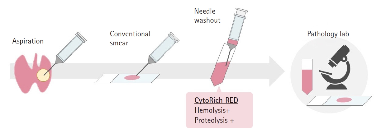
- In Japan, fine-needle aspiration (FNA) cytology is the most important diagnostic modality for triaging patients with thyroid nodules. A clinician (endocrinologist, endocrine surgeon, or head and neck surgeon) generally performs FNA cytology at the outpatient clinic, and ultrasound (US)-guided FNA is widespread because US is extremely common and most clinicians are familiar with it. Although almost all FNA thyroid samples are examined by certified cytopathologists and pathologists, some clinicians assess cytological specimens themselves. In Japan, there are two clinical guidelines regarding the management of thyroid nodules. One is the General Rules for the Description of Thyroid Cancer (GRDTC) published by the Japanese Society of Thyroid Surgery (JSTS) in 2005, and the other is the national reporting system for thyroid FNA cytology published by the Japan Thyroid Association in 2013 (Japanese system). Although the Bethesda System for Reporting Thyroid Cytopathology (Bethesda system) is rarely used in Japan, both the GRDTC and Japanese system tried to incorporate the Bethesda system so that the cytological diagnoses would be compatible with each other. The essential point of the Japanese system is stratification of follicular neoplasm (FN) into three subgroups based on cytological features in order to reduce unnecessary diagnostic thyroidectomy, and this system has been successful in stratifying the risk of malignancy in FN patients at several high-volume thyroid surgery centers. In Japan, the measurement of thyroglobulin and/or calcitonin in FNA needle washings is often used as an adjunct for diagnosis of possible cervical lymph node metastasis when FNA cytology is performed.
-
Citations
Citations to this article as recorded by- Impact of Molecular Testing on Surgical Decision-Making in Indeterminate Thyroid Nodules: A Global Meta-Analysis Across Test Generations
Truong Phan-Xuan Nguyen, Andrey Bychkov, Chan Kwon Jung, Kennichi Kakudo, Chanchal Rana
Endocrine Pathology.2026;[Epub] CrossRef - Thyroid Nodules with a Cytological Diagnosis of Follicular Neoplasm (Bethesda IV): Exploring Approaches to Risk Stratification
Boris M. Shifman, Nadezhda M. Platonova, Fatima M. Abdulkhabirova, Ekaterina V. Bondarenko, Liliya S. Urusova, Tatyana V. Soldatova, Alina A. Matrosova, Marina V. Utkina, Sergey V. Popov, Ekaterina A. Troshina
Acta Cytologica.2026; : 1. CrossRef - High Rates of Unnecessary Surgery for Indeterminate Thyroid Nodules in the Absence of Molecular Test and the Cost-Effectiveness of Utilizing Molecular Test in an Asian Population: A Decision Analysis
Man Him Matrix Fung, Ching Tang, Gin Wai Kwok, Tin Ho Chan, Yan Luk, David Tak Wai Lui, Carlos King Ho Wong, Brian Hung Hin Lang
Thyroid®.2025; 35(2): 166. CrossRef - Recent topics on thyroid cytopathology: reporting systems and ancillary studies
Mitsuyoshi Hirokawa, Ayana Suzuki
Journal of Pathology and Translational Medicine.2025; 59(4): 214. CrossRef - Risk Stratification of Thyroid Nodules Diagnosed as Follicular Neoplasm on Core Needle Biopsy
Byeong-Joo Noh, Won Jun Kim, Jin Yub Kim, Ha Young Kim, Jong Cheol Lee, Myoung Sook Shim, Yong Jin Song, Kwang Hyun Yoon, In-Hye Jung, Hyo Sang Lee, Wooyul Paik, Dong Gyu Na
Endocrinology and Metabolism.2025; 40(4): 610. CrossRef - Fine needle aspiration cytology diagnoses of follicular thyroid carcinoma: results from a multicenter study in Asia
Hee Young Na, Miyoko Higuchi, Shinya Satoh, Kaori Kameyama, Chan Kwon Jung, Su-Jin Shin, Shipra Agarwal, Jen-Fan Hang, Yun Zhu, Zhiyan Liu, Andrey Bychkov, Kennichi Kakudo, So Yeon Park
Journal of Pathology and Translational Medicine.2024; 58(6): 331. CrossRef - Thyroid FNA cytology: The Eastern versus Western perspectives
Mitsuyoshi Hirokawa, Manon Auger, Chan Kwon Jung, Fabiano Mesquita Callegari
Cancer Cytopathology.2023; 131(7): 415. CrossRef - The Asian Thyroid Working Group, from 2017 to 2023
Kennichi Kakudo, Chan Kwon Jung, Zhiyan Liu, Mitsuyoshi Hirokawa, Andrey Bychkov, Huy Gia Vuong, Somboon Keelawat, Radhika Srinivasan, Jen-Fan Hang, Chiung-Ru Lai
Journal of Pathology and Translational Medicine.2023; 57(6): 289. CrossRef - Criteria for follow‐up of thyroid nodules diagnosed as follicular neoplasm without molecular testing – The experience of a high‐volume thyroid centre in Japan
Mitsuyoshi Hirokawa, Ayana Suzuki, Makoto Kawakami, Takumi Kudo, Akira Miyauchi
Diagnostic Cytopathology.2022; 50(5): 223. CrossRef - The Significance of RAS-Like Mutations and MicroRNA Profiling in Predicting Malignancy in Thyroid Biopsy Specimens
Nicole A. Cipriani, Daniel N. Johnson, David H. Sarne, Peter Angelos, Ward Reeves, Tatjana Antic
Endocrine Pathology.2022; 33(4): 446. CrossRef - Molecular Testing for Thyroid Nodules: The Experience at McGill University Teaching Hospitals in Canada
Mohannad Rajab, Richard J. Payne, Véronique-Isabelle Forest, Marc Pusztaszeri
Cancers.2022; 14(17): 4140. CrossRef - Impact of Molecular Testing on the Management of Indeterminate Thyroid Nodules Among Western and Asian Countries: a Systematic Review and Meta-analysis
Hanh Thi Tuyet Ngo, Truong Phan Xuan Nguyen, Trang Huyen Vu, Chan Kwon Jung, Lewis Hassell, Kennichi Kakudo, Huy Gia Vuong
Endocrine Pathology.2021; 32(2): 269. CrossRef - The Incidence of Noninvasive Follicular Thyroid Neoplasm with Papillary-Like Nuclear Features: A Meta-Analysis Assessing Worldwide Impact of the Reclassification
Chanchal Rana, Huy Gia Vuong, Thu Quynh Nguyen, Hoang Cong Nguyen, Chan Kwon Jung, Kennichi Kakudo, Andrey Bychkov
Thyroid.2021;[Epub] CrossRef - Ultrasound-guided Fine Needle Aspiration Cytological Examination of Thyroid Nodules: A Practical Guideline (2019 edition)
ADVANCED ULTRASOUND IN DIAGNOSIS AND THERAPY.2021; 5(2): 134. CrossRef - Differences in surgical resection rate and risk of malignancy in thyroid cytopathology practice between Western and Asian countries: A systematic review and meta‐analysis
Huy Gia Vuong, Hanh Thi Tuyet Ngo, Andrey Bychkov, Chan Kwon Jung, Trang Huyen Vu, Kim Bach Lu, Kennichi Kakudo, Tetsuo Kondo
Cancer Cytopathology.2020; 128(4): 238. CrossRef - Exploring the Inter-observer Agreement Among the Members of the Italian Consensus for the Classification and Reporting of Thyroid Cytology
Anna Crescenzi, Pierpaolo Trimboli, Fulvio Basolo, Andrea Frasoldati, Fabio Orlandi, Lucio Palombini, Enrico Papini, Alfredo Pontecorvi, Paolo Vitti, Michele Zini, Francesco Nardi, Guido Fadda
Endocrine Pathology.2020; 31(3): 301. CrossRef - The Current Histologic Classification of Thyroid Cancer
Sylvia L. Asa
Endocrinology and Metabolism Clinics of North America.2019; 48(1): 1. CrossRef - Clinical Impact of Non-Invasive Follicular Thyroid Neoplasm with Papillary-Like Nuclear Features on the Risk of Malignancy in the Bethesda System for Reporting Thyroid Cytopathology: A Meta-Analysis of 14,153 Resected Thyroid Nodules
Huy Gia Vuong, Thao T.K. Tran, Andre y. Bychkov, Chan Kwon Jung, Tadao Nakazawa, Kennichi Kakudo, R yohei Katoh, Tetsuo Kondo
Endocrine Practice.2019; 25(5): 491. CrossRef - A Multi-institutional Study of Prevalence and Clinicopathologic Features of Non-invasive Follicular Thyroid Neoplasm with Papillary-like Nuclear Features (NIFTP) in Korea
Ja Yeong Seo, Ji Hyun Park, Ju Yeon Pyo, Yoon Jin Cha, Chan Kwon Jung, Dong Eun Song, Jeong Ja Kwak, So Yeon Park, Hee Young Na, Jang-Hee Kim, Jae Yeon Seok, Hee Sung Kim, Soon Won Hong
Journal of Pathology and Translational Medicine.2019; 53(6): 378. CrossRef - Noninvasive follicular thyroid neoplasm with papillary‐like nuclear features (NIFTP) in thyroid tumor classification
Kennichi Kakudo, Adel K. El‐Naggar, Steven P. Hodak, Elham Khanafshar, Yuri E Nikiforov, Vania Nosé, Lester D. R. Thompson
Pathology International.2018; 68(6): 327. CrossRef - Diagnostic value of HBME‐1, CK19, Galectin 3, and CD56 in the subtypes of follicular variant of papillary thyroid carcinoma
Haeyon Cho, Ji‐Ye Kim, Young Lyun Oh
Pathology International.2018; 68(11): 605. CrossRef - The Usefulness of Immunocytochemistry of CD56 in Determining Malignancy from Indeterminate Thyroid Fine-Needle Aspiration Cytology
Hyunseo Cha, Ju Yeon Pyo, Soon Won Hong
Journal of Pathology and Translational Medicine.2018; 52(6): 404. CrossRef - Impact of the modification of the diagnostic criteria in the 2017 Bethesda System for Reporting Thyroid Cytopathology: a report of a single institution in Japan
Miyoko Higuchi, Mitsuyoshi Hirokawa, Risa Kanematsu, Aki Tanaka, Ayana Suzuki, Naoki Yamao, Toshitetsu Hayashi, Seiji Kuma, Akira Miyauchi
Endocrine Journal.2018; 65(12): 1193. CrossRef - The Use of Fine-Needle Aspiration (FNA) Cytology in Patients with Thyroid Nodules in Asia: A Brief Overview of Studies from the Working Group of Asian Thyroid FNA Cytology
Chan Kwon Jung, SoonWon Hong, Andrey Bychkov, Kennichi Kakudo
Journal of Pathology and Translational Medicine.2017; 51(6): 571. CrossRef
- Impact of Molecular Testing on Surgical Decision-Making in Indeterminate Thyroid Nodules: A Global Meta-Analysis Across Test Generations
- Thyroid Fine-Needle Aspiration Cytology Practice in Korea
- Yoon Jin Cha, Ju Yeon Pyo, SoonWon Hong, Jae Yeon Seok, Kyung-Ju Kim, Jee-Young Han, Jeong Mo Bae, Hyeong Ju Kwon, Yeejeong Kim, Kyueng-Whan Min, Soonae Oak, Sunhee Chang
- J Pathol Transl Med. 2017;51(6):521-527. Published online October 11, 2017
- DOI: https://doi.org/10.4132/jptm.2017.09.26
- 11,565 View
- 248 Download
- 22 Web of Science
- 11 Crossref
-
Abstract
PDF
- We reviewed the current status of thyroid fine-needle aspiration cytology (FNAC) in Korea. Thyroid aspiration biopsy was first introduced in Korea in 1977. Currently, radiologists aspirate the thyroid nodule under the guidance of ultrasonography, and cytologic interpretation is only legally approved when a cytopathologist makes the diagnosis. In 2008, eight thyroid-related societies came together to form the Korean Thyroid Association. The Korean Society for Cytopathology and the endocrine pathology study group of the Korean Society for Pathologists have been updating the cytologic diagnostic guidelines. The Bethesda System for Reporting Thyroid Cytopathology was first introduced in 2009, and has been used by up to 94% of institutions by 2016. The average diagnosis rates are as follows for each category: I (12.4%), II (57.9%), III (10.4%), IV (2.9%), V (3.7%), and VI (12.7%). The malignancy rates in surgical cases are as follows for each category: I (28.7%), II (27.8%), III (50.6%), IV (52.3%), V (90.7%), and VI (100.0%). Liquid-based cytology has been used since 2010, and it was utilized by 68% of institutions in 2016. The categorization of thyroid lesions into “atypia of undetermined significance” or “follicular lesion of undetermined significance” is necessary to draw consensus in our society. Immunocytochemistry for galectin-3 and BRAF is used. Additionally, a molecular test for BRAF in thyroid FNACs is actively used. Core biopsies were performed in only 44% of institutions. Even the institutions that perform core biopsies only perform them for less than 3% of all FNACs. However, only 5% of institutions performed core biopsies up to three times more than FNAC.
-
Citations
Citations to this article as recorded by- Advantages of thyroid core needle biopsy: an emerging selective first-line biopsy modality
Jae Ho Shin, Yeseul Kim, Min Kyoung Lee, Jung Hwan Baek, So Lyung Jung
Ultrasonography.2026; 45(3): 205. CrossRef - State of the art of thyroid pathology: FNA diagnostic accuracy in an intermediate center in Ibagué
Daniel Javier Velez Bohorquez, Nohora Bibiana Varon Arce, Sandra Milena Tellez Olaya, Sebastian Camilo Mora Garcia, Anggi Margarita Velez Bohorquez, Mabel Elena Bohorquez Lozano
Universitas Médica.2023;[Epub] CrossRef - The Asian Thyroid Working Group, from 2017 to 2023
Kennichi Kakudo, Chan Kwon Jung, Zhiyan Liu, Mitsuyoshi Hirokawa, Andrey Bychkov, Huy Gia Vuong, Somboon Keelawat, Radhika Srinivasan, Jen-Fan Hang, Chiung-Ru Lai
Journal of Pathology and Translational Medicine.2023; 57(6): 289. CrossRef - Diagnostic value of thyroid imaging reporting and data system combined with BRAFV600E mutation analysis in Bethesda categories III–V thyroid nodules
Liuxi Wu, Hua Shu, Wenqin Chen, Yingqian Gao, Ya Yuan, Xiao Li, Wenjuan Lu, Xinhua Ye, Hongyan Deng
Scientific Reports.2022;[Epub] CrossRef - Contribution of cytologic examination to diagnosis of poorly differentiated thyroid carcinoma
Na Rae Kim, Jae Yeon Seok, Yoo Seung Chung, Joon Hyop Lee, Dong Hae Chung
Journal of Pathology and Translational Medicine.2020; 54(2): 171. CrossRef - Systematic thyroid screening in myotonic dystrophy: link between thyroid volume and insulin resistance
Adrien Ben Hamou, Stéphanie Espiard, Christine Do Cao, Miriam Ladsous, Camille Loyer, Alexandre Moerman, Samuel Boury, Maéva Kyheng, Claire-Marie Dhaenens, Vincent Tiffreau, Pascal Pigny, Gilles Lebuffe, Robert Caiazzo, Sébastien Aubert, Marie Christine V
Orphanet Journal of Rare Diseases.2019;[Epub] CrossRef - The History of Korean Thyroid Pathology
Soon Won Hong, Chan Kwon Jung
International Journal of Thyroidology.2018; 11(1): 15. CrossRef - BRAFV600E Mutation is a Strong Preoperative Indicator for Predicting Malignancy in Thyroid Nodule Patients with Atypia of Undetermined Significance Identified by Fine Needle Aspiration
Hye Rang Choi, Bo-Yoon Choi, Jae Hoon Cho, Young Chang Lim
Korean Journal of Otorhinolaryngology-Head and Neck Surgery.2018; 61(11): 600. CrossRef - The Usefulness of Immunocytochemistry of CD56 in Determining Malignancy from Indeterminate Thyroid Fine-Needle Aspiration Cytology
Hyunseo Cha, Ju Yeon Pyo, Soon Won Hong
Journal of Pathology and Translational Medicine.2018; 52(6): 404. CrossRef - Thyroid FNA cytology in Asian practice—Active surveillance for indeterminate thyroid nodules reduces overtreatment of thyroid carcinomas
K. Kakudo, M. Higuchi, M. Hirokawa, S. Satoh, C. K. Jung, A. Bychkov
Cytopathology.2017; 28(6): 455. CrossRef - The Use of Fine-Needle Aspiration (FNA) Cytology in Patients with Thyroid Nodules in Asia: A Brief Overview of Studies from the Working Group of Asian Thyroid FNA Cytology
Chan Kwon Jung, SoonWon Hong, Andrey Bychkov, Kennichi Kakudo
Journal of Pathology and Translational Medicine.2017; 51(6): 571. CrossRef
- Advantages of thyroid core needle biopsy: an emerging selective first-line biopsy modality
- Thyroid Cytology in India: Contemporary Review and Meta-analysis
- Shipra Agarwal, Deepali Jain
- J Pathol Transl Med. 2017;51(6):533-547. Published online October 5, 2017
- DOI: https://doi.org/10.4132/jptm.2017.08.04
- 15,540 View
- 260 Download
- 19 Web of Science
- 22 Crossref
-
Abstract
PDF
- Fine-needle aspiration cytology (FNAC) is a screening test for triaging thyroid nodules, aiding in subsequent clinical management. However, the advantages have been overshadowed by the multiplicity of reporting systems and a wide range of nomenclature used. The Bethesda System for Reporting Thyroid Cytopathology (TBSRTC) was formulated in 2007, to give the world a uniform thyroid cytology reporting system, facilitating easy interpretation by the clinicians. Here, we review the status of thyroid FNAC in India in terms of various reporting systems used including a meta-analysis of the previously published data. An extensive literature search was performed using internet search engines. The reports with detailed classification system used in thyroid cytology were included. The meta-analysis of published data was compared with the implied risk of malignancy by TBSRTC. More than 50 studies were retrieved and evaluated. TBSRTC is currently the most widely used reporting system with different studies showing good efficacy and interobserver concordance. Ancillary techniques have, as of now, limited applicability and acceptability in thyroid cytology in India. Twenty-eight published articles met the criteria for inclusion in the meta-analysis. When compared with TBSRTC recommendations, the meta-analysis showed a higher risk of malignancy for categories I and III. Thyroid FNAC is practiced all over India. TBSRTC has found widespread acceptance, with most institutions using this system for routine thyroid cytology reporting. However, reasons for a high malignancy risk for categories I and III need to be looked into. Various possible contributing factors are discussed in the review.
-
Citations
Citations to this article as recorded by- Guidelines for the safe performance, training, and adoption of ablation techniques for benign thyroid nodules: An Indian Association of endocrine surgeons consensus statement
Sanjay Kumar Yadav, Siddhartha Chakravarthy, Sanjeet Kumar, Abhishek Krishna, Anjali Mishra, Anukriti Sood, Aromal Chekavar Suresh, Balaji srinivasan, Devashish Mishra, Manjunath Goroshi, Nishtha Yadav, Ritesh Agrawal, Sabaretnam M, Sarrah Idrees, Shikhil
Indian Journal of Surgery.2026;[Epub] CrossRef - Reassessing Bethesda III and IV Thyroid Nodules: Insights from an Institution-based Malignancy Risk Analysis in a Cancer Center Setting
Raveena R. Nair, Sandeep Vijay, Anoop Attakkil, Sairu Philip, Karthickeyan Duraisamy, Kalpita Shringarpure, Vanithapriya Deenathayalan, Mohandoss Murugesan
Journal of Head & Neck Physicians and Surgeons.2026; 14(1): 45. CrossRef - Spinal metastases in primary thyroid malignancies: Single center experience of 44 cases
Basir Ahmed, Edmond Jonathan, M. J. Paul, Krishna Prabhu
World Journal of Surgery.2025; 49(2): 409. CrossRef - Cytopathology in India: Past, Present, and Future
Jitendra Singh Nigam, Jyotsna Naresh Bharti, Ashutosh Rath, Immanuel Pradeep, Biswajit Dey, Ravi Mehrotra
Diagnostic Cytopathology.2025; 53(9): 466. CrossRef - Evaluation of concordance between the Bethesda System for Reporting Thyroid Cytopathology 2023 (TBSRTC) and ACR-TIRADS at a tertiary care center in Gujarat
Sushrita Biswas, Ina Shah, Hansa Goswami, Avik Chaudhuri
Indian Journal of Pathology and Microbiology.2025; 68(2): 338. CrossRef - Thermal imaging based pre-diagnostics tool for Graves’ disease
Vaishali Sharma, Vandana K Dhingra, Snehlata Shakya, Ashok Kumar, Mayank Goswami
Measurement Science and Technology.2024; 35(3): 035702. CrossRef - High Malignancy Risk and Its Predictors in South Indian Patients With Bethesda II Thyroid Nodules
Sunanda Tirupati, Pradeep Puthenveetil, Shilpa Lakkundi, Anudeep Gaddam, Vijaya Sarathi
Cureus.2024;[Epub] CrossRef - Nuclear features in thyroid cytology: features helpful for a morphological diagnosis in routine practice
Priya Bhagwat, Sabine Pomplun
Diagnostic Histopathology.2024; 30(6): 312. CrossRef - DIAGNOSTIC EFFICACY OF FNAC IN THYROID LESIONS, CLASSIFIED ACCORDING TO BETHESDA SYSTEM WITH CYTOHISTOLOGICAL CORRELATION
KIRAN KUMARI MEENA, SANDHYA BORDIA, POOJA KANWAT, SEEMA MEENA, PRAGYA JAKHAR
Asian Journal of Pharmaceutical and Clinical Research.2024; : 125. CrossRef - Evaluation of Thyroid Lesions by the Bethesda System for Reporting Thyroid Cytopathology
Syed Asif Hashmi, Monika Aggrawal, Rahul Pandey, Deepika Gulati, Inam Danish Khan
Journal of Marine Medical Society.2023; 25(1): 73. CrossRef - Incidence and Malignancy Rates in Thyroid Nodules in North-East Indian Population by Bethesda System: A Single Institutional Experience of 3 Years
Suvamoy Chakraborty, Manu C. Balakrishnan, Vandana Raphael, Prachurya Tamuli, Anuradha Deka
South Asian Journal of Cancer.2023; 12(02): 166. CrossRef - Evaluation of Concordance of Ultrasound, Cytology, and Histopathology in Solitary Thyroid Nodules
Sunil Chumber, Surabhi Vyas, Kamal Kataria, Shipra Agarwal, Yashwant S Rathore, Gopal Puri, Sushma Yadav, Kanika Sharma, Amit Patidar
Indian Journal of Endocrine Surgery and Research.2023; 18(1): 17. CrossRef - Cytomorphological Spectrum of Head and Neck Lesions by Fine Needle Aspiration Cytology in a Tertiary Care Center
Amandeep Kaur, Sonali Poonia, Karandeep Singh, Dalbir Kaur, Mohit Madhukar, Ravish Godara
Journal of Pharmacy and Bioallied Sciences.2023; 15(Suppl 1): S315. CrossRef - The Asian Thyroid Working Group, from 2017 to 2023
Kennichi Kakudo, Chan Kwon Jung, Zhiyan Liu, Mitsuyoshi Hirokawa, Andrey Bychkov, Huy Gia Vuong, Somboon Keelawat, Radhika Srinivasan, Jen-Fan Hang, Chiung-Ru Lai
Journal of Pathology and Translational Medicine.2023; 57(6): 289. CrossRef - Cytomorphological Categorization of Thyroid Lesions according to The Bethesda System for Reporting Thyroid Cytology and Correlation with their Histological Outcome
Meenakshi Kamboj, Anurag Mehta, Sunil Pasricha, Gurudutt Gupta, Anila Sharma, Garima Durga
Journal of Cytology.2022; 39(1): 44. CrossRef - Is Surgery Necessary in Benign Thyroid Lesions?
Pushkar Chaudhary, Naseem Noorunnisa
Journal of Datta Meghe Institute of Medical Sciences University.2022; 17(3): 799. CrossRef - Effect of the Noninvasive Follicular Thyroid Neoplasm With Papillary-Like Nuclear Features (NIFTP) Nomenclature Revision on Indian Thyroid Fine-Needle Aspiration Practice
Chanchal Rana, Pooja Ramakant, Divya Goel, Akanksha Singh, KulRanjan Singh, Suresh Babu, Anand Mishra
American Journal of Clinical Pathology.2021; 156(2): 320. CrossRef - The combination of ACR‐Thyroid Imaging Reporting and Data system and The Bethesda System for Reporting Thyroid Cytopathology in the evaluation of thyroid nodules—An institutional experience
Shanmugasundaram Sakthisankari, Sreenivasan Vidhyalakshmi, Sivanandam Shanthakumari, Balalakshmoji Devanand, Udayasankar Nagul
Cytopathology.2021; 32(4): 472. CrossRef - Differentiated Thyroid Cancer
Anita M. Borges
Journal of Head & Neck Physicians and Surgeons.2021; 9(2): 69. CrossRef - Risk of malignancy in Thyroid “Atypia of undetermined significance/Follicular lesion of undetermined significance” and its subcategories – A 5-year experience
Abha Thakur, Haimanti Sarin, Dilpreet Kaur, Deepak Sarin
Indian Journal of Pathology and Microbiology.2019; 62(4): 544. CrossRef - Thyroid FNA cytology in Asian practice—Active surveillance for indeterminate thyroid nodules reduces overtreatment of thyroid carcinomas
K. Kakudo, M. Higuchi, M. Hirokawa, S. Satoh, C. K. Jung, A. Bychkov
Cytopathology.2017; 28(6): 455. CrossRef - The Use of Fine-Needle Aspiration (FNA) Cytology in Patients with Thyroid Nodules in Asia: A Brief Overview of Studies from the Working Group of Asian Thyroid FNA Cytology
Chan Kwon Jung, SoonWon Hong, Andrey Bychkov, Kennichi Kakudo
Journal of Pathology and Translational Medicine.2017; 51(6): 571. CrossRef
- Guidelines for the safe performance, training, and adoption of ablation techniques for benign thyroid nodules: An Indian Association of endocrine surgeons consensus statement
- Thyroid Fine-Needle Aspiration Practice in the Philippines
- Agustina D. Abelardo
- J Pathol Transl Med. 2017;51(6):555-559. Published online October 5, 2017
- DOI: https://doi.org/10.4132/jptm.2017.07.14
- 11,315 View
- 146 Download
- 7 Web of Science
- 7 Crossref
-
Abstract
PDF
- Fine-needle aspiration (FNA) is a well accepted initial approach in the management of thyroid lesions. It has come a long way since its introduction for nearly a century ago. In the Philippines, FNA of the thyroid was first introduced 30 years ago and has been utilized until now as a mainstay in the diagnosis of thyroid malignancy. The procedure is performed by pathologists, endocrinologists, surgeons, and radiologists. Most pathologists report the cytodiagnosis using a combination of the aspiration biopsy cytology method that closely resembles the histopathologic diagnosis of thyroid disorders and the six-tier nomenclature of The Bethesda System for Reporting Thyroid Cytopathology. Local endocrinologists and surgeons follow the guidelines of the 2015 American Thyroid Association in the management of thyroid disorders. There is still a paucity of local research studies but available data deal with cytohistologic correlations, sensitivity, specificity, and accuracy rates as well as usefulness of ultrasound-guided FNA. Cytohistologic correlations have a wide range of sensitivity from 30.7% to 73% and specificity from 83% to 100%. The low sensitivity can be attributed to poor tissue sampling since a majority of the thyroid FNA is done by palpation only. The reliability can be improved if FNA is guided by ultrasound as attested in both international and local studies. Overall, FNA of the thyroid has enabled the diagnosis of thyroid disorders with an accuracy of 72.8% to 87.2% and it correlates well with histopathology.
-
Citations
Citations to this article as recorded by- Combining radiomics and molecular biomarkers: a novel economic tool to improve diagnostic ability in papillary thyroid cancer
Qingxuan Wang, Linghui Dai, Sisi Lin, Shuwei Zhang, Jing Wen, Endong Chen, Quan Li, Jie You, Jinmiao Qu, Chunjue Ni, Yefeng Cai
Frontiers in Endocrinology.2024;[Epub] CrossRef - Agreement between Sonographic Features and Fine Needle Aspiration Cytology in the Diagnosis of Thyroid Nodules in a Tertiary Hospital
Danette Pabalan, Ricardo Victorio Quimbo
PJP.2024; 9(1): 37. CrossRef - The Asian Thyroid Working Group, from 2017 to 2023
Kennichi Kakudo, Chan Kwon Jung, Zhiyan Liu, Mitsuyoshi Hirokawa, Andrey Bychkov, Huy Gia Vuong, Somboon Keelawat, Radhika Srinivasan, Jen-Fan Hang, Chiung-Ru Lai
Journal of Pathology and Translational Medicine.2023; 57(6): 289. CrossRef - Application of the Bethesda System for Reporting Thyroid Cytopathology in the Pediatric Population
Huy Gia Vuong, Ayana Suzuki, Hee Young Na, Pham Van Tuyen, Doan Minh Khuy, Hiep Canh Nguyen, Tikamporn Jitpasutham, Agustina Abelardo, Takashi Amano, So Yeon Park, Chan Kwon Jung, Mitsuyoshi Hirokawa, Ryohei Katoh, Kennichi Kakudo, Andrey Bychkov
American Journal of Clinical Pathology.2021; 155(5): 680. CrossRef - Improvement in Specimen Adequacy with Ultrasound-guided Fine-Needle Aspiration Biopsy (FNAB) of Thyroid Nodules Using Rapid On-site Evaluation (ROSE): A Cross-sectional Study
Pia Pamela P Dungca, Francis Bryant G Chua, Elizabeth Ann S Alcazaren
Philippine Journal of Internal Medicine.2021; 59(4): 296. CrossRef - Thyroid FNA cytology in Asian practice—Active surveillance for indeterminate thyroid nodules reduces overtreatment of thyroid carcinomas
K. Kakudo, M. Higuchi, M. Hirokawa, S. Satoh, C. K. Jung, A. Bychkov
Cytopathology.2017; 28(6): 455. CrossRef - The Use of Fine-Needle Aspiration (FNA) Cytology in Patients with Thyroid Nodules in Asia: A Brief Overview of Studies from the Working Group of Asian Thyroid FNA Cytology
Chan Kwon Jung, SoonWon Hong, Andrey Bychkov, Kennichi Kakudo
Journal of Pathology and Translational Medicine.2017; 51(6): 571. CrossRef
- Combining radiomics and molecular biomarkers: a novel economic tool to improve diagnostic ability in papillary thyroid cancer
- Diagnostic Accuracy of Endoscopic Ultrasound-Guided Fine Needle Aspiration Cytology of Pancreatic Lesions
- Hae Woon Baek, Min Jee Park, Ye-Young Rhee, Kyoung Bun Lee, Min A Kim, In Ae Park
- J Pathol Transl Med. 2015;49(1):52-60. Published online January 15, 2015
- DOI: https://doi.org/10.4132/jptm.2014.10.26
- 14,317 View
- 97 Download
- 35 Web of Science
- 34 Crossref
-
Abstract
PDF
- Background
Endoscopic ultrasound–guided fine needle aspiration cytology (EUS-FNAC) is currently the most commonly used procedure for obtaining cytologic specimens of the pancreas. It is accurate, minimally invasive, safe and cost-effective. However, there is discrepancy between cytological and surgical diagnoses. This study was aimed at evaluating the diagnostic accuracy of EUS-FNAC of the pancreas. Methods: We performed a retrospective review of 191 cases of pancreatic lesions initially diagnosed by EUS-FNAC with subsequent histological diagnosis between 2010 and 2012 in the Department of Pathology, Seoul National University Hospital. Cytologic and surgical diagnoses were categorized into five groups: negative, benign, atypical, malignant, and insufficient for diagnosis. Subsequently, 167 cases with satisfactory yield in both surgical and cytology specimens were statistically analyzed to determine correlations with diagnosis. Results: In comparison to surgical diagnoses, cytologic diagnoses were true-positive in 103 cases (61.7%), true-negative in 28 cases (16.8%), false-positive in 9 cases (5.4%), and false-negative in 27 cases (16.1%). The diagnostic accuracy was 78.4%, sensitivity was 79.2%, and specificity was 75.7%. The positive predictive value was 92.0%, and negative predictive value was 50.9%. Conclusions: EUS-FNAC has high accuracy, sensitivity, specificity and positive predictive value. Overcoming the limitations of EUS-FNAC will make it a useful and reliable diagnostic tool for accurate evaluation of pancreatic lesions. -
Citations
Citations to this article as recorded by- Diagnostic efficacy of endoscopic ultrasound-guided fine needle aspiration cytology in pancreatic lesions
Pranita Mohanty, Priyadarshini Dehuri, Jimmy Narayan
Scientific Reports.2026;[Epub] CrossRef - miR-6855-5p Enhances Radioresistance and Promotes Migration of Pancreatic Cancer by Inducing Epithelial-Mesenchymal Transition via Suppressing FOXA1: Potential of Plasma Exosomal miR-6855-5p as an Indicator of Radiosensitivity in Patients with Pancreatic
Hiroki Ueda, Hidenori Takahashi, Shogo Kobayashi, Masahiko Kubo, Kazuki Sasaki, Yoshifumi Iwagami, Daisaku Yamada, Yoshito Tomimaru, Tadafumi Asaoka, Takehiro Noda, Junzo Shimizu, Yuichiro Doki, Hidetoshi Eguchi
Annals of Surgical Oncology.2025; 32(2): 720. CrossRef - Combating false negatives in pancreatic cancer: A deep learning approach for aiding fine needle aspiration via accurate subregion identification
Ms Jasmine Chhikara, Nidhi Goel, Neeru Rathee
Engineering Applications of Artificial Intelligence.2025; 157: 111347. CrossRef - Grading pancreatic adenocarcinomas on fine needle aspiration cytology. The outstanding issues
Mrinmay Kumar Mallik, Laila Rafiq Qadan, Asit Kumar Mohanty, Ali Alali, Kusum Kapila
Cytopathology.2024; 35(2): 256. CrossRef - Reporting Pancreatic FNAC using the Papanicolaou System: Still a Diagnostic Challenge
Parul Verma, Saloni Goyal, Ruchita Tyagi, Mehar Ghuman, Ramit Mahajan, Arshneet Kaur Selhi, Harpreet Kaur, Pavneet Kaur Selhi
Journal of Cytology.2024; 41(2): 123. CrossRef - Preoperative treatment response prediction for pancreatic cancer by multiple microRNAs in plasma exosomes: Optimization using machine learning and network analysis
Hiroki Ueda, Hidenori Takahashi, Ryoto Sakaniwa, Tetsuhisa Kitamura, Shogo Kobayashi, Yoshito Tomimaru, Masahiko Kubo, Kazuki Sasaki, Yoshifumi Iwagami, Daisaku Yamada, Tadafumi Asaoka, Takehiro Noda, Junzo Shimizu, Yuichiro Doki, Hidetoshi Eguchi
Pancreatology.2024; 24(7): 1097. CrossRef - Comparison of Endoscopic Ultrasound-Guided Fine-Needle Aspiration with Fine-Needle Biopsy for Solid Gastrointestinal Lesions: A Randomized Crossover Single-Center study
Shivaraj Afzalpurkar, Vijay Kumar Rai, Nikhil Sonthalia, Gajanan Rodge, Awanesh Tewary, Mahesh Goenka
Journal of Digestive Endoscopy.2023; 14(01): 014. CrossRef - Diagnosing and monitoring pancreatic cancer through cell-free DNA methylation: progress and prospects
María Victoria García-Ortiz, Pablo Cano-Ramírez, Marta Toledano-Fonseca, Enrique Aranda, Antonio Rodríguez-Ariza
Biomarker Research.2023;[Epub] CrossRef - The impact of preoperative EUS-FNA for distal resectable pancreatic cancer: Is it really effective enough to take risks?
Jin-Seok Park, Jae Hoon Lee, Tae Jun Song, Joune Seup Lee, Seok Jung Jo, Dong Wook Oh, Ki Byung Song, Dae Wook Hwang, Do Hyun Park, Sang Soo Lee, Song Cheol Kim, Dong Wan Seo, Sung Koo Lee, Myung-Hwan Kim
Surgical Endoscopy.2022; 36(5): 3192. CrossRef - Magnetic Resonance Imaging Radiomics‐Based Nomogram From Primary Tumor for Pretreatment Prediction of Peripancreatic Lymph Node Metastasis in Pancreatic Ductal Adenocarcinoma: A Multicenter Study
Zhenshan Shi, Chengle Ma, Xinming Huang, Dairong Cao
Journal of Magnetic Resonance Imaging.2022; 55(3): 823. CrossRef - Role of Pathologist in the Era of Image-Guided and EUS-Guided Aspirations: A 10-Year Study at a Single Tertiary Care Oncology Institute in North India
Gurudutt Gupta, Anila Sharma, Meenakshi Kamboj, Anurag Sharma, Sunil Pasricha, Garima Durga, Anurag Mehta, Avinash Rao
Acta Cytologica.2022; 66(3): 187. CrossRef - Predicting Factors for Pancreatic Malignancy with Computed Tomography and Endoscopic Ultrasonography in Chronic Pancreatitis
Jian-Han Lai, Keng-Han Lee, Chen-Wang Chang, Ming-Jen Chen, Ching-Chung Lin
Diagnostics.2022; 12(4): 1004. CrossRef - Phenotypic profiling of pancreatic ductal adenocarcinoma plasma-derived small extracellular vesicles for cancer diagnosis and cancer stage prediction: a proof-of-concept study
Wei Zhang, Ling Wang, Dan Li, Douglas H. Campbell, Bradley J. Walsh, Nicolle H. Packer, Qing Dong, Erkang Wang, Yuling Wang
Analytical Methods.2022; 14(23): 2255. CrossRef - Cancer-derived small extracellular vesicles: emerging biomarkers and therapies for pancreatic ductal adenocarcinoma diagnosis/prognosis and treatment
Wei Zhang, Douglas H. Campbell, Bradley J. Walsh, Nicolle H. Packer, Dingbin Liu, Yuling Wang
Journal of Nanobiotechnology.2022;[Epub] CrossRef - Single Cell RNA Sequencing: A New Frontier in Pancreatic Ductal Adenocarcinoma
Maroun Bou Zerdan, Malek Shatila, Dhruv Sarwal, Youssef Bouferraa, Morgan Bou Zerdan, Sabine Allam, Merima Ramovic, Stephen Graziano
Cancers.2022; 14(19): 4589. CrossRef - Molecular Subtyping and Precision Medicine for Pancreatic Cancer
Fieke Froeling, Raffaella Casolino, Antonio Pea, Andrew Biankin, David Chang
Journal of Clinical Medicine.2021; 10(1): 149. CrossRef - Reshaping preoperative treatment of pancreatic cancer in the era of precision medicine
R. Casolino, C. Braconi, G. Malleo, S. Paiella, C. Bassi, M. Milella, S.B. Dreyer, F.E.M. Froeling, D.K. Chang, A.V. Biankin, T. Golan
Annals of Oncology.2021; 32(2): 183. CrossRef - Endoscopic Ultrasound-Guided Fine Needle Aspiration Cytology of Pancreatic Adenocarcinomas Revisited. A Detailed Cytological Analysis
Mrinmay Kumar Mallik, Kusum Kapila, Asit Kumar Mohanty, Shafi Ahmed Inamdar, Ali AlAli, Abdullah Al Naseer
Journal of Cytology.2021; 38(1): 31. CrossRef - Diagnostic value of various liquid biopsy methods for pancreatic cancer
Yuzhou Zhu, Hao Zhang, Nan Chen, Jianqi Hao, Hongyu Jin, Xuelei Ma
Medicine.2020; 99(3): e18581. CrossRef - Factors affecting cytological results of endoscopic ultrasound guided-fine needle aspiration during learning
Jian-Han Lai, Hsiang-Hung Lin, Ching-Chung Lin
Diagnostic Pathology.2020;[Epub] CrossRef - Comparison between Conventional Smear and Liquid-Based Preparation in Endoscopic Ultrasonography-Fine Needle Aspiration Cytology of Pancreatic Lesions
Soo Hee Ko, Jung-Soo Pyo, Byoung Kwan Son, Hyo Young Lee, Il Whan Oh, Kwang Hyun Chung
Diagnostics.2020; 10(5): 293. CrossRef - Evaluation of Pancreatic Lesions With Endoscopic Ultrasound and Fine Needle Aspiration
Yan Luk, Wong Hoi She, Felix Che Lok Chow, Ka Wing Ma, Simon Hing Yin Tsang, Wing Chiu Dai, Tan To Cheung, Chung Mau Lo
Surgical Innovation.2020; 27(5): 431. CrossRef - The applicability of Papanicolaou Society of Cytopathology system on reporting endoscopic ultrasound‐guided fine needle aspiration cytology specimens of pancreatic lesions in situations with limited availability of ancillary tests. Experience at a single
Mrinmay Kumar Mallik, Laila Rafiq Qadan, Abdullah Al Naseer, Ali AlAli, Taiba Al Ansari, Shafi Ahmed Inamdar Naquib, Dilip Kumar Das, Kusum Kapila
Cytopathology.2020; 31(6): 564. CrossRef - Circulating Cell-Free DNA-Based Liquid Biopsy Markers for the Non-Invasive Prognosis and Monitoring of Metastatic Pancreatic Cancer
Marta Toledano-Fonseca, M. Teresa Cano, Elizabeth Inga, Rosa Rodríguez-Alonso, M. Auxiliadora Gómez-España, Silvia Guil-Luna, Rafael Mena-Osuna, Juan R. de la Haba-Rodríguez, Antonio Rodríguez-Ariza, Enrique Aranda
Cancers.2020; 12(7): 1754. CrossRef - Risk of malignancy in the categories of the Papanicolaou Society of Cytopathology system for reporting pancreaticobiliary cytology
Raza S. Hoda, Elizabeth B. Finer, Ronald N. Arpin, Matthew Rosenbaum, Martha B. Pitman
Journal of the American Society of Cytopathology.2019; 8(3): 120. CrossRef - Endoscopic ultrasound guided fine‐needle aspiration vs core needle biopsy for solid pancreatic lesions: Comparison of diagnostic accuracy and procedural efficiency
Aslam Syed, Olivia Babich, Bharat Rao, Shailendra Singh, Neil Carleton, Abhishek Gulati, Archana Kulkarni, Mrinal Garg, Katie Farah, Gursimran Kochhar, Suzanne Morrissey, Marcia Mitre, Abhijit Kulkarni, Manish Dhawan, Jan F. Silverman, Majed Pharaon, Shya
Diagnostic Cytopathology.2019; 47(11): 1138. CrossRef - Liquid Biopsy as Surrogate for Tissue for Molecular Profiling in Pancreatic Cancer: A Meta-Analysis Towards Precision Medicine
Claudio Luchini, Nicola Veronese, Alessia Nottegar, Vera Cappelletti, Maria G. Daidone, Lee Smith, Christopher Parris, Lodewijk A. A. Brosens, Maria G. Caruso, Liang Cheng, Christopher L. Wolfgang, Laura D. Wood, Michele Milella, Roberto Salvia, Aldo Scar
Cancers.2019; 11(8): 1152. CrossRef - It is necessary to exam bottom and top slide smears of EUS-FNA for pancreatic cancer
Jong-chan Lee, Haeryoung Kim, Hyoung Woo Kim, Jongchan Lee, Kyu-hyun Paik, Jingu Kang, Jin-Hyeok Hwang, Jaihwan Kim
Hepatobiliary & Pancreatic Diseases International.2018; 17(6): 553. CrossRef - Preoperative EUS-guided FNA: effects on peritoneal recurrence and survival in patients with pancreatic cancer
Sun Hwa Kim, Young Sik Woo, Kwang Hyuck Lee, Jong Kyun Lee, Kyu Taek Lee, Joo Kyung Park, Soo Hoon Kang, Ji Won Kim, Jae Keun Park, Sung-Wook Park
Gastrointestinal Endoscopy.2018; 88(6): 926. CrossRef - Cytological characteristics of atypical cells in endoscopic ultrasound-guided fine-needle aspiration specimens obtained from the pancreas
Shikine ESAKA, Yoko MATSUDA, Yuri HAMASHIMA, Masayuki IMAIZUMI, Hiroya KOJIMA, Yuri KISO, Hiroto SHIRAHATA, Mayumi KINOSHITA, Akemi SUZUKI, Tomio ARAI
The Journal of the Japanese Society of Clinical Cytology.2018; 57(4): 199. CrossRef - Performance measures for endoscopic retrograde cholangiopancreatography and endoscopic ultrasound: A European Society of Gastrointestinal Endoscopy (ESGE) Quality Improvement Initiative
Dirk Domagk, Kofi W Oppong, Lars Aabakken, Laszlo Czakó, Tibor Gyökeres, Gianpiero Manes, Peter Meier, Jan-Werner Poley, Thierry Ponchon, Andrea Tringali, Cristina Bellisario, Silvia Minozzi, Carlo Senore, Cathy Bennett, Michael Bretthauer, Cesare Hassan,
United European Gastroenterology Journal.2018; 6(10): 1448. CrossRef - Performance measures for ERCP and endoscopic ultrasound: a European Society of Gastrointestinal Endoscopy (ESGE) Quality Improvement Initiative
Dirk Domagk, Kofi W. Oppong, Lars Aabakken, Laszlo Czakó, Tibor Gyökeres, Gianpiero Manes, Peter Meier, Jan-Werner Poley, Thierry Ponchon, Andrea Tringali, Cristina Bellisario, Silvia Minozzi, Carlo Senore, Cathy Bennett, Michael Bretthauer, Cesare Hassan
Endoscopy.2018; 50(11): 1116. CrossRef - Imaging modalities for characterising focal pancreatic lesions
Lawrence MJ Best, Vishal Rawji, Stephen P Pereira, Brian R Davidson, Kurinchi Selvan Gurusamy
Cochrane Database of Systematic Reviews.2017;[Epub] CrossRef - Percutaneous ultrasound‐guided core needle biopsy of solid pancreatic masses: Results in 250 patients
Guven Kahriman, Nevzat Ozcan, Serap Dogan, Soner Ozmen, Kemal Deniz
Journal of Clinical Ultrasound.2016; 44(8): 470. CrossRef
- Diagnostic efficacy of endoscopic ultrasound-guided fine needle aspiration cytology in pancreatic lesions
- A Rare Case of Mesothelioma Showing Micropapillary and Small Cell Differentiation with Aggressive Behavior
- Yoon Jin Cha, Binnari Kim, Joungho Han, Chin A Yi, Jae Ill Zo
- Korean J Pathol. 2014;48(6):466-468. Published online December 31, 2014
- DOI: https://doi.org/10.4132/KoreanJPathol.2014.48.6.466
- 11,018 View
- 40 Download
- 2 Crossref
-
PDF
-
Citations
Citations to this article as recorded by- Small cell mesothelioma: A rare entity and diagnostic pitfall mimicking small cell lung carcinoma on fine‐needle aspiration
Yanhong Zhang, Alaa Afify, Regina F. Gandour‐Edwards, John W. Bishop, Eric C. Huang
Diagnostic Cytopathology.2016; 44(6): 526. CrossRef - A Biphasic Pleural Tumor with Features of an Epithelioid and Small Cell Mesothelioma: Morphologic and Molecular Findings
Sarah Hackman, Richard D. Hammer, Lester Layfield
Case Reports in Pathology.2016; 2016: 1. CrossRef
- Small cell mesothelioma: A rare entity and diagnostic pitfall mimicking small cell lung carcinoma on fine‐needle aspiration
- Supratentorial Hemangioblastoma with Unusual Features
- Yooju Shin, Seokhwi Kim, Hyun-Woo Lee, Heejin Bang, Yeon-Lim Suh
- Korean J Pathol. 2014;48(6):462-465. Published online December 31, 2014
- DOI: https://doi.org/10.4132/KoreanJPathol.2014.48.6.462
- 13,626 View
- 81 Download
- 6 Crossref
-
PDF
-
Citations
Citations to this article as recorded by- Supratentorial Hemangioblastoma in Adults: A Systematic Review and Comparison of Infratentorial and Spinal Cord Locations
Dragan Jankovic, Kyna Vuong, Bruno Splavski, Kresimir Rotim, Kenan I. Arnautovic
World Neurosurgery.2023; 173: 48. CrossRef - Supratentorial hemangioblastoma: correlation between phenotype, gender and vascular territory affected
Yosef Laviv, David Saraf, Liat Oxman, Ido Ben Zvi
Neurosurgical Review.2023;[Epub] CrossRef - Neuropathologic features of central nervous system hemangioblastoma
Rebecca A. Yoda, Patrick J. Cimino
Journal of Pathology and Translational Medicine.2022; 56(3): 115. CrossRef - The loss of succinate dehydrogenase B expression is frequently identified in hemangioblastoma of the central nervous system
Tae Hoon Roh, Hyunee Yim, Jin Roh, Kyi Beom Lee, So Hyun Park, Seon-Yong Jeong, Se-Hyuk Kim, Jang-Hee Kim
Scientific Reports.2019;[Epub] CrossRef - Supratentorial hemangioblastomas in von Hippel–Lindau wild-type patients – case series and literature review
Luís Rocha, Carolina Noronha, Ricardo Taipa, Joaquim Reis, Mário Gomes, Ernesto Carvalho
International Journal of Neuroscience.2018; 128(3): 295. CrossRef - MR Imaging Findings of Supratentorial Meningeal Hemangioblastoma: A Case Report
Gi Hong Kim, Ho Kyu Lee, Myeong Ju Koh, Young Hee Maeng
Journal of the Korean Society of Radiology.2016; 75(1): 26. CrossRef
- Supratentorial Hemangioblastoma in Adults: A Systematic Review and Comparison of Infratentorial and Spinal Cord Locations
- Peritoneal Carcinosarcoma and Ovarian Papillary Serous Carcinoma Are the Same Origin: Analysis of TP53 Mutation and Microsatellite Suggests a Monoclonal Origin
- Chang Gok Woo, Dae Shik Suh, Joo Young Kim, Chang Ohk Sung, Jene Choi, Kyu-Rae Kim
- Korean J Pathol. 2014;48(6):449-453. Published online December 31, 2014
- DOI: https://doi.org/10.4132/KoreanJPathol.2014.48.6.449
- 14,541 View
- 56 Download
- 3 Crossref
-
PDF
-
Citations
Citations to this article as recorded by- A successfully treated primary peritoneal carcinosarcoma and serous carcinoma of stage IIIC rescued from hypovolemic shock due to tumor rupture
Che-Cheng Huang, Horng-Jyh Tsai, Shih Hsuan Huang, Victor C. Kok
Taiwanese Journal of Obstetrics and Gynecology.2019; 58(2): 296. CrossRef - Ovarian Carcinosarcoma and Concurrent Serous Tubal Intraepithelial Carcinoma With Next-Generation Sequencing Suggesting an Origin From the Fallopian Tube
Sharlene Helene C. See, Amir Behdad, Kruti P. Maniar, Luis Z. Blanco
International Journal of Surgical Pathology.2019; 27(5): 574. CrossRef - Progression inference for somatic mutations in cancer
Leif E. Peterson, Tatiana Kovyrshina
Heliyon.2017; 3(4): e00277. CrossRef
- A successfully treated primary peritoneal carcinosarcoma and serous carcinoma of stage IIIC rescued from hypovolemic shock due to tumor rupture
- A Case of Mixed Adenoneuroendocrine Carcinoma of the Common Bile Duct: Initially Diagnosed as Cholangiocarcinoma
- Soon Wook Lee, In Seok Lee, Yu Kyung Cho, Jae Myung Park, Sang Woo Kim, Myung-Gyu Choi, Kyu Yong Choi, Myung Ah Lee, Tae Ho Hong, Young Kyoung You, Eun-Sun Jung
- Korean J Pathol. 2014;48(6):445-448. Published online December 31, 2014
- DOI: https://doi.org/10.4132/KoreanJPathol.2014.48.6.445
- 11,079 View
- 44 Download
- 12 Crossref
-
PDF
-
Citations
Citations to this article as recorded by- Long-term survival after chemotherapy combined immunotherapy for recurrent mixed neuroendocrine–non-neuroendocrine neoplasms of the common bile duct
Jia Li, Chunyan Yuan, Yulin Pan, Yuanyuan Yang, Nannan Lai, Xia Sheng
Clinical Journal of Gastroenterology.2025; 18(4): 653. CrossRef - Neuroendocrine carcinoma of the common hepatic duct coexisting with distal cholangiocarcinoma: A case report and review of literature
Fei Chen, Wei-Wei Li, Juan-Fen Mo, Min-Jie Chen, Su-Hang Wang, Shu-Ying Yang, Zheng-Wei Song
World Journal of Gastrointestinal Surgery.2024; 16(5): 1449. CrossRef - Comparison of Metastatic Patterns Among Neuroendocrine Tumors, Neuroendocrine Carcinomas, and Nonneuroendocrine Carcinomas of Various Primary Organs
Hyung Kyu Park, Ghee Young Kwon
Journal of Korean Medical Science.2023;[Epub] CrossRef - Mixed adenoneuroendocrine carcinoma of the distal bile duct: a case report
Takashi Maeda, Kyohei Yugawa, Nao Kinjo, Hiroto Kayashima, Daisuke Imai, Koto Kawata, Shinichiro Ikeda, Keitaro Edahiro, Kazuki Takeishi, Tomohiro Iguchi, Noboru Harada, Mizuki Ninomiya, Shohei Yamaguchi, Kozo Konishi, Shinichi Tsutsui, Hiroyuki Matsuda
Surgical Case Reports.2020;[Epub] CrossRef - The clinical profiles, management, and prognostic factors of biliary mixed neuroendocrine nonneuroendocrine neoplasms
Li-Jia Wen, Jun-Hong Chen, Hong-Ji Xu, Qiong Yu, Yu Deng, Kai Liu
Medicine.2020; 99(50): e23271. CrossRef - Rapidly progressed neuroendocrine carcinoma in the extrahepatic bile duct: a case report and review of the literature
Mariko Kamiya, Naoto Yamamoto, Yuto Kamioka, Hirohide Inoue, Hirokazu Yotsumoto, Masaaki Murakawa, Toru Aoyama, Kota Washimi, Kae Kawachi, Takashi Oshima, Makoto Ueno, Norio Yukawa, Yasushi Rino, Munetaka Masuda, Soichiro Morinaga
Surgical Case Reports.2020;[Epub] CrossRef - Mixed adenoneuroendocrine carcinoma of the hepatic bile duct: a case report and review of the literature
Sulai Liu, Zhendong Zhong, Meng Xiao, Yinghui Song, Youye Zhu, Bo Hu, Zengpeng Sun, Weimin Yi, Chuang Peng
BMC Gastroenterology.2020;[Epub] CrossRef - Mixed adenoendocrine carcinoma in the extrahepatic biliary tract: A case report and literature review
Liang Zhang, Zhengtao Yang, Qing Chen, Mengxia Li, Xiaolu Zhu, Dalong Wan, Haiyang Xie, Shusen Zheng
Oncology Letters.2019;[Epub] CrossRef - Large Cell Neuroendocrine Carcinoma Coexisting with Adenocarcinoma in the Extrahepatic Bile Duct
Masami Yuda, Teruyuki Usuba, Shin Hagiwara, Masahisa Okuma, Hisatoshi Asano, Hitoshi Sakuda, Hiroaki Katagi, Yoshiyuki Furukawa
The Japanese Journal of Gastroenterological Surgery.2018; 51(3): 187. CrossRef - Mixed Adenoneuroendocrine Carcinoma of the Distal Bile Duct
Chiaki Uchida, Yoshikazu Toyoki, Keinosuke Ishido, Daisuke Kudo, Norihisa Kimura, Shinji Tsutsumi, Takuji Kagiya, Toshiro Kimura, Kenichi Hakamada
The Japanese Journal of Gastroenterological Surgery.2017; 50(1): 43. CrossRef - Mixed adenoneuroendocrine carcinoma of the distal bile duct: A case report
Toshiaki Komo, Toshihiko Kohashi, Akira Nakashima, Ichiro Ohmori, Jun Hihara, Hidenori Mukaida, Mayumi Kaneko, Naoki Hirabayashi
International Journal of Surgery Case Reports.2017; 39: 203. CrossRef - Common Hepatic Duct Mixed Adenoneuroendocrine Carcinoma Masquerading as Cholangiocarcinoma
Sali Priyanka Akhilesh, Yadav Kamal Sunder, Tampi Chandralekha, Parikh Samir, Wagle Prasad Kashinath
Case Reports in Gastrointestinal Medicine.2016; 2016: 1. CrossRef
- Long-term survival after chemotherapy combined immunotherapy for recurrent mixed neuroendocrine–non-neuroendocrine neoplasms of the common bile duct
- Periductal Stromal Tumor of Breast: A Case Report and A Review of Literature
- Salma L. Abbasi, Kate McNamara, Mohammed S. Absar, Alison Darlington, Francene Clucas, Sami Titi
- Korean J Pathol. 2014;48(6):442-444. Published online December 31, 2014
- DOI: https://doi.org/10.4132/KoreanJPathol.2014.48.6.442
- 14,526 View
- 89 Download
- 5 Crossref
-
PDF
-
Citations
Citations to this article as recorded by- Genetic Profiling of Mammary Periductal Stromal Tumors With Histologic Correlation Highlights High-Grade and Low-Grade Groups and Similarities to Phyllodes Tumors
Gregor Krings, Gregory R. Bean, Elizabeth M. Hosfield, J Jordi Rowe, Joseph Geradts, Yunn-Yi Chen
Modern Pathology.2026; 39(3): 100961. CrossRef - Survey of recurrent diagnostic challenges in breast phyllodes tumours
Benjamin Yongcheng Tan, Stephen B Fox, Sunil R Lakhani, Puay Hoon Tan
Histopathology.2023; 82(1): 95. CrossRef - Management of a periductal stromal tumor in a young woman: Our breast unit experience
Irene Valente, Adela Ristani, Cristina Mancini, Eugenia Martella, Leonardo Quartieri, Cecilia D'Aloia
The Breast Journal.2020; 26(7): 1375. CrossRef - A Diagnostic Approach to Fibroepithelial Breast Lesions
Benjamin Yongcheng Tan, Puay Hoon Tan
Surgical Pathology Clinics.2018; 11(1): 17. CrossRef - A case of local recurrence of periductal stromal sarcoma of the breast
Kana TERAMOTO, Yasuro DOI, Kayo YAMAMOTO, Kaname MATSUKAWA, Hisaka IWAIHARA, Rumi MOTOSHIMA, Noboru TAKATA, Ichiro YOSHINAKA, Kazunori HARADA
Choonpa Igaku.2018; 45(1): 61. CrossRef
- Genetic Profiling of Mammary Periductal Stromal Tumors With Histologic Correlation Highlights High-Grade and Low-Grade Groups and Similarities to Phyllodes Tumors
- A Case of Epithelioid Blue Nevus
- Chung Hun Lee, Hye Sook Min, Eon Sub Park, Kye Yong Song
- Korean J Pathol. 2014;48(6):434-437. Published online December 31, 2014
- DOI: https://doi.org/10.4132/KoreanJPathol.2014.48.6.434
- 13,081 View
- 80 Download
- 3 Crossref
-
PDF
-
Citations
Citations to this article as recorded by- A Curious Case of a Pigmented Lesion
Dilshad Sachedina, Marisa Taylor
Indian Journal of Dermatology.2024; 69(1): 104. CrossRef - Recurrence of a Cellular Blue Nevus with Satellitosis—A Diagnostic Pitfall with Clinical Consequences
Hermann Kneitz, Christian Rose, Valerie Glutsch, Matthias Goebeler
Dermatopathology.2022; 9(4): 361. CrossRef - Epithelioid Blue Nevus
Kai-Lv Sun, Jian-Min Chang
International Journal of Dermatology and Venereology.2020; 3(3): 186. CrossRef
- A Curious Case of a Pigmented Lesion
- Indolent CD56-Positive Clonal T-Cell Lymphoproliferative Disease of the Stomach Mimicking Lymphomatoid Gastropathy
- Mineui Hong, Won Seog Kim, Young Hyeh Ko
- Korean J Pathol. 2014;48(6):430-433. Published online December 31, 2014
- DOI: https://doi.org/10.4132/KoreanJPathol.2014.48.6.430
- 11,653 View
- 71 Download
- 3 Crossref
-
PDF
-
Citations
Citations to this article as recorded by- Indolent T- and Natural Killer-Cell Lymphomas and Lymphoproliferative Diseases—Entities in Evolution
Chi Sing Ng
Lymphatics.2025; 3(4): 41. CrossRef - Case Report: Primary Indolent Epstein-Barr Virus-Positive T-Cell Lymphoproliferative Disease Involving the Central Nervous System
Kun Wang, Jinjian Li, Xuehui Zhou, Junhui Lv, Yirong Wang, Xinwei Li
Frontiers in Surgery.2022;[Epub] CrossRef - Indolent NK cell proliferative lesion mimicking NK/T cell lymphoma in the gallbladder
Su Hyun Hwang, Joon Seong Park, Seong Hyun Jeong, Hyunee Yim, Jae Ho Han
Human Pathology: Case Reports.2016; 5: 39. CrossRef
- Indolent T- and Natural Killer-Cell Lymphomas and Lymphoproliferative Diseases—Entities in Evolution
- A Case of Type II Enteropathy-Associated T-Cell Lymphoma with Epstein-Barr Virus Positivity
- Min Jeong Song, Chan Sik Park, Hee Sang Hwang, Cheol Won Suh, Dok Hyun Yoon, Jooryung Huh
- Korean J Pathol. 2014;48(6):426-429. Published online December 31, 2014
- DOI: https://doi.org/10.4132/KoreanJPathol.2014.48.6.426
- 11,297 View
- 53 Download
- 6 Crossref
-
PDF
-
Citations
Citations to this article as recorded by- Various Endoscopic Features in Monomorphic Epitheliotropic Intestinal T-Cell Lymphoma
Yasuhiro Aoki, Tomohisa Sujino, Kaoru Takabayashi, Makoto Mutakuchi, Katsura Emoto, Naoki Hosoe, Haruhiko Ogata, Takanori Kanai
Case Reports in Gastroenterology.2021; 15(1): 312. CrossRef - A Case Report of Monomorphic Epithelial Intestinal T-Cell Lymphoma and Literature Review
Jing Deng, Yongguang Zou, Guangshu Li
International Journal of Clinical Medicine.2020; 11(02): 15. CrossRef - A viral map of gastrointestinal cancers
Natália R. Costa, Rui M. Gil da Costa, Rui Medeiros
Life Sciences.2018; 199: 188. CrossRef - Type II Enteropathy-Associated T-cell Lymphoma: A Rare Report from Iran
Neda Nozari
Middle East Journal of Digestive Diseases.2017; 9(1): 55. CrossRef - Unusual Cause of Dysphagia
Shahram Agah, Ramak Ghavam, Ahmad Darvishi Zeidabadi, Arash Sarveazad
Middle East Journal of Digestive Diseases.2017; 9(1): 58. CrossRef - Multiple lesions of gastrointestinal tract invasion by monomorphic epitheliotropic intestinal T-cell lymphoma, accompanied by duodenal and intestinal enteropathy-like lesions and microscopic lymphocytic proctocolitis: a case series
Hideki Ishibashi, Satoshi Nimura, Yoshiyuki Kayashima, Yasushi Takamatsu, Kunihiko Aoyagi, Naohiko Harada, Masanori Kadowaki, Takihiko Kamio, Shotaro Sakisaka, Morishige Takeshita
Diagnostic Pathology.2016;[Epub] CrossRef
- Various Endoscopic Features in Monomorphic Epitheliotropic Intestinal T-Cell Lymphoma
- Primary Leiomyosarcoma of Adrenal Gland with Tissue Eosinophilic Infiltration
- Seungkoo Lee, Gail Domecq C. Tanawit, Rolando A. Lopez, Jaime T. Zamuco, Betsy Grace G. Cheng, Menandro V. Siozon
- Korean J Pathol. 2014;48(6):423-425. Published online December 31, 2014
- DOI: https://doi.org/10.4132/KoreanJPathol.2014.48.6.423
- 10,756 View
- 48 Download
- 8 Crossref
-
PDF
-
Citations
Citations to this article as recorded by- Primary adrenal leiomyosarcoma
Syed Muhammad Nazim, Muhammad Hummam Siddique, Imran Khan Jalbani, Ayesha Nusrat
BMJ Case Reports.2025; 18(10): e266476. CrossRef - Outcomes and Follow-Up Trends in Adrenal Leiomyosarcoma: A Comprehensive Literature Review and Case Report
Federico Maria Mongardini, Maddalena Paolicelli, Antonio Catauro, Alessandra Conzo, Luigi Flagiello, Giusiana Nesta, Rosetta Esposito, Andrea Ronchi, Alessandro Romano, Renato Patrone, Ludovico Docimo, Giovanni Conzo
Journal of Clinical Medicine.2024; 13(12): 3499. CrossRef - Challenges in the diagnosis of the enigmatic primary adrenal leiomyosarcoma: two case reports and review of the literature
Sawako Suzuki, Naoya Takahashi, Masafumi Sugo, Kazuki Ishiwata, Akiko Ishida, Suzuka Watanabe, Katsushi Igarashi, Yutaro Ruike, Kumiko Naito, Masanori Fujimoto, Hisashi Koide, Yusuke Imamura, Shinichi Sakamoto, Tomohiko Ichikawa, Yoshihiro Kubota, Takeshi
BMC Endocrine Disorders.2023;[Epub] CrossRef - Primary adrenal leiomyosarcoma: clinical case and literature review
S. V. Lukyanov, K. M. Blikyan, S. S. Todorov, V. Y. Deribas, N. S. Lukyanov
Endocrine Surgery.2021; 15(1): 36. CrossRef Pleomorphic Leiomyosarcoma of the Adrenal Gland in a Young Woman: A Case Report and Review of the Literature
Yuanyuan Wang, Yongliang Teng, Shibo Na, Ye Yuan
OncoTargets and Therapy.2020; Volume 13: 4705. CrossRef- Primary Adrenal Leiomyosarcoma: Clinical, Radiological, and Histopathological Characteristics
Fatema Jabarkhel, Henri Puttonen, Lina Hansson, Andreas Muth, Oskar Ragnarsson
Journal of the Endocrine Society.2020;[Epub] CrossRef - Primary adrenal leiomyosarcoma with inferior vena cava extension in a 70-year-old man
Sai K Doppalapudi, Tejash Shah, Valerie A Fitzhugh, Vladislav Bargman
BMJ Case Reports.2019; 12(3): e227670. CrossRef - Primary Adrenal Leiomyosarcoma: An Extremely Rare Mesenchymal Tumor
D Lokanatha, Linu Abraham Jacob, MC Suresh Babu, KN Lokesh, Ram Krishna Sai, AH Rudresha, LK Rajeev, Smitha Saldanha, MN Suma, A Usha
Indian Journal of Medical and Paediatric Oncology.2019; 40(04): 559. CrossRef
- Primary adrenal leiomyosarcoma
- Combined Squamous Cell Carcinoma and Follicular Carcinoma of the Thyroid
- Da Hye Son, Jong-Lyel Roh, Kyung-Ja Cho
- Korean J Pathol. 2014;48(6):418-422. Published online December 31, 2014
- DOI: https://doi.org/10.4132/KoreanJPathol.2014.48.6.418
- 10,832 View
- 59 Download
- 4 Crossref
-
PDF
-
Citations
Citations to this article as recorded by- Efficacy of combined immunotherapy and targeted therapy in overcoming barriers to postoperative recurrence in squamous subtype anaplastic thyroid carcinoma with abscess: a case report and literature review
Shuyun Jiang, Xiaowu Wang, Zhijun Ma
Frontiers in Oncology.2025;[Epub] CrossRef - State of Knowledge About Thyroid Cancers in the Era of COVID-19—A Narrative Review
Agnieszka Bronowicka-Szydełko, Maciej Rabczyński, Ilias Dumas, Żanna Fiodorenko-Dumas, Beata Wojtczak, Łukasz Kotyra, Irena Kustrzeba-Wójcicka, Łukasz Lewandowski, Beata Ponikowska, Aleksandra Kuzan, Joanna Kluz, Andrzej Gamian, Katarzyna Madziarska
Biomedicines.2024; 12(12): 2829. CrossRef - Primary and metastatic squamous cell carcinoma of the thyroid gland: Two case reports
Xing Zhao, Pengyu Hao, Jiangbei Tian, Jirun Sun, Dawei Chen, Zhehui Cui, Libo Xin, Yanmin Song, Gang Zhang
Open Life Sciences.2022; 17(1): 1148. CrossRef - Aggressive Thyroid Gland Carcinoma: A Case Series
JP Dworkin-Valenti
Archives of Otolaryngology and Rhinology.2017; : 129. CrossRef
- Efficacy of combined immunotherapy and targeted therapy in overcoming barriers to postoperative recurrence in squamous subtype anaplastic thyroid carcinoma with abscess: a case report and literature review
- Myoepithelial Carcinoma of Soft Tissue: A Case Report and Review of the Literature
- Chang Hwan Choi, Young Chae Chu, Lucia Kim, Suk Jin Choi, In Suh Park, Jee Young Han, Joon Mee Kim
- Korean J Pathol. 2014;48(6):413-417. Published online December 31, 2014
- DOI: https://doi.org/10.4132/KoreanJPathol.2014.48.6.413
- 15,285 View
- 134 Download
- 9 Crossref
-
PDF
-
Citations
Citations to this article as recorded by- Hyalinizing Clear Cell Carcinoma of the Lung With Uncommon Distant Cutaneous Metastasis and Aggressive Clinical Course
David M. Gustafson, Sansar Tiwari, Catherine G. Chung
Journal of Cutaneous Pathology.2026;[Epub] CrossRef - Myoepithelial Carcinoma Mimicking Basal Cell Carcinoma: A Case Report
Farlin Asharaff, Neena Nayak, Roger Webb, Karwan Moutasim, Soogan Lalla
Cureus.2025;[Epub] CrossRef - Fine‐needle aspiration cytology of retroperitoneal myoepithelial carcinoma: A rare encounter with diagnostic dilemmas
Aadya Kerkar, Ajay Savlania, Reetu Kundu, Suvradeep Mitra, Manish Rohilla, Harmandeep Singh, Harish Bhujade
Diagnostic Cytopathology.2024;[Epub] CrossRef - EWSR1::NR4A3 gene fusion in a cutaneous atypical myoepithelial neoplasm
Ashley Rose Scholl, Evelyna Kliassov, Diana M. Cardona, Rex Bentley, Rami N. Al‐Rohil
Journal of Cutaneous Pathology.2023; 50(7): 601. CrossRef - Abdominal myoepithelial carcinoma: A rare abdominal wall entity of an uncommon tumor
Daania Shoaib, Saqib Raza Khan, Yasmin Abdul Rashid, Muhammad Nauman Zahir
International Journal of Surgery Case Reports.2022; 99: 107618. CrossRef - Adult soft tissue myoepithelial carcinoma: treatment outcomes and efficacy of chemotherapy
Florence Chamberlain, Elena Cojocaru, Mariana Scaranti, Jonathan Noujaim, Anastasia Constantinou, Khin Thway, Cyril Fisher, Christina Messiou, Dirk C. Strauss, Aisha Miah, Shane Zaidi, Charlotte Benson, Spyridon Gennatas, Robin L. Jones
Medical Oncology.2020;[Epub] CrossRef - Foot plantar soft tissue malignant myoepithelioma tumor: Case report and review of the literature
Manuel Trevino, Chetan Moorthy, Lisa Kafchinski, Daniel Bustamante
Clinical Imaging.2020; 61: 90. CrossRef - Presumed choroidal metastasis from soft tissue myoepithelial carcinoma
Michelle M. Hui, Rohan Merani, Fiona Bonar, Angela M. Hong, Adrian T. Fung
American Journal of Ophthalmology Case Reports.2019; 14: 55. CrossRef - Myoepithelial carcinoma of the elbow diagnosed by immunohistochemistry: Case report of an uncommon neoplasm with metastatic recurrence
Madhura Mahapatra, Travis Lambert, Abdal Rahman El-Mallah, Andressa Balbi, Mohamad Aziz
Case Reports International.2019; 8(2): 1. CrossRef
- Hyalinizing Clear Cell Carcinoma of the Lung With Uncommon Distant Cutaneous Metastasis and Aggressive Clinical Course
- Solid Form of Epithelioid Hemangioma: A Case Report
- Jin Roh, Min Jeong Song, Mi Woo Lee, Chan-Sik Park
- Korean J Pathol. 2014;48(5):394-397. Published online October 27, 2014
- DOI: https://doi.org/10.4132/KoreanJPathol.2014.48.5.394
- 12,917 View
- 93 Download
- 4 Crossref
-
PDF
-
Citations
Citations to this article as recorded by- Clinicopathologic and radio-genomic insights into hepatic epithelioid hemangioma: An illustrated review
Elijah Brown, Karen Schoedel, Adarsh Barwad, Alessandro Furlan, Paul J.L. Zhang, Nigar Anjuman Khurram
Human Pathology.2026; 170: 105955. CrossRef - Epithelioid Haemangioma of Bone: A Case Series and Comprehensive Literature Review Reappraising the Diagnostic Classification of All Epithelioid Vascular Neoplasms of Bone
Subramaniam Ramkumar
Cureus.2021;[Epub] CrossRef - Update on cutaneous epithelioid vascular tumours
Boštjan Luzar, Eduardo Calonje
Diagnostic Histopathology.2018; 24(8): 273. CrossRef - Epithelioid Hemangioma (Angiolymphoid Hyperplasia With Eosinophilia) of the Heart With Peripheral Eosinophilia and Nephrotic Syndrome
Isidro Machado, Agustín Chong, Anisia Serrano, Alfredo Mario Naranjo Ugalde, Damian Pineda, Laynes Savón, Ever Olivera, Antonio Llombart-Bosch
International Journal of Surgical Pathology.2016; 24(1): 59. CrossRef
- Clinicopathologic and radio-genomic insights into hepatic epithelioid hemangioma: An illustrated review
- Cytotoxic Variant of Mycosis Fungoides with CD8+ CD56+ Phenotype: A Case Report and Review of Literature
- Meeran Kim, Moon Il Park, Myung Lim, Jinman Kim
- Korean J Pathol. 2014;48(5):390-393. Published online October 27, 2014
- DOI: https://doi.org/10.4132/KoreanJPathol.2014.48.5.390
- 9,423 View
- 63 Download
- 8 Crossref
-
PDF
-
Citations
Citations to this article as recorded by- Chronic Radiation Dermatitis Secondary to Narrow-Band Ultraviolet B Therapy in a Patient With Primary Cutaneous CD8+ T-Cell Lymphoma With Cytotoxic Granules
Mia P. Edelson, Jane J. Gay, Robert W. Thiel, Douglas J. Grider
The American Journal of Dermatopathology.2024; 46(5): 312. CrossRef - Null T‐cell phenotype mycosis fungoides with aberrant CD20 and CD56 expression: A diagnostic dilemma
Brenna M. Aran, Regina Burton, Whitney A. High, Alejandro A. Gru
Journal of Cutaneous Pathology.2024; 51(8): 614. CrossRef - Primary cutaneous CD8+ cytotoxic T‐cell lymphoma of the face with intraoral involvement, resulting in facial nerve palsy after chemotherapy
Daphine Caxias Travassos, Heitor Albergoni Silveira, Evânio Vilela Silva, Beatriz Zamboni Martins Panucci, Nilson Coelho da Silva Filho, Paula Verona Ragusa Silva, Andreia Bufalino, Jorge Esquiche León
Journal of Cutaneous Pathology.2022; 49(6): 560. CrossRef - A Study of Antimicrobial Activity of Herbal Extracts on Clostridium difficile
Eunhak Seong, Sookyoung Lim, Myeongjong Lee, Hojun Kim
Journal of Korean Medicine Rehabilitation.2021; 31(1): 47. CrossRef - Rare case of CD8+ CD56+ cytotoxic variant of mycosis fungoides clinically presenting with a combination of hypopigmentation and poikiloderma
Min‐Young Park, Shinwon Hwang, Jemin Kim, Abdurrahman I. Almurayshid, Sun Och Yoon, Sang Ho Oh
International Journal of Dermatology.2020;[Epub] CrossRef - Mycosis fungoides in Taiwan shows a relatively high frequency of large cell transformation and CD56 expression
Ren Ching Wang, Seiji Sakata, Bo-Jung Chen, Sheng-Tsung Chang, Pin-Pen Hsieh, Chi-Shun Yang, Satoko Baba, Kengo Takeuchi, Shih-Sung Chuang
Pathology.2018; 50(7): 718. CrossRef - CD8+ mycosis fungoides: A low-grade lymphoproliferative disorder
Maria Estela Martinez-Escala, Robert W. Kantor, Ahuva Cices, Xiaolong A. Zhou, Jason B. Kaplan, Barbara Pro, Jaehyuk Choi, Joan Guitart
Journal of the American Academy of Dermatology.2017; 77(3): 489. CrossRef - Phenotypic Variation in Different Lesions of Mycosis Fungoides Biopsied Within a Short Period of Time From the Same Patient
Natalie Kash, Cesare Massone, Regina Fink-Puches, Lorenzo Cerroni
The American Journal of Dermatopathology.2016; 38(7): 541. CrossRef
- Chronic Radiation Dermatitis Secondary to Narrow-Band Ultraviolet B Therapy in a Patient With Primary Cutaneous CD8+ T-Cell Lymphoma With Cytotoxic Granules
- Pulmonary Hodgkin Lymphoma in a Patient with Crohn’s Disease
- Jae-Young Park, Juhie Lee
- Korean J Pathol. 2014;48(5):387-389. Published online October 27, 2014
- DOI: https://doi.org/10.4132/KoreanJPathol.2014.48.5.387
- 8,683 View
- 35 Download
- 2 Crossref
-
PDF
-
Citations
Citations to this article as recorded by- The Complex Relationship between Mechanisms Underlying Inflammatory Bowel Disease, Its Treatment, and the Risk of Lymphomas: A Comprehensive Review
Katarzyna Stasik, Rafał Filip
International Journal of Molecular Sciences.2024; 25(8): 4241. CrossRef - Extra-intestinal malignancies in inflammatory bowel diseases: An update with emphasis on MDCT and MR imaging features
A. Dohan, S.A. Faraoun, M. Barral, Y. Guerrache, M. Boudiaf, X. Dray, C. Hoeffel, M. Allez, O. Farges, L. Beaugerie, T. Aparicio, P. Marteau, E.K. Fishman, O. Lucidarme, C. Eveno, M. Pocard, R. Dautry, P. Soyer
Diagnostic and Interventional Imaging.2015; 96(9): 871. CrossRef
- The Complex Relationship between Mechanisms Underlying Inflammatory Bowel Disease, Its Treatment, and the Risk of Lymphomas: A Comprehensive Review
- One Case of a BRCA1 Germ Line Mutation Ovarian Carcinoma Patient Based on Abnormal Immunohistochemistry Finding
- Hyun Joo Kim, Jung Min Park, Hyoun Wook Lee, Eun Hee Lee, Min Kyu Kim
- Korean J Pathol. 2014;48(5):379-381. Published online October 27, 2014
- DOI: https://doi.org/10.4132/KoreanJPathol.2014.48.5.379
- 8,882 View
- 34 Download
- Cystic Brunner’s Gland Hamartoma in the Gastric Body: A Case Report
- Dong Hae Chung, Na Rae Kim, Hyun Yee Cho, Yoon Jae Kim
- Korean J Pathol. 2014;48(5):371-374. Published online October 27, 2014
- DOI: https://doi.org/10.4132/KoreanJPathol.2014.48.5.371
- 12,157 View
- 64 Download
- 1 Crossref
- Histologic Disorderliness in the Arrangement of Tumor Cells as an Objective Measure of Tumor Differentiation
- Sungwook Suh, Gyeongsin Park, Young Sub Lee, Yosep Chong, Youn Soo Lee, Yeong Jin Choi
- Korean J Pathol. 2014;48(5):339-345. Published online October 27, 2014
- DOI: https://doi.org/10.4132/KoreanJPathol.2014.48.5.339
- 8,276 View
- 68 Download
-
Abstract
PDF
- Background: Inter-observer and intra-observer variation in histologic tumor grading are well documented. To determine whether histologic disorderliness in the arrangement of tumor cells may serve as an objective criterion for grading, we tested the hypothesis the degree of disorderliness is related to the degree of tumor differentiation on which tumor grading is primarily based. Methods: Borrowing from the statistical thermodynamic definition of entropy, we defined a novel mathematical formula to compute the relative degree of histologic disorderliness of tumor cells. We then analyzed a total of 51 photomicrographs of normal colorectal mucosa and colorectal adenocarcinoma with varying degrees of differentiation using our formula. Results: A one-way analysis of variance followed by post hoc pairwise comparisons using Bonferroni correction indicated that the mean disorderliness score was the lowest for the normal colorectal mucosa and increased with decreasing tumor differentiation. Conclusions: Disorderliness, a pathologic feature of malignant tumors that originate from highly organized structures is useful as an objective tumor grading proxy in the field of digital pathology.
- Fine Needle Aspiration Cytology of Thyroid Follicular Neoplasm: Cytohistologic Correlation and Accuracy
- Changyoung Yoo, Hyun Joo Choi, Soyoung Im, Ji Han Jung, Kiouk Min, Chang Suk Kang, Young-Jin Suh
- Korean J Pathol. 2013;47(1):61-66. Published online February 25, 2013
- DOI: https://doi.org/10.4132/KoreanJPathol.2013.47.1.61
- 14,301 View
- 87 Download
- 21 Crossref
-
Abstract
PDF
Background This study evaluated the accuracy of fine needle aspiration cytology (FNAC) in cases of follicular neoplasm (FN) on the basis of histologic diagnosis, and reviewed the cytologic findings of FN according to the FNAC.
Methods Among the 66 cases diagnosed with thyroid FN by FNAC during the 7-year period from 2003 to 2009, 36 cases that had undergone thyroid surgery were available for review. Cytologic diagnosis was compared with the histologic diagnosis of each case.
Results Among the 36 cases with a cytologic diagnosis of thyroid FN, histologic diagnosis was as follows: 20 follicular adenomas (55.6%), 3 Hurthle cell adenomas (8.3%), 2 follicular carcinomas (5.6%), 8 nodular goiters (22.2%), 2 papillary carcinomas (5.6%), and 1 Hashimoto's thyroiditis (2.8%), resulting in a diagnostic accuracy of FNAC for thyroid FN of 69.5%.
Conclusions This study shows that FNAC for thyroid FN is a useful primary screening method because when FN is diagnosed by FNAC, the rate of FN histologic diagnosis is relatively high, however, adequate sampling and experience is a prerequisite for this procedure.
-
Citations
Citations to this article as recorded by- Practical and challenging issue in thyroid cytopathology
Qianqian Zhang, Belen Padial Urtueta, Elisabetta Merenda, Gabriele Rotondaro, Noemi Morelli, Alessia Piermattei, Patrizia Straccia, Federica Cianfrini, Angela Feraco, Alessia Granitto, Antonino Mule, Esther Diana Rossi
Human Pathology.2026; 169: 106019. CrossRef - Evaluation and Management of Thyroid Nodules: A Joint Consensus Statement From the British Thyroid Association (BTA), British Association of Endocrine and Thyroid Surgeons (BAETS) and Collaborating Bodies
Ram Moorthy, Saba P. Balasubramanian, Kate Farnell, Mairead Kelly, Gitta Madani, Mufaddal Moonim, Carla Moran, Julia Priestley, Michael Stechman, Emma Watts, Kristien Boelaert
Clinical Endocrinology.2026; 104(6): 682. CrossRef - Indications for Core Needle Biopsy in Low-Suspicion Thyroid Nodules on US
Myoung Kyoung Kim, Jung Hee Shin, Haejung Kim, Soo Yeon Hahn
Journal of the Korean Society of Radiology.2026;[Epub] CrossRef - Diagnostic approach to FNA biopsy of cystic lesions of the head and neck
Stefen Andrianus, Olivia Leung, Zubair Baloch
Cancer Cytopathology.2025;[Epub] CrossRef - Malignancy risk in AUS thyroid lesions: comparison between FNA and CNB with implications for NIFTP diagnosis
Yeseul Kim, Jae Ho Shin, You-Na Sung, Dawon Park, Harim Oh, Hyo Seon Ryu, Kyeong Jin Kim, Hyun Joo Kim, Sin Gon Kim, Hoon Yub Kim, Kwang Yoon Jung, Seung-Kuk Baek, Sangjeong Ahn
Frontiers in Endocrinology.2025;[Epub] CrossRef - Correlation of Bethesda Categories in Thyroid Lesions With Histopathology: A Single Tertiary Care Center Experience
Sreenidhi Sreeram, Leena Dennis Joseph, Subalakshmi Balasubramanian
Cureus.2025;[Epub] CrossRef - Prevalence and Predictors of Malignancy in Contralateral Thyroid Lobe in Patients Undergoing Completion Thyroidectomy
Pradipta Kumar Parida, Siddhartha Pradhan, Chapity Preetam, Pradeep Pradhan, Dillip Kumar Samal, Saurav Sarkar
Indian Journal of Otolaryngology and Head & Neck Surgery.2022; 74(S2): 2053. CrossRef - Ultrasonographic and cytologic assessments of follicular neoplasms of the thyroid: Predictive features differentiating follicular carcinoma from follicular adenoma
Hye Shin Ahn, Hee Sung Kim, Min Ji Hong, Paula Soares
PLOS ONE.2022; 17(7): e0271437. CrossRef - 2019 Practice guidelines for thyroid core needle biopsy: a report of the Clinical Practice Guidelines Development Committee of the Korean Thyroid Association
Chan Kwon Jung, Jung Hwan Baek, Dong Gyu Na, Young Lyun Oh, Ka Hee Yi, Ho-Cheol Kang
Journal of Pathology and Translational Medicine.2020; 54(1): 64. CrossRef - Preoperative diagnostic categories of fine needle aspiration cytology for histologically proven thyroid follicular adenoma and carcinoma, and Hurthle cell adenoma and carcinoma: Analysis of cause of under- or misdiagnoses
Hee Young Na, Jae Hoon Moon, June Young Choi, Hyeong Won Yu, Woo-Jin Jeong, Yeo Koon Kim, Ji-Young Choe, So Yeon Park, Paula Soares
PLOS ONE.2020; 15(11): e0241597. CrossRef - Core needle biopsy of thyroid nodules: outcomes and safety from a large single-center single-operator study
Jooae Choe, Jung Hwan Baek, Hye Sun Park, Young Jun Choi, Jeong Hyun Lee
Acta Radiologica.2018; 59(8): 924. CrossRef - Cytological Features That Differentiate Follicular Neoplasm from Mimicking Lesions
Kanghee Han, Hwa-Jeong Ha, Joon Seog Kong, Jung-Soon Kim, Jae Kyung Myung, Jae Soo Koh, Sunhoo Park, Myung-Soon Shin, Woo-Tack Song, Hye Sil Seol, Seung-Sook Lee
Journal of Pathology and Translational Medicine.2018; 52(2): 110. CrossRef - Comparison of the Diagnostic Efficacy of Ultrasound‐Guided Core Needle Biopsy With 18‐ Versus 20‐Gauge Needles for Thyroid Nodules
Hye Shin Ahn, Mirinae Seo, Su Min Ha, Hee Sung Kim
Journal of Ultrasound in Medicine.2018; 37(11): 2565. CrossRef - Subclassification of Bethesda Atypical and Follicular Neoplasm Categories According to Nuclear and Architectural Atypia Improves Discrimination of Thyroid Malignancy Risk
Joel Xue Yi Lim, Min En Nga, Dedrick Kok Hong Chan, Wee Boon Tan, Rajeev Parameswaran, Kee Yuan Ngiam
Thyroid.2018; 28(4): 511. CrossRef - The expression profile of integrin receptors and osteopontin in thyroid malignancies varies depending on the tumor progression rate and presence of BRAF V600E mutation
Galina Chernaya, Nina Mikhno, Tatiana Khabalova, Svetlana Svyatchenko, Lyudmila Mostovich, Sergey Shevchenko, Lyudmila Gulyaeva
Surgical Oncology.2018; 27(4): 702. CrossRef - The Usefulness of Immunocytochemistry of CD56 in Determining Malignancy from Indeterminate Thyroid Fine-Needle Aspiration Cytology
Hyunseo Cha, Ju Yeon Pyo, Soon Won Hong
Journal of Pathology and Translational Medicine.2018; 52(6): 404. CrossRef - Core Needle Biopsy of the Thyroid: 2016 Consensus Statement and Recommendations from Korean Society of Thyroid Radiology
Dong Gyu Na, Jung Hwan Baek, So Lyung Jung, Ji-hoon Kim, Jin Yong Sung, Kyu Sun Kim, Jeong Hyun Lee, Jung Hee Shin, Yoon Jung Choi, Eun Ju Ha, Hyun Kyung Lim, Soo Jin Kim, Soo Yeon Hahn, Kwang Hwi Lee, Young Jun Choi, Inyoung Youn, Young Joong Kim, Hye Sh
Korean Journal of Radiology.2017; 18(1): 217. CrossRef - Radiofrequency ablation of small follicular neoplasms: initial clinical outcomes
Su Min Ha, Jin Yong Sung, Jung Hwan Baek, Dong Gyu Na, Ji-hoon Kim, Hyunju Yoo, Ducky Lee, Dong Whan Choi
International Journal of Hyperthermia.2017; : 1. CrossRef - A meta‐analytic review of the Bethesda System for Reporting Thyroid Cytopathology: Has the rate of malignancy in indeterminate lesions been underestimated?
Patrizia Straccia, Esther Diana Rossi, Tommaso Bizzarro, Chiara Brunelli, Federica Cianfrini, Domenico Damiani, Guido Fadda
Cancer Cytopathology.2015; 123(12): 713. CrossRef - Impact of NRAS Mutations on the Diagnosis of Follicular Neoplasm of the Thyroid
Ja-Seong Bae, Seung Kyu Choi, Sora Jeon, Yourha Kim, Sohee Lee, Youn Soo Lee, Chan Kwon Jung
International Journal of Endocrinology.2014; 2014: 1. CrossRef - Diagnosis of Thyroid Follicular Neoplasm: Fine-Needle Aspiration Versus Core-Needle Biopsy
Ra Gyoung Yoon, Jung Hwan Baek, Jeong Hyun Lee, Young Jun Choi, Min Ji Hong, Dong Eun Song, Jae Kyun Kim, Jong Ho Yoon, Won Bae Kim
Thyroid.2014; 24(11): 1612. CrossRef
- Practical and challenging issue in thyroid cytopathology
- Fine Needle Aspiration Cytology of hepatocellular Carcinoma: A Study on 247 Cases.
- Kwang Gil Lee, Jong Tae Lee, Soo Im Choi, Chan II Park
- J Pathol Transl Med. 1990;1(1):1-17.
- 43,507 View
- 99 Download
-
Abstract
PDF
- Hepatocellular carcinoma(HCC) is malignant tumor frequently occurring in Koreans. There have been few reports regarding the cytologic findings of fine needle aspiration(FNA) of HCC. Most have suggested a diagnostic problem in the cytology distinguishing HCC from some benign hepatic lesion-for example, a regeneration nodule in cirrhosis and liver cell adenoma. In spite of its high frequency in Korea, no cytologic study has been reported, concerning the FNA of HCC. In an attempt to achieve cytologic criteria for the diagnosis of HCC, the authors studied retrospectively cytopathologic findings of 247 cases of HCC. These cases were confirmed either by histologic examination including lobectomy, biopsy, or cell block material, or, when tissue diagnosis was unavailable, by a high serum alpha-fetoprotein level(over 400 I. U.). All aspiration smears were stained by the Papainicolaou method. In each case, the smears were analyzed for cell patterns and various cytomorphology of the tumor cells. The smear background was assessed for the presence of tumor cell necrosis and inflammatory components and compared to that of metastatic carcinomas. The cell patterns were classified as trabecular, acinar, dispersed, and irregular. The cytologic parameters analyzed included the degree of nuclear atypia and the presence of mitoses, intranuclear cytoplasmic inclusions, nucleolar prominency, endothelial lining, multinucleated giant cells, eosinophilc, globules bile and Mallory body. Most of the FNA of HCC showed markedly cellular smears. The tumor cells were most frequently arranged in a trabecular pattern(80.3%). The irregular(12.6%), the acinar(5.5%), and the dispersed patterns(1.7%) followed in decreasing frequency. Individual hepatoma cells were larger than normal liver cells. However, they had morphologic features characteristic of the hepatic cells : the cells were round or polygonal, their cytoplasm was abundant and granular with eosinophilic or amphophilic stainability, and their nuclei were round to oval, located centrally, and tended to have prominent nucleoli. Anaplasia and pleomorphism of tumor cells were generally mild to moderate. These findings existed even in very well differentiated cases. Mitotic figures were present in about 85% of the cases. Prominent nucleoli were observed only in about half the cases. The frequency of other cytologic features was as follows : intranuclear cytoplasmic inclusion in 86.8% ; endothelial lining in 56.1% ; bile in 19.8% ; and giant cells in 60.1%. Clear cells were often present in 11.7%, Most aspiration smears of HCC displayed clean background without necrosis or inflammatory material in contrast to the dirty, necrotic background of metastatic cancers and cholangiocarcinomas. Based on the above mentioned features, it is suqqested that the cytologic critieria most important for the diagnosis of HCC include a markedly cellular smear, trabecular pattern. hepatocytoid appearance of tumor cells, endothelial lining, the presence of bile, giant cells, intranuclear cytoplasmic inclusions, and prominent nucleoli, Among these, trabecular pattern, endothelial lining, giant cells and clean smear background are points to be considered in differentiating HCC from metastatic and cholangiocellular carcinoma.
- Fine Needle Aspiration Biopsy Cytology of Breast Tumors.
- In Sook Kim, Jung Dal Lee
- J Pathol Transl Med. 1990;1(1):51-59.
- 2,253 View
- 18 Download
-
Abstract
PDF
- Fine needle aspiration biopsy cytology (FNA) for diagnosis of a variety of breast tumors has been proven to be a simple, sate, and cost saving diagnostic methodology with high accuracy. Cytologic specimens from 1,029 fine needle aspirations of the breast during last 3-year period were reviewed and subsequent biopsies from 107 breast lesions were reevaluated for cytohistological correlation. FNA had a sensitivity of 81.6% and a specificity of 98.3%. One out of 107 cases biopsied revealed a false positive result (0.9%) and the case was due to misinterpretation of apocrine metaplastic cells in necrotic background as malignant cells. A false negative rate was 8.4% (9 of 107 cases biopsied). Six of 9 false negative cases were resulted from insufficient aspirates for diagnosis, and remaining three of 9 false negative cases revealed extensive necrosis with no or scanty viable cells on smears. The results indicate that for reducing false positive and false negative rates of FNA, an experienced cytopathologist and a proficient aspirator are of great importance.
- Fine Needle Aspiration Cytology in the Diagnosis of Tuberculosis.
- Dong Wha Lee, So Young Jin, Eun Suk Koh, Chung Ja Kwak
- J Pathol Transl Med. 1990;1(1):68-73.
- 2,490 View
- 42 Download
-
Abstract
PDF
- A total 48 cases of tuberculous lesion in the lymph nodes(43 cases), lung (3 cases) and soft tissue(2 cases), was subjected to fine needle aspiration cytology(FNAC). The age of the patients ranged from 19 to 77 year-old(average 33.6 years in age) and the male to female ratio was 1:4. Thirty-four cases (70.8%) demonstrated distinct granulomatous reaction with or without caseastion necrosis, nine cases(18.8%) showed no granulomas, but large amount of necrotic debris with numerous polymorphonuclear cells and histiocytes, and five cases (10.4%) revealed acellular material only. The overall AFB positivity in smears was 62.5%. In areas associated with granulomatous reaction and necrosis, AFB positivity was 55.8%, while it was 80.0% in cases with acellular necrotic material. There were 2 cases of parasitic infestation which could not be easily differentiated from tuberculosis based on aspiration smears only.
- Fine Needle Aspiration Cytology of Solid and papillary Neoplasm of the Pancreas: Report of a Case.
- Mee Yon Cho, Kwang Gil Lee, Kyi Beom Lee, Hyeun Joo Jeong, Woo Hee Jung
- J Pathol Transl Med. 1990;1(1):85-92.
- 2,030 View
- 21 Download
-
Abstract
PDF
- We present the cytologic features of a case of solid and papillary neoplasm of the pancreas. Cytologically, the tumor was composed of a monotonous population of polygonal cells containing ecentrically located round nuclei with one or two distinct small nucleoli and a finely stippled chromatin pattern. The tumor cells were similar to those of the islet cell tumor and showed isolated loosety aggregated and solid sheedts or large cell clumps. The large cell clumps revealed a branching papillary structure containing fibrovascular central core, which is characteristic histologic feature of solid and papillary neoplasm of the pancreas. The case was confirmed by tissue examination including histochemical immunohistochemical and electron microscopical studies. Utrastructurally, the tumor cells contanined a few membrane-bound electron dense granules.
- Two Cases of Giant Cell Tumor of the Bone Diagnosed by Fine Needle Aspiration Cytology.
- Na Hye Myong, Chang Won Ha, Kyung Ja Cho, Ja June Jang, Goo Hyun Baek, Soo Yong Lee
- J Pathol Transl Med. 1990;1(1):93-97.
- 3,376 View
- 46 Download
-
Abstract
PDF
- Two cases of giant cell tumor of bone diagnosed by fine needle aspiration cytology are described. Case 1 was a 28-year -old male who had pain sense for one year at the right distal thigh. His radiologic finding revealed a destructive cortical lesion with soft tissue extension at medial side of epiphysis of the distal femur. Case 2 was a 21-year-old female complaining pain at left distal forearm for eight months and showed a well-demarcated expansile osteolytic lesion with multisepatation, and cortical destruction at epiphysis and metaphysis of the left distal radius on the X-ray. Fine needle aspiration of each lesion was performed. The aspirate of the case 1 reveated moderate cellularity, which was composed of scattered giant cells of osteoclastic type and small round to oval monotonous stromal cells in large areas. Giant cells were evenly distributed in single or small groups and had irregular but abundant cytoplasms with 10 to 20 nuclei in the center. The nuclei showed ovoid shape, fine granular chromatin, and a small but conspicuous nucleolus, Stromal cells were dispersed in isolated pattern or sometimes aggregated in clusters and showed the same nuclei as those of giant cells and scanty cytoplasms, Comparing to case 1, case 2 had a more translucent abundant cytoplasm in the giant cells and more spindled stromal cells, All two cases revealed neither nuclear atypism nor increased abnormal mitoses in both giant and stromal cells, suggesting no evidence of malignancy. Thereafter the lesions were treated with excision and curettage and histologically confirmed as giant cell tumors of the bone.
- Fine Needle Aspiration Cytology of Hepatoblastoma: Report of Two Cases.
- Young Nyun Park, Kwang Gil Lee, Chan II Park
- J Pathol Transl Med. 1990;1(1):98-102.
- 2,396 View
- 35 Download
-
Abstract
PDF
- Hepatoblastoma(HB) is a rare embryonic malignant tumor of the liver. Most morphological studies on HB have limited to the histological characteristics and only 3 cases of HB have been described in the cytology literature. We present 2 cases of HB occurring in children aged 1 year and 3 years, respectively. The distinctive cytologic features of fine needle aspiration of HB were clusters of tumor cells showing acinar and trabecular pattern, smaller tumor cells with a high nuclear-cytoplasmic ratio and hyperchromatic nuclei having prominent nucleoli, and the presence of extramedullary hematopoiesis and osteoid material. These features were also found in the cell block and the biopsy specimen, and appeared very useful in the differentiation of HB from hepatocellular carcinoma.
- Cytologic findings of Hodgkin's disease with special emphasis on Reed-Sternberg cells and their variants.
- Young Hyeh Ko, Chan Pil Park, Jung Dal Lee
- J Pathol Transl Med. 1991;2(1):1-7.
- 3,306 View
- 46 Download
-
Abstract
PDF
- Cytologic findings from five cases with variable types of Hodgkin' disease were reviewed with special emphasis on the Reed-Sternberg (R-S) cells and their variants. Typical R-S and Hodgkin's cells were mono- or binucleated, and nuclei had rounded smooth contour. Acidophilic prominent nucleoli with perinucleolar halo were conspicuous. In comparison to typical Reed-Sternberg cells, L & H (lymphocytic and histiocytic) cells in the lymphocyte predominant type tended to show pop-corn like irregular nuclear contour and to lack the prominent nucleoli. Lacunar cells in the nodular sclerosis type had multilobated nuclei with prominent acidophilic nucleoli. There was no prominent perinucleolar halo in L & H and lacunar cells. In conjuction with the number of Reed-Sternberg cells and back ground findings observed on the smears, the characteristic features of R-S cells and their variants allowed to make typing of Hodgkin's disease.
- Fine needle aspiration cytology of so-called sclerosing hemangioma of the lung: report of two cases.
- Na Hye Myong, Chang Won Ha, Kyung Ja Cho, Ja June Jang
- J Pathol Transl Med. 1991;2(1):28-35.
- 2,213 View
- 13 Download
-
Abstract
PDF
- So-called sclerosing hemangioma of the lung is a rare benign neoplasm which usually presents with a coin lesion detected through routine chest X-ray. We report two cases showing characteristic cytologic appearances which have been rarely reported. Both cases were young females with coin lesions in the lung. Fine needle aspiration of each case revealed unique but some different cytologic features. Case 1 showed bland-looking polygonal epithelial cells resembling bronchioloalveolar cells having predominantly papillary configurations with loosely arranged solid sheets or isolated cells. Cytoplasms were plump, cyanophilic, and finely granular, with eccentric nuclei. The nuclei were usually monotonous, round-to-ovoid, and vesicular with a small but conspicuous nucleolus. In comparison to case 1, case 2 revealed largely loose pavement-like solid sheets or clusters rather than papillary patterns in the hemorrhagic background. The size of tumor cells were a little smaller than that of case 1. Bronchiotoalveolar carcinoma and papillary adenocarcinoma of metastatic origin were considered to be one of the important differential diagnoses with these cytologic features. Histologically, both cases exhibited findings compatible with so-called sclerosing hemangioma of the lung.
- Fine needle aspiration cytology of malignant thymoma: two cases of invasive thymoma and thymic carcinoma.
- Mee Yon Cho, Young Nyun Park, Kwang Gil Lee
- J Pathol Transl Med. 1991;2(1):36-42.
- 2,409 View
- 18 Download
-
Abstract
PDF
- We report 4 cases of malignant thymoma which were composed of 2 cases of invasive thymoma and 2 cases of thymic carcinoma. The cytologic findings of invasive thymoma were similar to those of benign thymoma. The distinctive cytologic features of thymic carcinoma were necrotic background, irregular clusters and individually scattered arrangement of anaplastic epithelial cells, and some scattered mature small lymphocytes. These findings may be found in the Hodgkin'slymphoma, seminoma, and metastatic squamous cell carcinoma, undifferentiated carcinoma, and large cell carcinoma of the. lung. But, the feature of irregular clustering of anaplastic epithelial cell having scanty cytoplasm was different from Hodgkin'slymphoma and seminoma. Clinical and radiologic findings as well as cytologic finding were helpful in differential diagnosis of thymic carcinoma from metastatic carcinoma.
- Fine needle aspiration cytology of malignant epithelial mesothelioma of the peritoneum.
- Sun Hee Sung, Kwang Gil Lee
- J Pathol Transl Med. 1991;2(1):43-50.
- 2,195 View
- 14 Download
-
Abstract
PDF
- A case of malignant epithelial mesothelioma of the peritoneum diagnosed by fine needle aspiration cytology is described. The smear showed many individually scattered or clustered large round malignant epithelial cells intermingled with relatively small nonneoplastic mesothelial and mesenchymal cells. Papillary configurations with thick fibrous core were also seen. The malignant cells were virtually reminiscent of reactive mesothelial cells but they were larger in size and had more prominent nucleoli and more frequent binucleated or multinucleated cell formations than reactive mesothelial cells. The characteristic features of malignant cell of mesothelioma compared with the metastatic adenocarcinoma were relatively uniform cellular size, prominent round nucleoli, large round vesicular nuclei with finely granular chromatin pattern, smooth nuclear membrane, abundant glassy cytoplasm rather than bubbly mucin-containing cytoplasm and fuzzy cell border.
- Parotid Gland Carcinosarcoma with Osteosarcoma as a Sarcomatous Component: A Case Report with Fine Needle Aspiration Cytologic Findings.
- Se Min Jang, Young Jin Jun, Hulin Han, Ki Seok Jang, Seung Sam Paik
- Korean J Pathol. 2011;45(4):412-416.
- DOI: https://doi.org/10.4132/KoreanJPathol.2011.45.4.412
- 5,032 View
- 43 Download
- 3 Crossref
-
Abstract
PDF
- Carcinosarcoma of the salivary glands is a rare malignant tumor showing both malignant epithelial and mesenchymal components. Herein, we present a carcinosarcoma of the parotid gland in a 67-year-old man consisting of osteosarcoma and adenocarcinoma components with fine needle aspiration cytological findings. The tumor was composed predominantly of osteosarcoma and small areas of adenocarcinomatous components and a hyalinized nodule reminiscent of pleomorphic adenoma. The tumor showed infiltrative growth features with perineural, lymphatic, and vascular invasion. Despite postoperative adjuvant radiation therapy, multiple metastatic lesions occurred in the both lungs 5 months after surgery. As salivary gland carcinosarcoma has been known to demonstrate highly aggressive behavior, an accurate pathological diagnosis is prerequisite for appropriate treatment.
-
Citations
Citations to this article as recorded by- Carcinosarcoma of the Parotid With Osteosarcoma Component: A Case Report
Shubham Dokania, Ajay S. Krishnan, Ashutosh Mukherji, Pritam Mondal, Ipsita Dhal, Zachariah Chowdhury, Shreya Shukla, Akhil Kapoor, Sunayana R. Sarkar
Clinical Case Reports.2026;[Epub] CrossRef - Clinical and Morphological Aspects of Aggressive Salivary Gland Mixed Tumors: A Narrative Review
Alexandra Corina Faur, Alina Maria Șișu, Laura Andreea Ghenciu, Roxana Iacob, Emil Robert Stoicescu, Ovidiu Alin Hațegan, Mărioara Cornianu
Diagnostics.2024; 14(17): 1942. CrossRef - Carcinosarcoma of the parotid gland with abdominal metastasis: a case report and review of literature
Chang Gok Woo, Seung-Myoung Son
World Journal of Surgical Oncology.2018;[Epub] CrossRef
- Carcinosarcoma of the Parotid With Osteosarcoma Component: A Case Report
- Fine Needle Aspiration Cytologic Findings of Angiosarcoma: Report of Two Cases.
- Jin Xian Ji, Young Chae Chu, Lucia Kim, Suk Jin Choi, In Suh Park, Jee Young Han, Joon Mee Kim, Kyu Ho Kim, Ju Young Song
- Korean J Pathol. 2011;45(2):217-222.
- DOI: https://doi.org/10.4132/KoreanJPathol.2011.45.2.217
- 5,629 View
- 36 Download
- 2 Crossref
-
Abstract
PDF
- Angiosarcoma is a rare malignant vascular neoplasm which can arise in any part of the body. Specific recognition of this neoplasm in cytological specimens is difficult in the absence of an ancillary method. Herein, we present the cytologic findings of two cases of angiosarcomas diagnosed on fine needle aspiration cytology. One case is a recurred angiosarcoma in the left chest wall and the other case is a lymphedema-associated angiosarcoma in the left lower leg. The cytologic findings of both cases are similar. Cytologic features that identified this neoplasm as an angiosarcoma included arborizing microtissue fragments, irregular anastomosing vascular spaces lined by atypical cells, microacini, intracytoplasmic lumen, and intracellular red blood cells, marked cell discohesiveness, spindle to ovoid, irregular, hyperchromatic nuclei, and elongated cytoplasmic processes with indistinct borders. This report emphasizes that when aspiration smears show vasoformative features in a bloody background, angiosarcoma should be included in the differential diagnosis.
-
Citations
Citations to this article as recorded by- A Case Series and Literature Review of Angiosarcoma With Malignant Effusion—A Challenging Cytologic Diagnosis With Dire Prognostic Implications
Jamie C. Y. Lam, Iris Y. H. Liu, Joanna K. M. Ng, Joshua J. X. Li
Diagnostic Cytopathology.2025;[Epub] CrossRef - Four newly reported ophichthid leptocephali species revealed by mitochondrial 12S rDNA, with implications of their occurrence in Korea
Hwan Sung Ji, Hae Won Lee, Byung Kyu Hong, Jin Koo Kim
Animal Cells and Systems.2012; 16(5): 415. CrossRef
- A Case Series and Literature Review of Angiosarcoma With Malignant Effusion—A Challenging Cytologic Diagnosis With Dire Prognostic Implications
- The Fine Needle Aspiration Cytology of a Metastatic Pulmonary Adrenocortical Carcinoma Mimicking Primary Large Cell Carcinoma of the Lung.
- Na Rae Kim, Dong Hae Chung, Jae Ik Lee, Seung Yeon Ha
- Korean J Pathol. 2010;44(5):558-563.
- DOI: https://doi.org/10.4132/KoreanJPathol.2010.44.5.558
- 3,621 View
- 17 Download
-
Abstract
PDF
- Adrenocortical carcinoma is a rare neoplasm and it has an invariably lethal prognosis. We report here on the fine needle aspiration cytologic findings of a solitary metastatic pulmonary adrenocortical carcinoma in a 24-year-old woman. The aspirate smears were very cellular and they were composed of a monomorphic population of large polyhedral cells with abundant granular or vacuolated cytoplasm, and the cells were predominantly singly scattered in a necrotic background. Multinucleated pleomorphic tumor cells were also found. Pleomorphic nuclei with thickened nuclear membranes were impinging on the cell membranes. Mitotic activity was occasionally seen. The cytologic findings of pleomorphic cells with microvacuolated cytoplasm and the presence of vague gland-like sheets, as well as the patient's history of undergoing adrenalectomy for primary adrenocortical carcinoma helped the pathologist reach the diagnosis of metastatic adrenocortical carcinoma. Here, we focus on the cytologic differential points of metastastic pulmonary adrenocortical carcinoma and primary pulmonary carcinoma, especially large cell carcinoma.
- Aspiration Cytology of Cervical Thymoma: A Case Report.
- Xian Ji Jin, Ju Young Song, Suk Jin Choi, Lucia Kim, In Suh Park, Jee Young Han, Joon Mee Kim, Young Chae Chu
- Korean J Pathol. 2010;44(4):444-447.
- DOI: https://doi.org/10.4132/KoreanJPathol.2010.44.4.444
- 4,906 View
- 27 Download
- 1 Crossref
-
Abstract
PDF
- An ectopic cervical thymoma is an uncommon tumor of the neck displaying the same histologic features as a mediastinal thymoma. Because of its unusual location, this mass is often confused as originating from the thyroid. In this report we describe a case of cervical type AB thymoma. Aspiration cytologic smears were highly cellular and revealed irregular tissue fragments, cohesive sheets, and clusters of crowded epithelial cells intermingled with many small lymphocytes. The epithelial cells had oval and spindle shaped bland-looking nuclei and scant cytoplasm with indistinct cell borders. Mitosis, nuclear atypia, and necrosis were not observed. The cells were focally arranged in a nesting pattern. Many small lymphocytes, a few activated lymphocytes, and lymphoid tangles were seen in the background. A cytologic misdiagnosis of "possible carcinoma" was rendered. Therefore, ectopic thymoma should also be considered in fine needle aspiration cytology of a neck mass when the smear shows epithelial and lymphoid components.
-
Citations
Citations to this article as recorded by- Aspiration cytology of an ectopic cervical thymoma misinterpreted as a lymphoproliferative lesion of the thyroid: A case report
YI-YING LEE, WEN-CHING WANG, CHIEN-FENG LI
Oncology Letters.2015; 10(3): 1255. CrossRef
- Aspiration cytology of an ectopic cervical thymoma misinterpreted as a lymphoproliferative lesion of the thyroid: A case report
- Fine Needle Aspiration Cytology of Kuttner Tumor of the Submandibular Gland: A Case Report.
- Dong Hoon Kim, Jin Hee Sohn, Seoung Wan Chae, Min Kyung Kim, Kyungbun Lee, Seong Hee Kang, Young Hye Cho, Sang Hyuk Lee
- Korean J Pathol. 2009;43(2):195-198.
- DOI: https://doi.org/10.4132/KoreanJPathol.2009.43.2.195
- 4,790 View
- 53 Download
-
Abstract
PDF
- Kuttner tumor is a relatively uncommon benign tumor-like lesion of the salivary gland that clinically mimics neoplasm because of its presentation as a hard mass. It is also known as chronic sclerosing sialadenitis or cirrhosis of the submandibular gland. We present here the aspiration cytological findings of a case of Kuttner tumor in a 58-year old woman. The aspiration specimen of this lesion showed numerous lymphoid cells that were similar to lymph node aspirates, with several scattered benign ductal cells and eosinophilic fibrous tissue. The lymphoid cells were composed of mature bland lymphocytes and follicular center cells. Any acinar cells were not identified. The excised mass was ill-demarcated, hard and fibrotic, and it histologically exhibited an intense lymphocytic infiltration with irregular lymphoid follicle formation, dense periductal and stromal sclerosis, and loss of acini. No evidence of neoplasm or sialolithiasis was discovered, and this led to the diagnosis of Kuttner tumor.
- Fine Needle Aspiration Cytology of Metastatic Prostatic Adenocarcinoma, Pseudohyperplastic Variant.
- Youngmee Kwon, Won Seo Park, Geon Kook Lee, Eun Kyung Hong
- J Pathol Transl Med. 2008;19(2):183-187.
- DOI: https://doi.org/10.3338/kjc.2008.19.2.183
- 2,959 View
- 19 Download
-
Abstract
PDF
- Pseudohyperplastic prostatic adenocarcinoma is a rare histologic variant of prostatic adenocarcinoma that resembles benign nodular hyperplasia. Immunohistochemistry can verify the absence of basal cells, but it is frequently admixed with conventional adenocarcinoma. Because fine needle aspiration cytology is rarely performed in primary prostatic adenocarcinoma, the cytology of the pseudohyperplastic variant has not been described. We experienced a case of metastatic pseudohyperplastic adenocarcinoma in a pulmonary nodule of 75-year-old man. The cytologic smear was mostly composed of large, flat sheets with elongated branching papillae in a clean background. The sheets showed a well-defined honeycomb appearance of tall columnar, regularly arranged monotonous cells with little cytologic atypia. In subsequent prostatic biopsy, pseudohyperplastic variants were identified together with conventional adenocarcinoma of Gleason's grade 3 and 4. The cytologic features of pulmonary nodules were identical to those of pseudohyperplastic components of prostatic adenocarcinoma.
- Endoscopic Ultrasound-Guided Fine Needle Aspiration Cytology in the Diagnosis of a Gastrointestinal Stromal Tumor of the Stomach: A Case Report.
- Lucia Kim, Hyung Gil Kim, Young Chae Chu, In Suh Park, Suk Jin Choi, Jee Young Han, Sun Hee Kim, Don Haeng Lee, Joon Mee Kim
- J Pathol Transl Med. 2008;19(2):178-182.
- DOI: https://doi.org/10.3338/kjc.2008.19.2.178
- 3,095 View
- 24 Download
-
Abstract
PDF
- We report here a case of a gastrointestinal stromal tumor (GIST) in the stomach that was diagnosed by endoscopic ultrasound-guided fine needle aspiration cytology (EUS-FNA). A 67 year old male patient underwent regular check-ups for five years due to the presence of a submucosal tumor that was found in the fundus of the stomach incidentally. EUS-FNA was performed to evaluate the tumor, which had increased in size from 1cm to 2.8cm. A cytologic smear revealed cohesive sheets or clusters of spindle cells with elongated nuclei. Immunohistochemical staining revealed a strong positive reaction for c-kit and CD34, without any reaction for smooth muscle actin and Ki-67. Therefore, a diagnosis of GIST was made.
- Fine-Needle Aspiration Cytology of Epithelial-Myoepithelial Carcinoma of Parotid Gland: A Report of Two Cases.
- Hyekyung Lee, Eunjoo Jang, Jooheon Kim, Dongwook Kang, Hyunjin Son, Meeja Park
- J Pathol Transl Med. 2008;19(2):173-177.
- DOI: https://doi.org/10.3338/kjc.2008.19.2.173
- 3,127 View
- 33 Download
-
Abstract
PDF
- Epithelial-myoepithelial carcinoma (EMC) is a rare low grade malignant tumor of the salivary glands and it shows a characteristic biphasic population of epithelium and myoepithelium. It shows various cytologic and histologic features, so making an exact diagnosis is difficult. We report here on two cases of epithelial-myoepithelial carcinoma arising from the parotid gland and we compare the cytologic findings of the aspirated samples with the histologic findings of the tumors. We think the finding of mixed pattern of large, clear myoepithelia and small epithelia is the most valuable finding in the diagnosis of EMC.
- Correlation and Accuracy Between Fine Needle Aspiration Cytology of Thyroid Lesions and Histopathologic Diagnosis: Analysis of 322 Histopathologically Confirmed Cases.
- Jaseung Koo, Woo Hee Jung, Seokwoo Yang, SoonWon Hong
- J Pathol Transl Med. 2008;19(2):144-151.
- DOI: https://doi.org/10.3338/kjc.2008.19.2.144
- 3,407 View
- 19 Download
- 1 Crossref
-
Abstract
PDF
- Fine needle aspiration (FNA) cytology is the decisive test in the preoperative diagnostics of thyroid nodules. Here we share our institutional experience about thyroid aspiration and give suggestions for suspicious cytology results. Three hundred twenty-two cases in 270 patients (mean age 47.4 years, 243 women and 27 men) who underwent thyroidectomy were reviewed. Among the 322 cases, the FNA diagnosis of "positive for malignancy" was 87 cases (27.0%), "suggestive of malignancy" 30 cases (9.3%), "suspicious for malignancy" 61 cases (18.9%), "negative for malignancy" 102 cases (31.7%), and "unsatisfactory smear" 42 cases (13.0%). Eighty seven cases (100%) out of "positive for malignancy", 29 cases (96.7%) out of "suggestive of malignancy", and 39 cases (64.0%) out of "suspicious for malignancy" were papillary carcinoma (148 cases, 95.5%), or follicular carcinoma (2 cases, 1.3%), or metastatic carcinoma (1 case, 0.6%). Seventeen patients who had only negative or unsatisfactory cytology underwent thyroidectomy and nine cases (52.9%) were papillary carcinoma. We suggest that: the cytology diagnosis of "suggestive of malignancy" could be regarded as "positive for malignancy", the cytology diagnosis of "suspicious of malignancy" should be carefully correlated with clinico-radiologic manifestation, and even a negative or unsatisfactory smear should be carefully followed up.
-
Citations
Citations to this article as recorded by- Fine Needle Aspiration Cytology of Thyroid Follicular Neoplasm: Cytohistologic Correlation and Accuracy
Changyoung Yoo, Hyun Joo Choi, Soyoung Im, Ji Han Jung, Kiouk Min, Chang Suk Kang, Young-Jin Suh
Korean Journal of Pathology.2013; 47(1): 61. CrossRef
- Fine Needle Aspiration Cytology of Thyroid Follicular Neoplasm: Cytohistologic Correlation and Accuracy
 First
First Prev
Prev


