 E-submission
Search
- Page Path
- HOME > Search
Review Article
- Recent topics on thyroid cytopathology: reporting systems and ancillary studies
- Mitsuyoshi Hirokawa, Ayana Suzuki
- J Pathol Transl Med. 2025;59(4):214-224. Published online June 30, 2025
- DOI: https://doi.org/10.4132/jptm.2025.04.18
- 6,447 View
- 351 Download
- 1 Web of Science
- 1 Crossref
-
 Abstract
 PDF
PDF - As fine-needle aspiration techniques and diagnostic methodologies for thyroid nodules have continued to evolve and reporting systems have been updated accordingly, we need to be up to date with the latest information to achieve accurate diagnoses. However, the diagnostic approaches and therapeutic strategies for thyroid nodules vary across laboratories and institutions. Several differences exist between Western and Eastern practices regarding thyroid fine-needle aspiration. This review describes the reporting systems for thyroid cytopathology and ancillary studies. Updated reporting systems enhance the accuracy, consistency, and clarity of cytology reporting, leading to improved patient outcomes and management strategies. Although a single global reporting system is optimal, reporting systems tailored to each country is acceptable. In such cases, compatibility must be ensured to facilitate data sharing. Ancillary methods include liquid-based cytology, immunocytochemistry, biochemical measurements, flow cytometry, molecular testing, and artificial intelligence, all of which improve diagnostic accuracy. These methods continue to evolve, and cytopathologists should actively adopt the latest methods and information to achieve more accurate diagnoses. We believe this review will be useful to practitioners of routine thyroid cytology.
-
Citations
Citations to this article as recorded by
- Importance of clinical, ultrasound and cytological characteristics in predicting malignancy in thyroid nodules with indeterminate cytology
Gabriel Gonçalves dos Santos, Wendy Muller Tirapani, Cínthia Minatel Riguetto, Icleia Siqueira Barreto, Denise Engelbrecht Zantut-Wittmann
Annals of Medicine.2026;[Epub] CrossRef
- Importance of clinical, ultrasound and cytological characteristics in predicting malignancy in thyroid nodules with indeterminate cytology
Review
- Breast fine-needle aspiration cytology in the era of core-needle biopsy: what is its role?
- Ahrong Kim, Hyun Jung Lee, Jee Yeon Kim
- J Pathol Transl Med. 2025;59(1):26-38. Published online January 15, 2025
- DOI: https://doi.org/10.4132/jptm.2024.11.01
- Correction in: J Pathol Transl Med 2025;59(2):147
- 17,812 View
- 552 Download
- 6 Web of Science
- 7 Crossref
-
Abstract
PDF
- Fine-needle aspiration cytology (FNAC) has long been recognized as a minimally invasive, cost-effective, and reliable diagnostic tool for breast lesions. However, with the advent of core-needle biopsy (CNB), the role of FNAC has diminished in some clinical settings. This review aims to re-evaluate the diagnostic value of FNAC in the current era, focusing on its complementary use alongside CNB, the adoption of new approaches such as the International Academy of Cytology Yokohama System, and the implementation of rapid on-site evaluation to reduce inadequate sample rates. Advances in liquid-based cytology, receptor expression testing, molecular diagnostics, and artificial intelligence are discussed, highlighting their potential to enhance the diagnostic accuracy of FNAC. Despite challenges, FNAC remains a valuable diagnostic method, particularly in low-resource settings and specific clinical scenarios, and its role continues to evolve with technology.
-
Citations
Citations to this article as recorded by- Evaluation of Breast Lesions on Cytology Using International Academy of Cytology Yokohama Standardized Reporting System
Manish Jaiswal, Anurag Gupta, Tripti Verma, Pradyumn Singh, Rita Yadav, Akash Agarwal, Ashish Singhal, Nuzhat Husain, Shamrendra Narayan, Neha Singh
Diagnostic Cytopathology.2026; 54(3): 184. CrossRef - Personalizing therapies over the course of hormone receptor‐positive/HER2‐negative metastatic breast cancer
Akshara Singareeka Raghavendra, Senthil Damodaran, Carlos H. Barcenas, Suzanne A. Fuqua, Rachel M. Layman, Debu Tripathy
CA: A Cancer Journal for Clinicians.2026;[Epub] CrossRef - Transforming Breast Cancer Control in East Africa by Integrating Cytomorphology and Genetics Into National Policy
Josephine N Rioki, Mwangi Joseph, Rency Lel, Marshal Mweu, Lucy Muchiri
Cureus.2026;[Epub] CrossRef - Prélèvements mammaires percutanés
A. Ribrag, R. Foucher
EMC - Gynécologie.2026; 41(3): 1. CrossRef - Age and tumor size as independent predictors of malignancy in BI-RADS 4 and 5 breast lesions: A cross-sectional study in Vietnam
De Van Nguyen, Trung Van Pham, Tam Huu Dinh, Dung Ngoc Tran, Chung Thanh Dang, Dongling Wu
PLOS One.2026; 21(7): e0352690. CrossRef - Selective medical intelligence: Optimising AI-based breast cancer diagnosis classification through adaptive data filtering
Nicholas Christakis, Panagiotis Tirchas, Dimitris Drikakis
Neurocomputing.2026; 700: 134471. CrossRef - Bulk-lysis protocols as a sensitive method for investigation of circulating CK19 cells in the peripheral blood of patients with breast cancer by flow cytometry
Daniella Serafin Couto Vieira, Laura Otto Walter, Maria Eduarda Cunha da Silva, Lisandra de Oliveira Silva, Heloísa Zorzi Costa, Chandra Chiappin Cardoso, Fernando Carlos de Lander Schmitt, Maria Cláudia Santos-Silva
Analytical Methods.2025; 17(23): 4771. CrossRef
- Evaluation of Breast Lesions on Cytology Using International Academy of Cytology Yokohama Standardized Reporting System
Original Articles
- International Academy of Cytology standardized reporting of breast fine-needle aspiration cytology with cyto-histopathological correlation of breast carcinoma
- Shweta Pai
- J Pathol Transl Med. 2024;58(5):241-248. Published online September 13, 2024
- DOI: https://doi.org/10.4132/jptm.2024.07.14

- 11,459 View
- 555 Download
-
Abstract
PDF
- Background
The International Academy of Cytology (IAC) has developed a standardized approach for reporting the findings of breast fine-needle aspiration cytology (FNAC). Accordingly, there are five chief categories of breast lesions, C1 (insufficient material), C2 (benign), C3 (atypical), C4 (suspicious), and C5 (malignant). The prognostication and management of breast carcinoma can be performed readily on the basis of this classification system. The aim of this study was to classify various breast lesions into one of the above-named categories and to further grade the C5 lesions specifically using the Robinson system. The latter grades were then correlated with modified Scarff-Bloom-Richardson (SBR) grades.
Methods
This retrospective study was undertaken in the pathology department of a hospital located in the urban part of the city of Bangalore. All FNAC procedures performed on breast lumps spanning the year 2020 were included in the study.
Results
A total of 205 breast lesions was classified according to the IAC guidelines into C1 (6 cases, 2.9%), C2 (151 cases, 73.7%), C3 (13 cases, 6.3%), C4 (5 cases, 2.5%), and C5 (30 cases, 14.6%) groups. The C5 cases were further graded using Robinson’s system. The latter showed a significant correlation with the SBR system (concordance=83.3%, Spearman correlation=0.746, Kendall’s tau-b=0.736, kappa=0.661, standard error=0.095, p≤.001).
Conclusions
A standardized approach for FNAC reporting of breast lesions, as advocated for by the IAC, improves the quality and clarity of the reports and assures diagnostic reproducibility on a global scale. Further, the cytological grading of C5 lesions provides reliable cyto-prognostic scores that can help assess a tumor’s aggressiveness and predict its histological grade.
- Revisiting the utility of identifying nuclear grooves as unique nuclear changes by an object detector model
- Pedro R. F. Rende, Joel Machado Pires, Kátia Sakimi Nakadaira, Sara Lopes, João Vale, Fabio Hecht, Fabyan E. L. Beltrão, Gabriel J. R. Machado, Edna T. Kimura, Catarina Eloy, Helton E. Ramos
- J Pathol Transl Med. 2024;58(3):117-126. Published online April 30, 2024
- DOI: https://doi.org/10.4132/jptm.2024.03.07

- 6,705 View
- 281 Download
-
Abstract
PDF
- Background
Among other structures, nuclear grooves are vastly found in papillary thyroid carcinoma (PTC). Considering that the application of artificial intelligence in thyroid cytology has potential for diagnostic routine, our goal was to develop a new supervised convolutional neural network capable of identifying nuclear grooves in Diff-Quik stained whole-slide images (WSI) obtained from thyroid fineneedle aspiration.
Methods
We selected 22 Diff-Quik stained cytological slides with cytological diagnosis of PTC and concordant histological diagnosis. Each of the slides was scanned, forming a WSI. Images that contained the region of interest were obtained, followed by pre-formatting, annotation of the nuclear grooves and data augmentation techniques. The final dataset was divided into training and validation groups in a 7:3 ratio.
Results
This is the first artificial intelligence model based on object detection applied to nuclear structures in thyroid cytopathology. A total of 7,255 images were obtained from 22 WSI, totaling 7,242 annotated nuclear grooves. The best model was obtained after it was submitted 15 times with the train dataset (14th epoch), with 67% true positives, 49.8% for sensitivity and 43.1% for predictive positive value.
Conclusions
The model was able to develop a structure predictor rule, indicating that the application of an artificial intelligence model based on object detection in the identification of nuclear grooves is feasible. Associated with a reduction in interobserver variability and in time per slide, this demonstrates that nuclear evaluation constitutes one of the possibilities for refining the diagnosis through computational models.
Reviews
- The Asian Thyroid Working Group, from 2017 to 2023
- Kennichi Kakudo, Chan Kwon Jung, Zhiyan Liu, Mitsuyoshi Hirokawa, Andrey Bychkov, Huy Gia Vuong, Somboon Keelawat, Radhika Srinivasan, Jen-Fan Hang, Chiung-Ru Lai
- J Pathol Transl Med. 2023;57(6):289-304. Published online November 14, 2023
- DOI: https://doi.org/10.4132/jptm.2023.10.04

- 12,198 View
- 314 Download
- 13 Web of Science
- 11 Crossref
-
Abstract
PDF
 Supplementary Material
 - The Asian Thyroid Working Group was founded in 2017 at the 12th Asia Oceania Thyroid Association (AOTA) Congress in Busan, Korea. This group activity aims to characterize Asian thyroid nodule practice and establish strict diagnostic criteria for thyroid carcinomas, a reporting system for thyroid fine needle aspiration cytology without the aid of gene panel tests, and new clinical guidelines appropriate to conservative Asian thyroid nodule practice based on scientific evidence obtained from Asian patient cohorts. Asian thyroid nodule practice is usually designed for patient-centered clinical practice, which is based on the Hippocratic Oath, “First do not harm patients,” and an oriental filial piety “Do not harm one’s own body because it is a precious gift from parents,” which is remote from defensive medical practice in the West where physicians, including pathologists, suffer from severe malpractice climate. Furthermore, Asian practice emphasizes the importance of resource management in navigating the overdiagnosis of low-risk thyroid carcinomas. This article summarizes the Asian Thyroid Working Group activities in the past 7 years, from 2017 to 2023, highlighting the diversity of thyroid nodule practice between Asia and the West and the background reasons why Asian clinicians and pathologists modified Western systems significantly.
-
Citations
Citations to this article as recorded by- Performance of Two‐Tiered Subclassification of Atypia of Undetermined Significance in Thyroid Fine‐Needle Aspiration Without Routine Molecular Testing
Pocholo D. Santos, Chiung‐Ru Lai, Jen‐Fan Hang
Diagnostic Cytopathology.2026; 54(2): 78. CrossRef - Risk of Infertility in Reproductive-Age Patients With Thyroid Cancer Receiving or Not Receiving 131I Treatment
Chun-Yi Lin, Cheng-Li Lin, Chia-Hung Kao
Clinical Nuclear Medicine.2025; 50(3): 201. CrossRef - Association Between Metabolic Dysfunction-Associated Steatotic Liver Disease and Thyroid Cancer
Sang Yi Moon, Minkook Son, Jung-Hwan Cho, Hye In Kim, Ji Min Han, Ji Cheol Bae, Sunghwan Suh
Thyroid®.2025; 35(1): 79. CrossRef - Letter: “High Rates of Unnecessary Surgery for Indeterminate Thyroid Nodules in the Absence of Molecular Test and the Cost-Effectiveness of Utilizing Molecular Test in an Asian Population: A Decision Analysis” by Fung et al
Kennichi Kakudo, Andrey Bychkov, Jen-Fan Hang, Mitsuyoshi Hirokawa, Somboon Keelawat, Zhiyan Liu, Radhika Srinivasan, Chan Kwon Jung
Thyroid®.2025; 35(5): 595. CrossRef - Thyroid Nodules with Nuclear Atypia of Undetermined Significance (AUS-Nuclear) Hold a Two-Times-Higher Risk of Malignancy than AUS-Other Nodules Regardless of EU-TIRADS Class of the Nodule or Borderline Tumor Interpretation
Dorota Słowińska-Klencka, Bożena Popowicz, Joanna Duda-Szymańska, Mariusz Klencki
Cancers.2025; 17(8): 1365. CrossRef - Response to Kakudo et al.: “High Rates of Unnecessary Surgery for Indeterminate Thyroid Nodules in the Absence of Molecular Test and the Cost-Effectiveness of Utilizing Molecular Test in an Asian Population: A Decision Analysis”
Man Him Matrix Fung, Ching Tang, Gin Wai Kwok, Tin Ho Chan, Yan Luk, David Tak Wai Lui, Carlos King Ho Wong, Brian Hung Hin Lang
Thyroid®.2025; 35(5): 597. CrossRef - Molecular Testing Could Drive Smarter Decision-Marking for Indeterminate Thyroid Nodule if the Price was Right
Sarah C. Brennan, Matti L. Gild, Venessa Tsang
Clinical Thyroidology®.2025; 37(5): 165. CrossRef - Welcoming the new, revisiting the old: a brief glance at cytopathology reporting systems for lung, pancreas, and thyroid
Rita Luis, Balamurugan Thirunavukkarasu, Deepali Jain, Sule Canberk
Journal of Pathology and Translational Medicine.2024; 58(4): 165. CrossRef - Are we ready to bridge classification systems? A comprehensive review of different reporting systems in thyroid cytology
Esther Diana Rossi, Liron Pantanowitz
Cytopathology.2024; 35(6): 674. CrossRef - Aggressive Types of Malignant Thyroid Neoplasms
Maria Boudina, Eleana Zisimopoulou, Persefoni Xirou, Alexandra Chrisoulidou
Journal of Clinical Medicine.2024; 13(20): 6119. CrossRef - Fine needle aspiration cytology diagnoses of follicular thyroid carcinoma: results from a multicenter study in Asia
Hee Young Na, Miyoko Higuchi, Shinya Satoh, Kaori Kameyama, Chan Kwon Jung, Su-Jin Shin, Shipra Agarwal, Jen-Fan Hang, Yun Zhu, Zhiyan Liu, Andrey Bychkov, Kennichi Kakudo, So Yeon Park
Journal of Pathology and Translational Medicine.2024; 58(6): 331. CrossRef
- Performance of Two‐Tiered Subclassification of Atypia of Undetermined Significance in Thyroid Fine‐Needle Aspiration Without Routine Molecular Testing
- A stepwise approach to fine needle aspiration cytology of lymph nodes
- Yosep Chong, Gyeongsin Park, Hee Jeong Cha, Hyun-Jung Kim, Chang Suk Kang, Jamshid Abdul-Ghafar, Seung-Sook Lee
- J Pathol Transl Med. 2023;57(4):196-207. Published online July 11, 2023
- DOI: https://doi.org/10.4132/jptm.2023.06.12

- 56,353 View
- 2,562 Download
- 16 Web of Science
- 19 Crossref
-
Abstract
PDFSupplementary Material
- The cytological diagnosis of lymph node lesions is extremely challenging because of the diverse diseases that cause lymph node enlargement, including both benign and malignant or metastatic lymphoid lesions. Furthermore, the cytological findings of different lesions often resemble one another. A stepwise diagnostic approach is essential for a comprehensive diagnosis that combines: clinical findings, including age, sex, site, multiplicity, and ultrasonography findings; low-power reactive, metastatic, and lymphoma patterns; high-power population patterns, including two populations of continuous range, small monotonous pattern and large monotonous pattern; and disease-specific diagnostic clues including granulomas and lymphoglandular granules. It is also important to remember the histological features of each diagnostic category that are common in lymph node cytology and to compare them with cytological findings. It is also essential to identify a few categories of diagnostic pitfalls that often resemble lymphomas and easily lead to misdiagnosis, particularly in malignant small round cell tumors, poorly differentiated squamous cell carcinomas, and nasopharyngeal undifferentiated carcinoma. Herein, we review a stepwise approach for fine needle aspiration cytology of lymphoid diseases and suggest a diagnostic algorithm that uses this approach and the Sydney classification system.
-
Citations
Citations to this article as recorded by- From smear to diagnosis: the impact of ancillary techniques in lymph node fine-needle cytology
Elisabetta Maffei, Giuseppe Di Motta, Angela D’Ardia, Riccardo Ruotolo, Valentina Giudice, Alessandro Caputo, Pio Zeppa
Journal of the American Society of Cytopathology.2026; 15(2): 120. CrossRef - The Critical Role of Ancillary Testing in Fine-Needle Aspiration of Lymph Nodes, Thymus, and Spleen
Diana A. Baptista, Helena Barroca, Fernando C. Schmitt
Acta Cytologica.2026; : 1. CrossRef - Primary cutaneous squamous cell carcinoma of the neck mimicking tuberculous lymphadenitis
Lin Zhang, Zhenjiang Zhang, Huichun Ji
Discover Oncology.2026;[Epub] CrossRef - When Lymph Nodes Don’t Lie: Report of Three Unusual Presentations of Thoracic Tumors
Stefano Lucà, Francesco Barbato, Amedeo Di Maio, Liliana Montella, Stefano Farese, Gaetano Di Guida, Beatrice Leonardi, Rosa Giannatiempo, Rosario Salvi, Marco Montella, Carminia Maria Della Corte, Morena Fasano, Michele Orditura, Alfonso Fiorelli, Floria
Diagnostics.2026; 16(11): 1618. CrossRef - Differences in ultrasonographic features and diagnostic value of biopsy between tuberculous lymphadenitis and reactive lymphadenopathy
Jun Peng, Zengpeng Chi, Jun Zhang, Weifei Li, Juanjuan Ren, Yajun Bi, Wenjing Lu, Meijia Hao
Medicine.2026; 105(30): e49848. CrossRef - Development and Validation of Explainable Artificial Intelligence System for Automatic Diagnosis of Cervical Lymphadenopathy From Ultrasound Images
Ming Xu, Yubiao Yue, Zhenzhang Li, Yinhong Li, Guoying Li, Haihua Liang, Di Liu, Xiaohong Xu, Mohamadreza (Mohammad) Khosravi
International Journal of Intelligent Systems.2025;[Epub] CrossRef - Application of the Sydney system for classification and reporting lymph node cytopathology: a retrospective analysis at a tertiary centre
Ashok Teja Kummari, Pramod Kumar Pamu, Krishna Kiran Ganna, Param Jyothi, Sadashivudu
International Journal of Research in Medical Sciences.2025;[Epub] CrossRef - Diagnostic approach to FNA biopsy of cystic lesions of the head and neck
Stefen Andrianus, Olivia Leung, Zubair Baloch
Cancer Cytopathology.2025;[Epub] CrossRef - Applicability of Fine-Needle Aspiration Biopsy of Lymph Nodes Using WHO Reporting System: Comparison between Pediatric and Adult Brazilian Populations
Leonardo Fávaro Ficoto, Deolino João Camilo Júnior, Gustavo Resende Nora, Vitor Bonetti Valente, Daniel Galera Bernabé, José Cândido Caldeira Xavier-Júnior
Acta Cytologica.2025; 70(3): 289. CrossRef - Intraoperative cytological assessment of sentinel lymph nodes in gynecologic cancer: diagnostic accuracy and limitations
O. V. Pankova, S. V. Vtorushin, M. V. Klimova, D. S. Pismenny, M. O. Ochirov, L. A. Kolomiets, V. M. Perelmuter
Siberian journal of oncology.2025; 24(5): 72. CrossRef - Cytopathologic Diagnosis of Non-Neoplastic Inflammatory Disorders of Lymphoid Organs
Joy M. Hoang, Havva Gokce Terzioglu, Matthew L. Kleinjan, Tatjana Antic, Nalini Gupta, Manish Rohilla, Radhika Srinivasan, Arvind Rajwanshi, Eva M. Wojcik, Güliz A. Barkan, Mark A. Russell, Swati Mehrotra
Acta Cytologica.2025; : 1. CrossRef - Cytological spectrum of suppurative cutaneous and subcutaneous nodules: a one-year retrospective observational study at a tertiary care hospital.
Dr. Lakshmi Saraswathi Boni, Dr. Sasikala Salikanti, Dr. Sandhya Pitla, Dr. Satyanarayana Polisetty
Student's Journal of Health Research Africa.2025; 6(12): 10. CrossRef - Immunocytochemical markers, molecular testing and digital cytopathology for aspiration cytology of metastatic breast carcinoma
Joshua J. X. Li, Gary M. Tse
Cytopathology.2024; 35(2): 218. CrossRef - Response to comment on “A stepwise approach to fine needle aspiration cytology of lymph nodes”
Yosep Chong, Gyeongsin Park, Hee Jeong Cha, Hyun-Jung Kim, Chang Suk Kang, Jamshid Abdul-Ghafar, Seung-Sook Lee
Journal of Pathology and Translational Medicine.2024; 58(1): 43. CrossRef - Comment on “A stepwise approach to fine needle aspiration cytology of lymph nodes”
Elisabetta Maffei, Valeria Ciliberti, Pio Zeppa, Alessandro Caputo
Journal of Pathology and Translational Medicine.2024; 58(1): 40. CrossRef - The Incidence of Thyroid Cancer in Bethesda III Thyroid Nodules: A Retrospective Analysis at a Single Endocrine Surgery Center
Iyad Hassan, Lina Hassan, Nahed Balalaa, Mohamad Askar, Hussa Alshehhi, Mohamad Almarzooqi
Diagnostics.2024; 14(10): 1026. CrossRef - Efficiency of Fine-Needle Aspiration (FNA) in Relation to Tru-Cut Biopsy of Lateral Neck Swellings
Mohammed S Al Olaimat, Fahad S Al Qooz, Zaid R Alzoubi, Elham M Alsharaiah, Ali S Al Murdif, Mohammad O Alanazi
Cureus.2024;[Epub] CrossRef - Pitfalls in the Cytological Diagnosis of Nodal Hodgkin Lymphoma
Uma Handa, Rasheeda Mohamedali, Rajpal Singh Punia, Simrandeep Singh, Ranjeev Bhagat, Phiza Aggarwal, Manveen Kaur
Diagnostic Cytopathology.2024; 52(12): 715. CrossRef - Rapid 3D imaging at cellular resolution for digital cytopathology with a multi-camera array scanner (MCAS)
Kanghyun Kim, Amey Chaware, Clare B. Cook, Shiqi Xu, Monica Abdelmalak, Colin Cooke, Kevin C. Zhou, Mark Harfouche, Paul Reamey, Veton Saliu, Jed Doman, Clay Dugo, Gregor Horstmeyer, Richard Davis, Ian Taylor-Cho, Wen-Chi Foo, Lucas Kreiss, Xiaoyin Sara J
npj Imaging.2024;[Epub] CrossRef
- From smear to diagnosis: the impact of ancillary techniques in lymph node fine-needle cytology
- Noninvasive follicular thyroid neoplasm with papillary-like nuclear features: its updated diagnostic criteria, preoperative cytologic diagnoses and impact on the risk of malignancy
- Hee Young Na, So Yeon Park
- J Pathol Transl Med. 2022;56(6):319-325. Published online November 9, 2022
- DOI: https://doi.org/10.4132/jptm.2022.09.29
- 14,795 View
- 333 Download
- 9 Web of Science
- 9 Crossref
-
Abstract
PDF
- Due to the extremely indolent behavior, a subset of noninvasive encapsulated follicular variant papillary thyroid carcinomas has been classified as “noninvasive follicular thyroid neoplasm with papillary-like nuclear features (NIFTP)” since 2016 and is no longer considered carcinoma. Since the introduction of this new terminology, changes and refinements have been made in diagnostic criteria. Initially, the incidence of NIFTP was estimated substantial. However, the reported incidence of NIFTP varies greatly among studies and regions, with higher incidence in North American and European countries than in Asian countries. Thus, the changes in the risk of malignancy (ROM) in the Bethesda System for Reporting Thyroid Cytopathology (TBSRTC) differ inevitably among regions. Because more conservative surgery is recommended for NIFTPs, distinguishing NIFTPs from papillary thyroid carcinomas in preoperative fine-needle aspiration cytology became one of the major concerns. This review will provide comprehensive overview of updates on diagnostic criteria, actual incidence and preoperative cytologic diagnoses of NIFTP, and its impact on the ROM in TBSRTC.
-
Citations
Citations to this article as recorded by- Diagnosis of invasive encapsulated follicular variant papillary thyroid carcinoma by protein-based machine learning
Truong Phan-Xuan Nguyen, Minh-Khang Le, Sittiruk Roytrakul, Shanop Shuangshoti, Nakarin Kitkumthorn, Somboon Keelawat
Journal of Pathology and Translational Medicine.2025; 59(1): 39. CrossRef - Papillae, psammoma bodies, and/or many nuclear pseudoinclusions are helpful criteria but should not be required for a definitive cytologic diagnosis of papillary thyroid carcinoma: An institutional experience of 207 cases with surgical follow up
Tarik M. Elsheikh, Matthew Thomas, Jennifer Brainard, Jessica Di Marco, Erica Manosky, Bridgette Springer, Dawn Underwood, Deborah J. Chute
Cancer Cytopathology.2024; 132(6): 348. CrossRef - ThyroSeq overview on indeterminate thyroid nodules: An institutional experience
Sam Sirotnikov, Christopher C. Griffith, Daniel Lubin, Chao Zhang, Nabil F. Saba, Dehong Li, Amanda Kornfield, Amy Chen, Qiuying Shi
Diagnostic Cytopathology.2024; 52(7): 353. CrossRef - Oncocytic Noninvasive Follicular Thyroid Neoplasm with Papillary-Like Nuclear Features: A Case Report
Kaveripakam Ajay Joseph, Sana Ahuja, Sufian Zaheer
Indian Journal of Surgical Oncology.2024; 15(S4): 606. CrossRef - Cytologic hallmarks and differential diagnosis of papillary thyroid carcinoma subtypes
Agnes Stephanie Harahap, Chan Kwon Jung
Journal of Pathology and Translational Medicine.2024; 58(6): 265. CrossRef - Preoperative evaluation of thyroid nodules – Diagnosis and management strategies
Tapoi Dana Antonia, Lambrescu Ioana Maria, Gheorghisan-Galateanu Ancuta-Augustina
Pathology - Research and Practice.2023; 246: 154516. CrossRef - Reevaluating diagnostic categories and associated malignancy risks in thyroid core needle biopsy
Chan Kwon Jung
Journal of Pathology and Translational Medicine.2023; 57(4): 208. CrossRef - Strategies for Treatment of Thyroid Cancer
Deepika Yadav, Pramod Kumar Sharma, Rishabha Malviya, Prem Shankar Mishra
Current Drug Targets.2023; 24(5): 406. CrossRef - Identification of NIFTP-Specific mRNA Markers for Reliable Molecular Diagnosis of Thyroid Tumors
So-Yeon Lee, Jong-Lyul Park, Kwangsoon Kim, Ja Seong Bae, Jae-Yoon Kim, Seon-Young Kim, Chan Kwon Jung
Endocrine Pathology.2023; 34(3): 311. CrossRef
- Diagnosis of invasive encapsulated follicular variant papillary thyroid carcinoma by protein-based machine learning
- The application of high-throughput proteomics in cytopathology
- Ilias P. Nikas, Han Suk Ryu
- J Pathol Transl Med. 2022;56(6):309-318. Published online November 9, 2022
- DOI: https://doi.org/10.4132/jptm.2022.08.30

- 9,170 View
- 158 Download
- 2 Web of Science
- 2 Crossref
-
Abstract
PDF
- High-throughput genomics and transcriptomics are often applied in routine pathology practice to facilitate cancer diagnosis, assess prognosis, and predict response to therapy. However, the proteins rather than nucleic acids are the functional molecules defining the cellular phenotype in health and disease, whereas genomic profiling cannot evaluate processes such as the RNA splicing or posttranslational modifications and gene expression does not necessarily correlate with protein expression. Proteomic applications have recently advanced, overcoming the issue of low depth, inconsistency, and suboptimal accuracy, also enabling the use of minimal patient-derived specimens. This review aims to present the recent evidence regarding the use of high-throughput proteomics in both exfoliative and fine-needle aspiration cytology. Most studies used mass spectrometry, as this is associated with high depth, sensitivity, and specificity, and aimed to complement the traditional cytomorphologic diagnosis, in addition to identify novel cancer biomarkers. Examples of diagnostic dilemmas subjected to proteomic analysis included the evaluation of indeterminate thyroid nodules or prediction of lymph node metastasis from thyroid cancer, also the differentiation between benign and malignant serous effusions, pancreatic cancer from autoimmune pancreatitis, non-neoplastic from malignant biliary strictures, and benign from malignant salivary gland tumors. A few cancer biomarkers—related to diverse cancers involving the breast, thyroid, bladder, lung, serous cavities, salivary glands, and bone marrow—were also discovered. Notably, residual liquid-based cytology samples were suitable for satisfactory and reproducible proteomic analysis. Proteomics could become another routine pathology platform in the near future, potentially by using validated multi-omics protocols.
-
Citations
Citations to this article as recorded by- Mass spectrometry-based proteomics of FFPE tissues: progress, limitations, and clinical translation barriers
Sara Abdulmohsen AlHammadi, Lamar Nabil Nagshabandi, Huzaifa Muhammad, Hatouf H. Sukkarieh, Ahmad Aljada
Clinical Proteomics.2025;[Epub] CrossRef - Identification of NIFTP-Specific mRNA Markers for Reliable Molecular Diagnosis of Thyroid Tumors
So-Yeon Lee, Jong-Lyul Park, Kwangsoon Kim, Ja Seong Bae, Jae-Yoon Kim, Seon-Young Kim, Chan Kwon Jung
Endocrine Pathology.2023; 34(3): 311. CrossRef
- Mass spectrometry-based proteomics of FFPE tissues: progress, limitations, and clinical translation barriers
Case Study
- Papillary and medullary thyroid carcinomas coexisting in the same lobe, first suspected based on fine-needle aspiration cytology: a case report
- Hyun Hee Koh, Young Lyun Oh
- J Pathol Transl Med. 2022;56(5):301-308. Published online September 13, 2022
- DOI: https://doi.org/10.4132/jptm.2022.08.03

- 7,708 View
- 120 Download
- 6 Crossref
-
Abstract
PDF
- Because different types of thyroid malignancies have distinct embryological origins, coexisting tumors are rarely observed. We describe a coexisting papillary thyroid carcinoma (PTC) and medullary thyroid carcinoma (MTC) first suspected by fine-needle aspiration cytology (FNAC). A 57-year-old female presented with an irregular mass in the right thyroid lobe. The cytopathologic findings of fine-needle aspiration showed two components: a papillary-like arrangement consisting of cells with pale enlarged nuclei indicative of PTC and loose clusters comprised of oval cells with granular chromatin indicative of MTC. The diagnosis of a coexisting PTC and MTC was initially confirmed by calcitonin immunocytochemistry and later after total thyroidectomy. Although some surgical case reports of PTC and MTC coexisting in either the same or different lobes have been documented, a case suspected by FNAC before the surgery has rarely been reported. Because appropriate treatment and prognosis of PTC and MTC are different, cytopathologists should be aware of this rare entity.
-
Citations
Citations to this article as recorded by- Evaluation of Diagnostic Accuracy of Medullary Thyroid Carcinoma Using Fine‐Needle Aspiration Cytology—Based on a Single Tertiary Centre Experience
Si‐Yi Chen, Dong‐Mei Gu
Cytopathology.2026; 37(3): 255. CrossRef - Synchronous Presence of Papillary, Medullary, and Anaplastic Thyroid Tumors in a Single Patient: A Rare Case Report
Mohammed Al Essa, Reema Aldawish, Abdullah Alkhaldi, Ghaidaa Aljbli, Thamer Althunayan, Abdullah Alkarni, Abdullah Alsalamah
American Journal of Case Reports.2026;[Epub] CrossRef - Synchronous papillary and medullary thyroid carcinoma with distinct genetic mutations: A case report
Huanyu Jiang, Lijuan Zhou, Gang Zou, Haidong Zhang, Zhenkun Yu
Oral Oncology.2025; 161: 107191. CrossRef - Coexisting papillary and medullary thyroid carcinomas in a 60 year old male: a case report
Allahdad Khan, Anam Malik, Abdul Ahad Riaz, Muhammad Hussnain Sadiq, Muhammad Shahzaib Arshad, Alka Rani, Ibrahim Nagmeldin Hassan
Annals of Medicine & Surgery.2025; 87(10): 6740. CrossRef - Dedifferentiated Leiomyosarcoma of the Uterine Corpus with Heterologous Component: Clinicopathological Analysis of Five Consecutive Cases from a Single Institution and Comprehensive Literature Review
Suyeon Kim, Hyunsik Bae, Hyun-Soo Kim
Diagnostics.2024; 14(2): 160. CrossRef - Coexisting Medullary and Papillary Thyroid Carcinomas: A Case of Dual Neoplasia With a High Risk of Misdiagnosis
Santiago Sierra Castillo, Maria A Henao Rincón, David Aristizabal Colorado, David Alexander Vernaza Trujillo, Alin Abreu Lomba
Cureus.2024;[Epub] CrossRef
- Evaluation of Diagnostic Accuracy of Medullary Thyroid Carcinoma Using Fine‐Needle Aspiration Cytology—Based on a Single Tertiary Centre Experience
Original Article
- Causes of necrotic features in fine-needle aspirates from cervical lymph nodes
- Young Jin Seo, Hyeongchan Shin, Hye Won Lee, Hye Ra Jung
- J Pathol Transl Med. 2021;55(1):60-67. Published online November 27, 2020
- DOI: https://doi.org/10.4132/jptm.2020.09.28

- 21,852 View
- 212 Download
- 5 Web of Science
- 7 Crossref
-
Abstract
PDF
- Background
Lymph node fine-needle aspiration (LN FNA) cytology indicates necrosis in various diseases. Dominant necrotic features make the diagnosis of underlying conditions very difficult.
Methods
We retrospectively reviewed 460 patients who underwent cervical LN aspiration cytology that revealed necrotic findings at Keimyung University Dongsan Hospital in Daegu, Korea, from 2003–2017. Each specimen was evaluated and analyzed in association with the clinical findings, biopsy findings, and/or other ancillary tests, including acid-fast bacilli staining and molecular testing for Mycobacterium tuberculosis.
Results
When necrotic features were noted upon cervical LN FNA cytology, the most common pathologic LN FNA category was necrosis alone (31.5%). The second most common category was granulomatous inflammation (31.3%), followed by Kikuchi disease (20.0%) and malignant neoplasm (8.7%). In cases where the cervical LN FNA revealed necrosis alone, the most common final diagnosis was tuberculosis. In young patients, Kikuchi disease should be considered as one cervical LN FNA category, while metastatic carcinoma should be suspected in older patients.
Conclusions
Even when necrosis alone is observed in LN FNA cytology, it is important to determine the cause through further evaluation. -
Citations
Citations to this article as recorded by- Necrosis of a Metastatic Axillary Lymph Node in Breast Cancer Possibly Induced by Fine-Needle Aspiration Cytology: A Case Report
Yoshiko Masuda, Mikiko Aoki, Masumi Tanaka, Yukari Koga, Kyoko Eto, Yasuteru Yoshinaga, Toshihiko Sato
Case Reports in Oncology.2026; 19(1): 349. CrossRef - Telecytology in head and neck cytopathology: current applications and practical considerations
Yeongjoon Kim
Kosin Medical Journal.2026; 41(2): 126. CrossRef - The Potential Use of Peptides in the Fight against Chagas Disease and Leishmaniasis
Hayelom Berhe, Mahesh Kumar Cinthakunta Sridhar, Mulate Zerihun, Nir Qvit
Pharmaceutics.2024; 16(2): 227. CrossRef - Tips and tricks for a proper radiological assessment of abdominal and pelvic lymph nodes
Ana Laura Lopes Potente, Cynthia Lopes Pereira de Borborema, Iza Campos Pedra Vieira, Aley Talans, Eduardo Oliveira Pacheco, Lucas Rios Torres, Serli Kiyomi Nakao Ueda, Fernanda Lopez Mazzucato, Andrei Saraiva Purysko, Daniel Lahan Martins, Ulysses Santos
Abdominal Radiology.2024; 49(11): 4057. CrossRef - Does the Necrotic Portion of Metastatic Lymphadenopathy from Squamous Cell Carcinoma Still Have Tumoral Oncologic Information? Differential Diagnosis of Benign Necrotic Lymphadenopathy Using microRNA
Eun Shin, Seung Hoon Han, Il-Seok Park, Jee Hye Wee, Joong Seob Lee, Heejin Kim
Biomedicines.2023; 11(9): 2407. CrossRef - Impact of HPV status in T1–2 oropharyngeal squamous cell carcinoma with bulky N3 nodes: a multicenter GETTEC study
Charles Hurel, Florent Carsuzaa, Julia Salleron, Philippe Gorphe, Christian Righini, Maximilien Rogé, Erwan de Mones, Sylvain Morinière, Sébastien Vergez, Juliette Thariat, Xavier Dufour
European Archives of Oto-Rhino-Laryngology.2023; 280(2): 847. CrossRef - Lymph nodes in health and disease – A pathologist's perspective
N S Priya
Journal of Oral and Maxillofacial Pathology.2023; 27(1): 6. CrossRef
- Necrosis of a Metastatic Axillary Lymph Node in Breast Cancer Possibly Induced by Fine-Needle Aspiration Cytology: A Case Report
Reviews
- Pathologic interpretation of endoscopic ultrasound–guided fine needle aspiration cytology/biopsy for pancreatic lesions
- Haeryoung Kim, Kee-Taek Jang
- J Pathol Transl Med. 2020;54(5):367-377. Published online August 31, 2020
- DOI: https://doi.org/10.4132/jptm.2020.07.21

- 11,496 View
- 235 Download
- 5 Web of Science
- 5 Crossref
-
Abstract
PDF
- Pathologic interpretation of endoscopic ultrasound–guided fine needle aspiration (EUS-FNA) cytology/biopsy specimens is one of the most challenging tasks in cytology and surgical pathology practice, as the procedure often yields minimal amounts of diagnostic material and contains contaminants, such as blood cells and normal intestinal mucosa. EUS-FNA cytology/biopsy will nevertheless become a more popular procedure for evaluation of various pancreatic lesions because they are difficult to approach with conventional endoscopic procedures. Pathologists should understand the structural differences and limitations of EUS-FNA that make pathologic diagnosis difficult. Ancillary tests are available for differential diagnosis of EUS-FNA for various pancreatic lesions. Immunostains are the most commonly used ancillary tests, and pathologists should able to choose the necessary panel for differential diagnosis. Pathologists should review clinical history and radiologic and/or EUS findings before selecting an immunostain panel and making a pathologic diagnosis. In addition, one’s threshold of malignancy should be adjusted according to the appropriate clinical setting to avoid under-evaluation of pathologic diagnoses. Clinico-pathologic correlation is essential in pathologic evaluation of EUS-FNA for pancreatic lesions. Pathologists can reduce errors by correlating clinical and radiologic findings when evaluating EUS-FNA. Some molecular tests can be applied in differential diagnosis of pancreatic neoplastic and cystic lesions. Molecular data should be used as supportive evidence of a specific disease entity, rather than direct evidence, and should be correlated with clinico-pathologic findings to avoid errors in pathologic diagnosis.
-
Citations
Citations to this article as recorded by- Diagnostic Performance of EUS‐FNA for Pancreatic Lesions at Tertiary Centers in Iran Without Rapid On‐Site Evaluation
Maryam Bazmandegan, Gholam Reza Sivandzadeh, Kamran Bagheri Lankarani, Zahra Beyzaei, Bita Geramizadeh
Cytopathology.2026; 37(3): 263. CrossRef - Endoscopic Ultrasound-Guided Pancreatic Tissue Sampling: Lesion Assessment, Needles, and Techniques
Jahnvi Dhar, Jayanta Samanta, Zaheer Nabi, Manik Aggarwal, Maria Cristina Conti Bellocchi, Antonio Facciorusso, Luca Frulloni, Stefano Francesco Crinò
Medicina.2024; 60(12): 2021. CrossRef - A prospective randomized noninferiority trial comparing conventional smears and SurePathTM liquid-based cytology in endoscopic ultrasound-guided sampling of esophageal, gastric, and duodenal lesions
Jae Chang Jun, Sang Hyub Lee, Han Myung Lee, Sang Gyun Kim, Hyunsoo Chung, Joo Seong Kim, Namyoung Park, Jin Ho Choi, Yoonjin Kwak, Soo-Jeong Cho
Medicine.2023; 102(29): e34321. CrossRef - Double Ki-67 and synaptophysin labeling in pancreatic neuroendocrine tumor biopsies
Bokyung Ahn, Jin Kying Jung, HaeSung Jung, Yeon-Mi Ryu, Yeon Wook Kim, Tae Jun Song, Do Hyun Park, Dae wook Hwang, HyungJun Cho, Sang-Yeob Kim, Seung-Mo Hong
Pancreatology.2022; 22(3): 427. CrossRef - Comparison of Endoscopic Ultrasound-Guided Fine Needle Aspiration with 19-Gauge and 22-Gauge Needles for Solid Pancreatic Lesions
Changjuan Li, Jianwei Mi, Fulai Gao, Xinying Zhu, Miao Su, Xiaoli Xie, Dongqiang Zhao
International Journal of General Medicine.2021; Volume 14: 10439. CrossRef
- Diagnostic Performance of EUS‐FNA for Pancreatic Lesions at Tertiary Centers in Iran Without Rapid On‐Site Evaluation
- Thyroid fine-needle aspiration cytology in Taiwan: a nationwide survey and literature update
- Chien-Chin Chen, Jen-Fan Hang, Chih-Yi Liu, Yeh-Han Wang, Chiung-Ru Lai
- J Pathol Transl Med. 2020;54(5):361-366. Published online August 31, 2020
- DOI: https://doi.org/10.4132/jptm.2020.07.17
- 7,831 View
- 159 Download
- 8 Web of Science
- 7 Crossref
-
Abstract
PDF
- In Taiwan, thyroid fine-needle aspiration cytology is easily accessible and reliable for evaluating thyroid nodules. The sonographic pattern plays a major role and is the deciding factor for aspiration. We conducted a nationwide survey in 2017 and it revealed that 31% of laboratories had adopted The Bethesda System for Reporting Thyroid Cytopathology. There was a relatively high unsatisfactory rate (24.04%) and low rates of indeterminate diagnoses, including atypia of undetermined significance/follicular lesions of undetermined significance: 4.87%, and follicular neoplasm/suspicious for a follicular neoplasm: 0.35%. Moreover, the risks of malignancy in benign, atypia of undetermined significance, and suspicious for a follicular neoplasm were relatively high. These may reflect strict diagnostic criteria for indeterminate categories and better patient selection for surgery. Improvements in specimen sampling and continuing education programs are crucial. Newly-developed thyroid cytology technologies, such as immunocytochemistry, molecular testing, and computerized cytomorphometry, may further facilitate cytology diagnoses.
-
Citations
Citations to this article as recorded by- Cytopathology in Taiwan: Historical Perspectives, Current Practices, and Future Directions
Yeh‐Han Wang, Wen‐Ying Lee, Jen‐Fan Hang
Diagnostic Cytopathology.2026; 54(1): 54. CrossRef - Validation of preoperative BRAF V600E testing by ThyroSCAN PanelChip in thyroid nodules
Po-Sheng Lee, Jui-Yu Chen, Chia-Chin Lee, Li-Hsin Pan, Jen-Fan Hang, Po-Chung Kuo, Shan-Fan Yao, Chii-Min Hwu, Chin-Sung Kuo
Endocrine Connections.2025;[Epub] CrossRef - Diagnostic values of SurePath liquid‐based cytology versus conventional smear in thyroid aspiration samples: A 13‐year experience at a single institution
Wen‐Ying Lee, Hsiu‐Chu Wang, Lee‐E Huang, Min‐Hui Tseng, Shu‐Hui Chiang, Ching‐Chien Lee
Diagnostic Cytopathology.2024; 52(7): 369. CrossRef - Comparison of fine-needle aspiration with fine-needle capillary cytology in thyroid nodules
H Hatami, M Samsami, S Movahedinia, B Salehi, M Movahedinia, M Ardeshir
The Annals of The Royal College of Surgeons of England.2023; 105(2): 162. CrossRef - Identification of NIFTP-Specific mRNA Markers for Reliable Molecular Diagnosis of Thyroid Tumors
So-Yeon Lee, Jong-Lyul Park, Kwangsoon Kim, Ja Seong Bae, Jae-Yoon Kim, Seon-Young Kim, Chan Kwon Jung
Endocrine Pathology.2023; 34(3): 311. CrossRef - Noninvasive follicular thyroid neoplasm with papillary-like nuclear features: its updated diagnostic criteria, preoperative cytologic diagnoses and impact on the risk of malignancy
Hee Young Na, So Yeon Park
Journal of Pathology and Translational Medicine.2022; 56(6): 319. CrossRef - Thyroid malignancy rates according to the Bethesda reporting system in Israel - A multicenter study
Ory Madgar, Galit Avior, Isaac Shochat, Ben-Zion Joshua, Lior Baraf, Yuval Avidor, Avi khafif, Niddal Assadi, Eran E. Alon
European Journal of Surgical Oncology.2021; 47(6): 1370. CrossRef
- Cytopathology in Taiwan: Historical Perspectives, Current Practices, and Future Directions
Original Articles
- A retrospective cytohistological correlation of fine-needle aspiration cytology with classification by the Milan System for Reporting Salivary Gland Cytopathology
- Ji Hyun Park, Yoon Jin Cha, Ja Yeong Seo, Jae Yol Lim, Soon Won Hong
- J Pathol Transl Med. 2020;54(5):419-425. Published online July 8, 2020
- DOI: https://doi.org/10.4132/jptm.2020.06.09

- 8,226 View
- 208 Download
- 12 Web of Science
- 15 Crossref
-
Abstract
PDF
- Background
Before publication of the new classification system named the Milan System for Reporting Salivary Gland Cytopathology (MSRSGC) in 2018, there was no standard classification for salivary gland lesions obtained by fine-needle aspiration (FNA). We therefore aimed to evaluate the diagnostic utility of this system by retrospectively reviewing FNA samples using the MSRSGC and to determine their risk of developing into neoplasms and becoming malignant.
Methods
Retrospective slide review and classification of salivary gland FNAs obtained over a 6-year period (2013–2018) at a single center were performed by two pathologists. The risks of neoplasm and malignancy for each category also were calculated.
Results
This study surveyed 374 FNAs (371 patients) performed over a six-year period and selected 148 cases that included documented surgical follow-up (39.6%). Among the surgically treated cases, the distributions of FNA categories were as follows: non-diagnostic (ND; 16.9%), non-neoplastic (NN; 2.7%), atypia of undetermined significance (AUS; 3.4%), benign (BN; 54.7%), salivary gland neoplasm of uncertain malignant potential (SUMP; 10.1%), suspicious for malignancy (SM; 6.8%), and malignant (M; 5.4%). The risk of malignancy (ROM) was 24.0% for ND, 0% for NN, 40.0% for AUS, 2.5% for BN, 46.7% for SUMP, 100% for SM, and 87.5% for M. The overall diagnostic accuracy was 95.9% (142/148 cases).
Conclusions
The newly proposed MSRSGC appears to be a reliable system for classification of salivary gland lesions according to the associated ROM. -
Citations
Citations to this article as recorded by- Optimizing preoperative assessment of salivary gland lesions: updates and practical implications in the Milan System 2nd edition
Patrizia Straccia, Vincenzo Fiorentino, Alessia Piermattei, Qianqian Zhang, Belen Padial-Urtueta, Federica Cianfrini, Antonino Mule’, Esther Diana Rossi
Virchows Archiv.2026; 488(6): 1203. CrossRef - The Impact of Lesion-Specific and Sampling-Related Factors on Success of Salivary Gland Fine-Needle Aspiration Cytology
Marcel Mayer, Mohammad Marwan Alfarra, Kathrin Möllenhoff, Marianne Engels, Christoph Arolt, Alexander Quaas, Philipp Wolber, Louis Jansen, Lisa Nachtsheim, Maria Grosheva, Jens Peter Klussmann, Sami Shabli
Head and Neck Pathology.2025;[Epub] CrossRef - The Myriad Spectrum of Salivary Gland Lesions: Cytohistological Correlation on Fine Needle Aspiration Cytology, Core Needle Biopsy, and Resections in a 5‐Year Single Institutional Experience of North India
Zachariah Chowdhury, Pallavi Majumdar, Sumeet Narain, Komal Lamba
Diagnostic Cytopathology.2025; 53(8): 391. CrossRef - Diagnostic Performance of the Milan System for Reporting Salivary Gland Cytopathology and a Proposed Algorithm for Fine-Needle Aspiration Cytology of Salivary Gland Lesions
Norihide Mochizuki, Hirotaka Fujita, Takuma Tajiri, Masataka Ueda, Makiko Kurata, Chie Inomoto, Tomoko Sugiyama, Daisuke Maki, Shuichi Shiraishi, Tomohisa Machida, Hitoshi Ito, Yohei Masugi, Naoya Nakamura
Acta Cytologica.2025; 69(4): 324. CrossRef - A study of fine needle aspiration cytology and histopathology correlation of salivary gland neoplasms in a tertiary care hospital: an observational study
Asima Malik, Ahlam Mushtaq, Salma Bhat, Suhail Naik
International Journal of Contemporary Pediatrics.2025; 13(1): 23. CrossRef - The Milan system for reporting salivary gland cytopathology – Assessment of utility and the risk of malignancy
Annu E. Prakash, Renu Sukumaran, Nileena Nayak, K. Lakshmi, Anitha Mathews, Jayasree Kattoor
Indian Journal of Cancer.2024; 61(3): 575. CrossRef - Salivary gland fine-needle aspiration biopsy: quality assurance results from a tertiary cancer center
Fanni Ratzon, Dominique L. Feliciano, Nora Katabi, Bin Xu, Oscar Lin, Xiao-Jun Wei
Journal of the American Society of Cytopathology.2023; 12(3): 206. CrossRef - Cytohistological correlation and risk stratification of salivary gland lesions using the Milan System for Reporting Salivary Gland Cytopathology: A tertiary care centre experience
Tarun Kumar, Prerna Tewari, Jitendra Singh Nigam, Shreekant Bharti, Surabhi, Ruchi Sinha, Punam Prasad Bhadani
Cytopathology.2023; 34(3): 225. CrossRef - Assessment of Risk of Malignancy of Fine-needle Aspiration Cytology in Salivary Gland Lesions Using the Milan System for Reporting Salivary Gland Cytopathology Categorization: A Systematic Review and Meta-analysis
Amit Kumar, Subhash Chandra, Bishnupati Singh, Swati Sharma, Ankita Tandon, Ajoy Kumar Shahi
The Journal of Contemporary Dental Practice.2023; 23(10): 1039. CrossRef - Milan Sınıflandırma Sistemi’ne Göre Değerlendirilen Tükürük Bezi İnce İğne Aspirasyon Sitolojilerinin Histopatolojik Tanı Uyumu
Özlem SARAYDAROĞLU, Selin YİRMİBEŞ
Uludağ Üniversitesi Tıp Fakültesi Dergisi.2023; 49(3): 285. CrossRef - Milan system for reporting salivary gland cytopathology: Adoption and outcomes in a community setting
Samih J. Nassif, Ali R. Sasani, Garrey T. Faller, Jennifer L. Harb, Jagdish K. Dhingra
Head & Neck.2022; 44(6): 1462. CrossRef - Nondiagnostic salivary gland FNAs are associated with decreased risk of malignancy compared with “all‐comer” patients: Analysis of the Milan System for Reporting Salivary Gland Cytopathology with a focus on Milan I: Nondiagnostic
Shu K. Lui, Troy Tenney, Patrick C. Mullane, Kartik Viswanathan, Daniel J. Lubin
Cancer Cytopathology.2022; 130(10): 800. CrossRef - Application of the Milan System for Reporting Salivary Gland Cytopathology: A systematic review and meta‐analysis
Zhaoyang Wang, Huan Zhao, Huiqin Guo, Changming An
Cancer Cytopathology.2022; 130(11): 849. CrossRef - Multiplexed single‐cell analysis of FNA allows accurate diagnosis of salivary gland tumors
Juhyun Oh, Tae Yeon Yoo, Talia M. Saal, Lisa Tsay, William C. Faquin, Jonathan C.T. Carlson, Daniel G. Deschler, Sara I. Pai, Ralph Weissleder
Cancer Cytopathology.2022; 130(8): 581. CrossRef - Cytologic analysis of vitreous fluids: A retrospective review of our 24 years of experience
Gabriel L. Collins, Elizabeth W. Hubbard, Christopher T. Clark, Lisa D. Duncan, Laurentia Nodit
Diagnostic Cytopathology.2021; 49(10): 1122. CrossRef
- Optimizing preoperative assessment of salivary gland lesions: updates and practical implications in the Milan System 2nd edition
- The Usefulness of Immunocytochemistry of CD56 in Determining Malignancy from Indeterminate Thyroid Fine-Needle Aspiration Cytology
- Hyunseo Cha, Ju Yeon Pyo, Soon Won Hong
- J Pathol Transl Med. 2018;52(6):404-410. Published online October 15, 2018
- DOI: https://doi.org/10.4132/jptm.2018.09.20
- 10,799 View
- 178 Download
- 8 Web of Science
- 7 Crossref
-
Abstract
PDF
- Background
Fine-needle aspiration cytology serves as a safe, economical tool in evaluating thyroid nodules. However, about 30% of the samples are categorized as indeterminate. Hence, many immunocytochemistry markers have been studied, but there has not been a single outstanding marker. We studied the efficacy of CD56 with human bone marrow endothelial cell marker-1 (HBME-1) in diagnosis in the Bethesda System for Reporting Thyroid Cytopathology (TBSRTC) category III.
Methods
We reviewed ThinPrep liquid-based cytology (LBC) samples with Papanicolaou stain from July 1 to December 31, 2016 (2,195 cases) and selected TBSRTC category III cases (n = 363). Twenty-six cases were histologically confirmed as benign (six cases, 23%) or malignant (20 cases, 77%); we stained 26 LBC slides with HBME-1 and CD56 through the cell transfer method. For evaluation of reactivity of immunocytochemistry, we chose atypical follicular cell clusters.
Results
CD56 was not reactive in 18 of 20 cases (90%) of malignant nodules and showed cytoplasmic positivity in five of six cases (83%) of benign nodules. CD56 showed high sensitivity (90.0%) and relatively low specificity (83.3%) in detecting malignancy (p = .004). HBME-1 was reactive in 17 of 20 cases (85%) of malignant nodules and was not reactive in five of six cases (83%) of benign nodules. HBME-1 showed slightly lower sensitivity (85.0%) than CD56. The specificity in detecting malignancy by HBME-1 was similar to that of CD56 (83.3%, p = .008). CD56 and HBME-1 tests combined showed lower sensitivity (75.0% vs 90%) and higher specificity (93.8% vs 83.3%) in detecting malignancy compared to using CD56 alone.
Conclusions
Using CD56 alone showed relatively low specificity despite high sensitivity for detecting malignancy. Combining CD56 with HBME-1 could increase the specificity. Thus, we suggest that CD56 could be a useful preoperative marker for differential diagnosis of TBSRTC category III samples. -
Citations
Citations to this article as recorded by- An In‐Depth Evaluation of CD15 Expression in Thyroid Carcinoma: A Comprehensive Systematic Review
Amir Ebrahimzadeh Pasha, Amirhossein Ehsani, Sara Ashtari, Mohammad Sadeq Najafi, Bardia Baik, Mohammad Mahdi Mehrabi
Cancer Reports.2026;[Epub] CrossRef - Adequacy Criteria for Thyroid Fine-Needle Aspiration in the Era of the Bethesda Reporting System
Jin Young Kwak, Sangwoo Cho, Hye Sun Lee, Jung Hyun Yoon, Syed Z. Ali, Soon Won Hong
Yonsei Medical Journal.2026; 67(4): 314. CrossRef - The Effects of Immunocytochemistry on Diagnostic Accuracy in Thyroid Fine Needle Aspiration Cytology: A Retrospective Study
Ali Mızrak, Cansu Benli Işık, Esen Gül Uzuner, Saime Gül Barut
Clinical and Experimental Health Sciences.2025; 15(3): 531. CrossRef - Preoperative evaluation of thyroid nodules – Diagnosis and management strategies
Tapoi Dana Antonia, Lambrescu Ioana Maria, Gheorghisan-Galateanu Ancuta-Augustina
Pathology - Research and Practice.2023; 246: 154516. CrossRef - Immunocytochemistry in thyroid cytology and its multiple roles: a systematic review
Federica Policardo, Pietro Tralongo, Angela Feraco, Federica Vegni, Angela Carlino, Alfredo Pontecorvi, Celestino Pio Lombardi, Marco Raffaelli, Francesco Pierconti, Luigi Maria Larocca, Esther Diana Rossi
Diagnostic Histopathology.2023; 29(8): 386. CrossRef - Paving the path toward multi-omics approaches in the diagnostic challenges faced in thyroid pathology
Isabella Piga, Vincenzo L’Imperio, Giulia Capitoli, Vanna Denti, Andrew Smith, Fulvio Magni, Fabio Pagni
Expert Review of Proteomics.2023; 20(12): 419. CrossRef - CD56 Expression in Papillary Thyroid Carcinoma Is Highly Dependent on the Histologic Subtype: A Potential Diagnostic Pitfall
Uiju Cho, Yourha Kim, Sora Jeon, Chan Kwon Jung
Applied Immunohistochemistry & Molecular Morphology.2022; 30(5): 389. CrossRef
- An In‐Depth Evaluation of CD15 Expression in Thyroid Carcinoma: A Comprehensive Systematic Review
- Cytologic Diagnosis of Noninvasive Follicular Thyroid Neoplasm with Papillary-like Nuclear Features and Its Impact on the Risk of Malignancy in the Bethesda System for Reporting Thyroid Cytopathology: An Institutional Experience
- Milim Kim, Joung Eun Kim, Hyun Jeong Kim, Yul Ri Chung, Yoonjin Kwak, So Yeon Park
- J Pathol Transl Med. 2018;52(3):171-178. Published online April 3, 2018
- DOI: https://doi.org/10.4132/jptm.2018.04.03
- 12,903 View
- 210 Download
- 24 Web of Science
- 19 Crossref
-
Abstract
PDF
- Background
This study was performed to analyze cytologic diagnosis of noninvasive follicular thyroid neoplasm with papillary-like nuclear features (NIFTP) and its impact on the risk of malignancy (ROM) in the Bethesda System for Reporting Thyroid Cytopathology (TBSRTC).
Methods
Five thousand five hundred and forty-nine cases of thyroid fine-needle aspiration cytology (FNAC) diagnosed between 2012 and 2014 were included in this study. Diagnostic categories based on TBSRTC were compared with final surgical diagnoses, and the ROM in each category was calculated both when NIFTP was included in malignant lesions and when excluded from malignant lesions.
Results
Of the 5,549 thyroid FNAC cases, 1,891 cases underwent surgical resection. In final diagnosis, 1,700 cases were revealed as papillary thyroid carcinoma (PTC), and 25 cases were reclassified as NIFTP. The cytologic diagnoses of NIFTP were non-diagnostic in one, benign in five, atypia of undetermined significance (AUS) in 14, follicular neoplasm in two, and suspicious for malignancy in three cases. Collectively, NIFTP/encapsulated follicular variant of PTC (EFVPTC) were more frequently classified as benign, AUS, or follicular neoplasm and less frequently categorized as malignant compared to conventional PTCs. Exclusion of NIFTP from malignant diagnoses resulted in a slight decrease in malignancy rates in non-diagnostic, benign, AUS, follicular neoplasm, and suspicious for malignancy categories without any statistical significance.
Conclusions
The decrease in the ROM was not significant when NIFTP was excluded from malignant lesions. In thyroid FNACs, NIFTP/EFVPTCs were mostly classified into indeterminate categories. Therefore, it might be feasible to separate NIFTP/EFVPTC from conventional PTC on FNAC to guide clinicians to conservative management for patients with NIFTP/EFVPTC. -
Citations
Citations to this article as recorded by- High Rates of Unnecessary Surgery for Indeterminate Thyroid Nodules in the Absence of Molecular Test and the Cost-Effectiveness of Utilizing Molecular Test in an Asian Population: A Decision Analysis
Man Him Matrix Fung, Ching Tang, Gin Wai Kwok, Tin Ho Chan, Yan Luk, David Tak Wai Lui, Carlos King Ho Wong, Brian Hung Hin Lang
Thyroid®.2025; 35(2): 166. CrossRef - Spatial transcriptomics reveals prognosis‐associated cellular heterogeneity in the papillary thyroid carcinoma microenvironment
Kai Yan, Qing‐Zhi Liu, Rong‐Rong Huang, Yi‐Hua Jiang, Zhen‐Hua Bian, Si‐Jin Li, Liang Li, Fei Shen, Koichi Tsuneyama, Qing‐Ling Zhang, Zhe‐Xiong Lian, Haixia Guan, Bo Xu
Clinical and Translational Medicine.2024;[Epub] CrossRef - Cytological Features of “Non-invasive Follicular Tumour with Papillary Like Nuclear Features” – A Single Institutional Experience in India
K Amita, HB Rakshitha, M Sanjay, Prashantha Kalappa
Journal of Cytology.2023; 40(1): 28. CrossRef - Detailed fine needle aspiration cytopathology findings of noninvasive follicular thyroid neoplasm with papillary‐like nuclear features with nuclear grading correlated to that of biopsy and Bethesda category and systematic review
Sevgiye Kaçar Özkara, Gupse Turan
Diagnostic Cytopathology.2023; 51(12): 758. CrossRef - Non-Invasive Follicular Thyroid Neoplasm with Papillary-Like Nuclear Features Is Not a Cytological Diagnosis, but It Influences Cytological Diagnosis Outcomes: A Systematic Review and Meta-Analysis
Elina Haaga, David Kalfert, Marie Ludvíková, Ivana Kholová
Acta Cytologica.2022; 66(2): 85. CrossRef - Noninvasive follicular thyroid neoplasm with papillary-like nuclear features: its updated diagnostic criteria, preoperative cytologic diagnoses and impact on the risk of malignancy
Hee Young Na, So Yeon Park
Journal of Pathology and Translational Medicine.2022; 56(6): 319. CrossRef - Usage and Diagnostic Yield of Fine-Needle Aspiration Cytology and Core Needle Biopsy in Thyroid Nodules: A Systematic Review and Meta-Analysis of Literature Published by Korean Authors
Soon-Hyun Ahn
Clinical and Experimental Otorhinolaryngology.2021; 14(1): 116. CrossRef - Comprehensive DNA Methylation Profiling Identifies Novel Diagnostic Biomarkers for Thyroid Cancer
Jong-Lyul Park, Sora Jeon, Eun-Hye Seo, Dong Hyuck Bae, Young Mun Jeong, Yourha Kim, Ja Seong Bae, Seon-Kyu Kim, Chan Kwon Jung, Yong Sung Kim
Thyroid®.2020; 30(2): 192. CrossRef - Differences in surgical resection rate and risk of malignancy in thyroid cytopathology practice between Western and Asian countries: A systematic review and meta‐analysis
Huy Gia Vuong, Hanh Thi Tuyet Ngo, Andrey Bychkov, Chan Kwon Jung, Trang Huyen Vu, Kim Bach Lu, Kennichi Kakudo, Tetsuo Kondo
Cancer Cytopathology.2020; 128(4): 238. CrossRef - Noninvasive follicular neoplasm with papillary like nuclear features: A comprehensive analysis with a diagnostic algorithm
Chanchal Rana, Shreyamsa Manjunath, Pooja Ramakant, Kulranjan Singh, Suresh Babu, Anand Mishra
Diagnostic Cytopathology.2020; 48(4): 330. CrossRef - Noninvasive follicular thyroid neoplasm with papillary‐like nuclear features and the risk of malignancy in The Bethesda System for the Reporting of Thyroid Cytopathology
Danielle Elliott Range, Xiaoyin “Sara” Jiang
Diagnostic Cytopathology.2020; 48(6): 531. CrossRef - Did Introducing a New Category of Thyroid Tumors (Non-invasive Follicular Thyroid Neoplasm with Papillary-like Nuclear Features) Decrease the Risk of Malignancy for the Diagnostic Categories in the Bethesda System for Reporting Thyroid Cytopathology?
Janusz Kopczyński, Agnieszka Suligowska, Kornelia Niemyska, Iwona Pałyga, Agnieszka Walczyk, Danuta Gąsior-Perczak, Artur Kowalik, Kinga Hińcza, Ryszard Mężyk, Stanisław Góźdź, Aldona Kowalska
Endocrine Pathology.2020; 31(2): 143. CrossRef - High risk of malignancy in cases with atypia of undetermined significance on fine needle aspiration of thyroid nodules even after exclusion of NIFTP
Sevgiye Kaçar Özkara, Büşra Yaprak Bayrak, Gupse Turan
Diagnostic Cytopathology.2020; 48(11): 986. CrossRef - The importance of risk of neoplasm as an outcome in cytologic‐histologic correlation studies on thyroid fine needle aspiration
Yu‐Hsin Chen, Kristen L. Partyka, Rae Dougherty, Harvey M. Cramer, Howard H. Wu
Diagnostic Cytopathology.2020; 48(12): 1237. CrossRef - Preoperative diagnostic categories of fine needle aspiration cytology for histologically proven thyroid follicular adenoma and carcinoma, and Hurthle cell adenoma and carcinoma: Analysis of cause of under- or misdiagnoses
Hee Young Na, Jae Hoon Moon, June Young Choi, Hyeong Won Yu, Woo-Jin Jeong, Yeo Koon Kim, Ji-Young Choe, So Yeon Park, Paula Soares
PLOS ONE.2020; 15(11): e0241597. CrossRef - How is noninvasive follicular thyroid neoplasm with papillary-like nuclear features (NIFTP) shaping the way we interpret thyroid cytology?
Michiya Nishino
Journal of the American Society of Cytopathology.2019; 8(1): 1. CrossRef - Cytological Diagnoses Associated with Noninvasive Follicular Thyroid Neoplasms with Papillary-Like Nuclear Features According to the Bethesda System for Reporting Thyroid Cytopathology: A Systematic Review and Meta-Analysis
Massimo Bongiovanni, Luca Giovanella, Francesco Romanelli, Pierpaolo Trimboli
Thyroid.2019; 29(2): 222. CrossRef - Preoperative Diagnostic Categories of Noninvasive Follicular Thyroid Neoplasm with Papillary-Like Nuclear Features in Thyroid Core Needle Biopsy and Its Impact on Risk of Malignancy
Hee Young Na, Ji Won Woo, Jae Hoon Moon, June Young Choi, Woo-Jin Jeong, Yeo Koon Kim, Ji-Young Choe, So Yeon Park
Endocrine Pathology.2019; 30(4): 329. CrossRef - Papillary Thyroid Microcarcinoma: Reclassification to Non-Invasive Follicular Thyroid Neoplasm with Papillary-Like Nuclear Features (NIFTP): a Retrospective Clinicopathologic Study
Khurram Shafique, Virginia A. LiVolsi, Kathleen Montone, Zubair W. Baloch
Endocrine Pathology.2018; 29(4): 339. CrossRef
- High Rates of Unnecessary Surgery for Indeterminate Thyroid Nodules in the Absence of Molecular Test and the Cost-Effectiveness of Utilizing Molecular Test in an Asian Population: A Decision Analysis
- Cytological Features That Differentiate Follicular Neoplasm from Mimicking Lesions
- Kanghee Han, Hwa-Jeong Ha, Joon Seog Kong, Jung-Soon Kim, Jae Kyung Myung, Jae Soo Koh, Sunhoo Park, Myung-Soon Shin, Woo-Tack Song, Hye Sil Seol, Seung-Sook Lee
- J Pathol Transl Med. 2018;52(2):110-120. Published online January 29, 2018
- DOI: https://doi.org/10.4132/jptm.2018.01.17
- 18,260 View
- 234 Download
- 6 Web of Science
- 7 Crossref
-
Abstract
PDF
- Background
It is difficult to correctly diagnose follicular neoplasms (FNs) on fine-needle aspiration cytology (FNAC) because it shares many cytological features with other mimicking lesions. The aim of this study was to identify the cytological features that differentiate FNs from mimicking lesions.
Methods
We included the cytological slides from 116 cases of thyroid FN diagnosed on FNAC, and included their subsequent histological diagnoses. We evaluated the cytological architectural pattern and nuclear features of the lesions according to their histological groups.
Results
The final histological diagnoses of the 116 cases varied, and included 51 FNs (44%), 47 papillary thyroid carcinomas (40%) including follicular variant, and seventeen cellular nodular hyperplasias (15%). Regardless of the final histological diagnosis, microfollicular pattern was observed in most cases. On the other hand, trabecular pattern was identified in 34% of FNs, but not in any other lesions. Additionally, elongated nuclei and ground glass chromatin were found in only some papillary thyroid carcinomas.
Conclusions
This study shows that the trabecular pattern is a representative cytological feature of FNs that can be used to distinguish FNs from mimicking lesions. In addition, nuclear shape and chromatin pattern can be used to further confirm the diagnosis of FNs from mimicking lesions through FNAC. -
Citations
Citations to this article as recorded by- АКТУАЛЬНІ ТЕНДЕНЦІЇ ДІАГНОСТИКИ ТА ЛІКУВАННЯ ФОЛІКУЛЯРНИХ НЕОПЛАЗІЙ ЩИТОПОДІБНОЇ ЗАЛОЗИ
А. Я. Пасько, В. Д. Скрипко
Art of Medicine.2025; : 82. CrossRef - Diagnostic implication of thyroid spherules for cytological diagnosis of thyroid nodules
Heeseung Sohn, Kennichi Kakudo, Chan Kwon Jung
Cytopathology.2024; 35(3): 383. CrossRef - Fine needle aspiration cytology diagnoses of follicular thyroid carcinoma: results from a multicenter study in Asia
Hee Young Na, Miyoko Higuchi, Shinya Satoh, Kaori Kameyama, Chan Kwon Jung, Su-Jin Shin, Shipra Agarwal, Jen-Fan Hang, Yun Zhu, Zhiyan Liu, Andrey Bychkov, Kennichi Kakudo, So Yeon Park
Journal of Pathology and Translational Medicine.2024; 58(6): 331. CrossRef - Using Deep Convolutional Neural Networks for Enhanced Ultrasonographic Image Diagnosis of Differentiated Thyroid Cancer
Wai-Kin Chan, Jui-Hung Sun, Miaw-Jene Liou, Yan-Rong Li, Wei-Yu Chou, Feng-Hsuan Liu, Szu-Tah Chen, Syu-Jyun Peng
Biomedicines.2021; 9(12): 1771. CrossRef - The Role of Fine Needle Aspiration Biopsy with Bethesda System in the Evaluation of Thyroid Nodules
Gizem AKKAŞ AKGÜN, Figen ASLAN
Anadolu Kliniği Tıp Bilimleri Dergisi.2021; 26(1): 23. CrossRef - Comprehensive DNA Methylation Profiling Identifies Novel Diagnostic Biomarkers for Thyroid Cancer
Jong-Lyul Park, Sora Jeon, Eun-Hye Seo, Dong Hyuck Bae, Young Mun Jeong, Yourha Kim, Ja Seong Bae, Seon-Kyu Kim, Chan Kwon Jung, Yong Sung Kim
Thyroid®.2020; 30(2): 192. CrossRef - Preoperative diagnostic categories of fine needle aspiration cytology for histologically proven thyroid follicular adenoma and carcinoma, and Hurthle cell adenoma and carcinoma: Analysis of cause of under- or misdiagnoses
Hee Young Na, Jae Hoon Moon, June Young Choi, Hyeong Won Yu, Woo-Jin Jeong, Yeo Koon Kim, Ji-Young Choe, So Yeon Park, Paula Soares
PLOS ONE.2020; 15(11): e0241597. CrossRef
- АКТУАЛЬНІ ТЕНДЕНЦІЇ ДІАГНОСТИКИ ТА ЛІКУВАННЯ ФОЛІКУЛЯРНИХ НЕОПЛАЗІЙ ЩИТОПОДІБНОЇ ЗАЛОЗИ
- Importance of Individual Ghost Cells in Fine-Needle Aspiration Cytology Diagnosis of Pilomatricoma
- Kanghee Han, Hwa-Jeong Ha, Joon Seog Kong, Jae Kyung Myung, Sunhoo Park, Jung-Soon Kim, Myung-Soon Shin, Hye Sil Seol, Jae Soo Koh, Seung-Sook Lee
- J Pathol Transl Med. 2018;52(1):45-50. Published online January 15, 2018
- DOI: https://doi.org/10.4132/jptm.2017.10.18
- 11,622 View
- 163 Download
- 2 Web of Science
- 3 Crossref
-
Abstract
PDF
- Background
Although histological diagnosis of pilomatricoma is not difficult because of its unique histological features, cytological diagnosis through fine-needle aspiration cytology (FNAC) is often problematic due to misdiagnoses as malignancy.
Methods
We reviewed the cytological features of 14 cases of histologically-proven pilomatricoma from Korea Cancer Center Hospital, with a discussion on the diagnostic pitfalls of FNAC.
Results
Among 14 cases of pilomatricoma, 10 (71.4%) were correctly diagnosed through FNAC, and two (14.3%) were misdiagnosed as carcinoma. Cytologically, all cases had easily recognizable clusters of basaloid cells and foreign body-type multinucleated cells. Although ghost cells were also found in all cases, some were inconspicuous and hardly recognizable due to their small numbers.
Conclusions
An accurate diagnosis of pilomatricoma in FNAC is feasible with consideration of clinical information and close examination of ghost cells. -
Citations
Citations to this article as recorded by- Eyelid Pilomatrixomas: A Case Report and Comprehensive Literature Review
Georgia L Schafer, Krista Thompson, Katie Topping
Cureus.2025;[Epub] CrossRef - Caracterización epidemiológica e histopatológica del pilomatrixoma en un centro dermatológico en Bogotá
Manuela Vargas-Osorno, Jesús Daniel Fierro-Lozada, Yensi Lorena Romero-Díaz, Isabella Lozano-Mora, David Alfredo Castillo-Molina, Samuel David Morales-Naranjo
Piel.2025; 40(10): 633. CrossRef - A case of pilomatricoma with suspected malignancy diagnosed by intraoperative rapid cytological diagnosis
Miho YOSHIDA-TANAKA, Kazuya KURAOKA, Naoko YASUMURA, Arisa KAN, Yumi SAIKI, Akihiro KAGAWA, Akihisa SAITO, Kiyomi TANIYAMA
The Journal of the Japanese Society of Clinical Cytology.2019; 58(3): 133. CrossRef
- Eyelid Pilomatrixomas: A Case Report and Comprehensive Literature Review
Reviews
- Current Status of Thyroid Fine-Needle Aspiration Practice in Thailand
- Somboon Keelawat, Samreung Rangdaeng, Supinda Koonmee, Tikamporn Jitpasutham, Andrey Bychkov
- J Pathol Transl Med. 2017;51(6):565-570. Published online November 15, 2017
- DOI: https://doi.org/10.4132/jptm.2017.08.12
- 12,145 View
- 150 Download
- 9 Web of Science
- 6 Crossref
-
Abstract
PDF
- Thyroid carcinoma is one of the leading malignancies in Thailand increasingly prevalent in the female population. Fine-needle aspiration (FNA) cytology is a widely used diagnostic tool for evaluation of thyroid nodules and thyroid cancer. Thyroid FNA is a routine procedure universally performed in Thai hospitals by a variety of clinical specialists. Manual guidance is the first-line choice complemented by ultrasound assistance in selected cases. Despite national guidelines recommendations, the diagnostic criteria and terminology of the Bethesda System for Reporting Thyroid Cytopathology (TBSRTC) was slowly adopted in the local settings. Currently, the Bethesda system is actively promoted by the local professional societies as a uniform reporting system. Experience with thyroid FNA has been rarely reported to date—only a handful of publications are available in local journals. Our review, in addition to presenting various aspects of thyroid FNA in Thailand, established for the first time national references for a certain statistical outputs of TBSRTC based on the original multi-institutional cohort. The risk of malignancy in 2,017 operated thyroid nodules collected from three tertiary thyroid cancer centers was 21.7%, 14.7%, 35.9%, 44.4%, 76.7%, and 92.6% for categories I to VI, respectively. The malignancy risk in several diagnostic categories (II to IV) was higher than the risk estimated by TBSRTC and recent meta-analysis studies. We endorse the use of uniform terminology of the Bethesda system in Thailand, which will help facilitate communication among diverse medical professionals involved in the management of patients with thyroid nodules, to share local experience with the international audience.
-
Citations
Citations to this article as recorded by- The Asian Thyroid Working Group, from 2017 to 2023
Kennichi Kakudo, Chan Kwon Jung, Zhiyan Liu, Mitsuyoshi Hirokawa, Andrey Bychkov, Huy Gia Vuong, Somboon Keelawat, Radhika Srinivasan, Jen-Fan Hang, Chiung-Ru Lai
Journal of Pathology and Translational Medicine.2023; 57(6): 289. CrossRef - Application of the Bethesda system for reporting thyroid cytopathology for classification of thyroid nodules: A clinical and cytopathological characteristics in Bhutanese population
Sonam Choden, Chimi Wangmo, Sushna Maharjan
Diagnostic Cytopathology.2021; 49(11): 1179. CrossRef - Patient Discomfort in Relation to Thyroid Nodule Fine-Needle Aspiration (FNA) Performed with or without Parenteral and/or Topical Anesthetic
Chenxiang Cao, Sina Jasim, Amrita Cherian, Aziza Nassar, Ana-Maria Chindris, Ana Marcella Rivas, Stephanie Bonnett, Melanie Caserta, Marius N. Stan, Victor J. Bernet
Endocrine Practice.2020; 26(12): 1497. CrossRef - Incidence and malignancy rates classified by The Bethesda System for Reporting Thyroid Cytopathology (TBSRTC) – An 8-year tertiary center experience in Thailand
Yotsapon Thewjitcharoen, Siriwan Butadej, Soontaree Nakasatien, Phawinpon Chotwanvirat, Sriurai Porramatikul, Sirinate Krittiyawong, Nampetch Lekpittaya, Thep Himathongkam
Journal of Clinical & Translational Endocrinology.2019; 16: 100175. CrossRef - The Use of Fine-Needle Aspiration (FNA) Cytology in Patients with Thyroid Nodules in Asia: A Brief Overview of Studies from the Working Group of Asian Thyroid FNA Cytology
Chan Kwon Jung, SoonWon Hong, Andrey Bychkov, Kennichi Kakudo
Journal of Pathology and Translational Medicine.2017; 51(6): 571. CrossRef - Thyroid FNA cytology in Asian practice—Active surveillance for indeterminate thyroid nodules reduces overtreatment of thyroid carcinomas
K. Kakudo, M. Higuchi, M. Hirokawa, S. Satoh, C. K. Jung, A. Bychkov
Cytopathology.2017; 28(6): 455. CrossRef
- The Asian Thyroid Working Group, from 2017 to 2023
- The Use of Fine-Needle Aspiration (FNA) Cytology in Patients with Thyroid Nodules in Asia: A Brief Overview of Studies from the Working Group of Asian Thyroid FNA Cytology
- Chan Kwon Jung, SoonWon Hong, Andrey Bychkov, Kennichi Kakudo
- J Pathol Transl Med. 2017;51(6):571-578. Published online October 27, 2017
- DOI: https://doi.org/10.4132/jptm.2017.10.19
- 14,311 View
- 194 Download
- 21 Web of Science
- 23 Crossref
-
Abstract
PDF
- Ultrasound-guided fine-needle aspiration (FNA) cytology is the most widely used screening and diagnostic method for thyroid nodules. Although Western guidelines for managing thyroid nodules and the Bethesda System for Reporting Thyroid Cytopathology are widely available throughout Asia, the clinical practices in Asia vary from those of Western countries. Accordingly, the Working Group of Asian Thyroid FNA Cytology encouraged group members to publish their works jointly with the same topic. The articles in this special issue focused on the history of thyroid FNA, FNA performers and interpreters, training programs of cytopathologists and cytotechnicians, staining methods, the reporting system of thyroid FNA, quality assurance programs, ancillary testing, and literature review of their own country’s products. Herein, we provide a brief overview of thyroid FNA practices in China, India, Japan, Korea, the Philippines, Taiwan, and Thailand.
-
Citations
Citations to this article as recorded by- Evaluating the diagnostic performance of multimodal large language models in thyroid cytology
Thiyaphat Laohawetwanit, Sonali Dixit, Shipra Agarwal, Akira Leon Yoshikawa, Andrey Bychkov
Human Pathology.2026; 175: 106156. CrossRef - An online nomogram based on bimodal ultrasound images for preoperative diagnosis of cytologically indeterminate thyroid nodules
Wenwu Lu, Di Zhang, Xin Wu, Wenbo Ding, Xiang Xie, Lei Hu, Lei Ye, Wang Zhou, Chaoxue Zhang
Frontiers in Endocrinology.2026;[Epub] CrossRef - Trends in Thyroid Fine-Needle Aspiration Cytology - Results from the Italian Cytopathology Committee National Practice Survey
Gennaro Acanfora, Mariantonia Nacchio, Carla Baronchelli, Amedeo Boscaino, Carolina Buriani, Elisabetta Carico, Andrea Cavazzana, Anna Maria Cesinaro, Doglioni Claudio, Immacolata Cozzolino, Anna Crescenzi, Stefania Damiani, Giovanni De Chiara, Gisele de
Pathologica.2025; 117(4): 309. CrossRef - Impact of Thyroid Cancers on Thyroid Hormones among Patients Attended Tripoli University Hospital
Salah Elbaruni, Magdoline Almehdawi, Lubna Badi, Najua Ferrara, Nidal Bilkhier
AlQalam Journal of Medical and Applied Sciences.2024; : 107. CrossRef - Differentiating BRAF V600E- and RAS-like alterations in encapsulated follicular patterned tumors through histologic features: a validation study
Chankyung Kim, Shipra Agarwal, Andrey Bychkov, Jen-Fan Hang, Agnes Stephanie Harahap, Mitsuyoshi Hirokawa, Kennichi Kakudo, Somboon Keelawat, Chih-Yi Liu, Zhiyan Liu, Truong Phan-Xuan Nguyen, Chanchal Rana, Huy Gia Vuong, Yun Zhu, Chan Kwon Jung
Virchows Archiv.2024; 484(4): 645. CrossRef - Fine needle aspiration cytology diagnoses of follicular thyroid carcinoma: results from a multicenter study in Asia
Hee Young Na, Miyoko Higuchi, Shinya Satoh, Kaori Kameyama, Chan Kwon Jung, Su-Jin Shin, Shipra Agarwal, Jen-Fan Hang, Yun Zhu, Zhiyan Liu, Andrey Bychkov, Kennichi Kakudo, So Yeon Park
Journal of Pathology and Translational Medicine.2024; 58(6): 331. CrossRef - Cytological evaluation of thyroid nodules in children and young adults: a multi-institutional experience
Chanchal Rana, Neha Nigam, Shipra Agarwal, Prabhakar Mishra, Akanksha Singh, Andrey Bychkov
Endocrine.2023; 80(3): 580. CrossRef - The Asian Thyroid Working Group, from 2017 to 2023
Kennichi Kakudo, Chan Kwon Jung, Zhiyan Liu, Mitsuyoshi Hirokawa, Andrey Bychkov, Huy Gia Vuong, Somboon Keelawat, Radhika Srinivasan, Jen-Fan Hang, Chiung-Ru Lai
Journal of Pathology and Translational Medicine.2023; 57(6): 289. CrossRef - Core Needle Biopsy in Suspicious Malignant Thyroid Nodules with Repeated Nondiagnostic Fine Needle Aspiration
Farrokh Heidari, Firouzeh Heidari, Mohammad Sadeq Najafi, Reza Ansari, Kayvan Aghazadeh, Saeed Sohrabpour, Ebrahim Karimi
Indian Journal of Otolaryngology and Head & Neck Surgery.2022; 74(S2): 2071. CrossRef - Molecular Testing for Thyroid Nodules: The Experience at McGill University Teaching Hospitals in Canada
Mohannad Rajab, Richard J. Payne, Véronique-Isabelle Forest, Marc Pusztaszeri
Cancers.2022; 14(17): 4140. CrossRef - Cytologic diagnosis of medullary thyroid carcinoma in the Asia‐Pacific region
Chih‐Yi Liu, Andrey Bychkov, Shipra Agarwal, Yun Zhu, Jen‐Fan Hang, Chiung‐Ru Lai, Hee Young Na, Weiwei Li, Zhiyan Liu, Deepali Jain, Ayana Suzuki, Mitsuyoshi Hirokawa, Noel Chia, Min En Nga, Tikamporn Jitpasutham, Somboon Keelawat, So Yeon Park, Shinya S
Diagnostic Cytopathology.2021; 49(1): 60. CrossRef - Constitutive Cytomorphologic Features of Medullary Thyroid Carcinoma Using Different Staining Methods
Chih-Yi Liu, Chien-Chin Chen, Andrey Bychkov, Shipra Agarwal, Yun Zhu, Jen-Fan Hang, Chiung-Ru Lai, Hee Young Na, So Yeon Park, Weiwei Li, Zhiyan Liu, Deepali Jain, Ayana Suzuki, Mitsuyoshi Hirokawa, Noel Chia, Min En Nga, Tikamporn Jitpasutham, Somboon K
Diagnostics.2021; 11(8): 1396. CrossRef - Molecular Correlates and Nuclear Features of Encapsulated Follicular-Patterned Thyroid Neoplasms
Chan Kwon Jung, Andrey Bychkov, Dong Eun Song, Jang-Hee Kim, Yun Zhu, Zhiyan Liu, Somboon Keelawat, Chiung-Ru Lai, Mitsuyoshi Hirokawa, Kaori Kameyama, Kennichi Kakudo
Endocrinology and Metabolism.2021; 36(1): 123. CrossRef - Clinical Outcome of Fine Needle Aspiration Cytology and Washout Thyroglobulin in Suspicious Lymph Nodes in Differentiated Thyroid Carcinoma: Discordant Results in Real-World Practice
Jeongmin Lee, Hye Lim Park, Kwanhoon Jo, Min-Hee Kim, Ja Seong Bae, Sohee Lee, Chan Kwon Jung, So-Lyung Jung, Dong-Jun Lim
International Journal of Thyroidology.2021; 14(1): 18. CrossRef - Fine-needle Aspiration Washout Precipitation Specimens: An Acceptable Supplement to Genetic Mutation Detection of Thyroid Nodules
Yongmei Cui, Xiangqi Huang, Jinrui Guo, Nana Zhang, Jing Liang, Yiwang Zhang, Yueting Liao, Dan He
Technology in Cancer Research & Treatment.2021;[Epub] CrossRef - Emerging Biomarkers in Thyroid Practice and Research
Shipra Agarwal, Andrey Bychkov, Chan-Kwon Jung
Cancers.2021; 14(1): 204. CrossRef - Differences in surgical resection rate and risk of malignancy in thyroid cytopathology practice between Western and Asian countries: A systematic review and meta‐analysis
Huy Gia Vuong, Hanh Thi Tuyet Ngo, Andrey Bychkov, Chan Kwon Jung, Trang Huyen Vu, Kim Bach Lu, Kennichi Kakudo, Tetsuo Kondo
Cancer Cytopathology.2020; 128(4): 238. CrossRef - Thyroid fine-needle aspiration cytology in Taiwan: a nationwide survey and literature update
Chien-Chin Chen, Jen-Fan Hang, Chih-Yi Liu, Yeh-Han Wang, Chiung-Ru Lai
Journal of Pathology and Translational Medicine.2020; 54(5): 361. CrossRef - Pathological diagnosis of thyroid nodules based on core needle biopsies: comparative study between core needle biopsies and resected specimens in 578 cases
Yan Xiong, Limin Yan, Lin Nong, Yalin Zheng, Ting Li
Diagnostic Pathology.2019;[Epub] CrossRef - Noninvasive Follicular Thyroid Neoplasm with Papillary-Like Nuclear Features in Asian Practice: Perspectives for Surgical Pathology and Cytopathology
Andrey Bychkov, Chan Kwon Jung, Zhiyan Liu, Kennichi Kakudo
Endocrine Pathology.2018; 29(3): 276. CrossRef - The History of Korean Thyroid Pathology
Soon Won Hong, Chan Kwon Jung
International Journal of Thyroidology.2018; 11(1): 15. CrossRef - The Usefulness of Immunocytochemistry of CD56 in Determining Malignancy from Indeterminate Thyroid Fine-Needle Aspiration Cytology
Hyunseo Cha, Ju Yeon Pyo, Soon Won Hong
Journal of Pathology and Translational Medicine.2018; 52(6): 404. CrossRef - Recent Advances in Core Needle Biopsy for Thyroid Nodules
Chan Kwon Jung, Jung Hwan Baek
Endocrinology and Metabolism.2017; 32(4): 407. CrossRef
- Evaluating the diagnostic performance of multimodal large language models in thyroid cytology
- History and Practice of Thyroid Fine-Needle Aspiration in China, Based on Retrospective Study of the Practice in Shandong University Qilu Hospital
- Zhiyan Liu, Dongge Liu, Bowen Ma, Xiaofang Zhang, Peng Su, Li Chen, Qingdong Zeng
- J Pathol Transl Med. 2017;51(6):528-532. Published online October 19, 2017
- DOI: https://doi.org/10.4132/jptm.2017.09.12
- 11,928 View
- 105 Download
- 16 Web of Science
- 13 Crossref
-
Abstract
PDF
- Cytology in China developed from nothing and underwent a long journey from gynecologic cytology to that of all organs, laying a solid foundation for new developments in the 21st century. Thyroid fine-needle aspiration (FNA) was primarily developed in an endocrinology department and then in the clinical laboratory department or pathology department in the 1970–80s. Wrights staining is popular in endocrine and clinical laboratory departments, while hematoxylin and eosin staining is common in pathology. Liquid based cytology is not common in thyroid FNA cytology, while BRAFV600E mutation analysis has been the most popular molecular test. The history and practice of thyroid FNA practice in China were reviewed based on retrospective study of the practice in Qilu Hospital of Shandong University.
-
Citations
Citations to this article as recorded by- Dual-plane taller-than-wide definition for thyroid nodules: comparative analysis across TIRADS
Yu Zhuang, Jiamin Chen, Jing Zhong, Qixin Wen, Shanshan Ran, Bixue Deng, Jiayi Hong, Qingan Zeng, Lin Liu, Xin Wen
European Radiology.2026; 36(7): 5269. CrossRef - High Rates of Unnecessary Surgery for Indeterminate Thyroid Nodules in the Absence of Molecular Test and the Cost-Effectiveness of Utilizing Molecular Test in an Asian Population: A Decision Analysis
Man Him Matrix Fung, Ching Tang, Gin Wai Kwok, Tin Ho Chan, Yan Luk, David Tak Wai Lui, Carlos King Ho Wong, Brian Hung Hin Lang
Thyroid®.2025; 35(2): 166. CrossRef - Construction of a Multimodal Machine Learning Model for Papillary Thyroid Carcinoma Based on Pathomics and Ultrasound Radiomics Dataset
Yu-Yan Pang, Zhong-Qing Tang, Chang Song, Ning Qu, Jing-Yu Chen, Dan-Dan Xiong, Zhen-Bo Feng, Gang Chen
Data in Brief.2025; 60: 111583. CrossRef - Clinical Implications and Application of Molecular Testing in the Diagnosis and Management of Thyroid Nodules in the Chinese Population
Min Ding, Gaosong Wu, Songtao Zhang, Rongli Xie, Jianming Yuan, Guohui Xiao, Xiaoyue Zhang, Jiaqi Dai, Jian Chen, Sanming Wang, Rui Zhou, Xingchen Li, Jiuzhou Zhao, Fangzheng Ning, Zeyu Wu, Zhiqiang Yin, Jian Fei
Clinical Cancer Research.2025; 31(23): 4996. CrossRef - Deep learning to predict cervical lymph node metastasis from intraoperative frozen section of tumour in papillary thyroid carcinoma: a multicentre diagnostic study
Yihao Liu, Fenghua Lai, Bo Lin, Yunquan Gu, Lili Chen, Gang Chen, Han Xiao, Shuli Luo, Yuyan Pang, Dandan Xiong, Bin Li, Sui Peng, Weiming Lv, Erik K. Alexander, Haipeng Xiao
eClinicalMedicine.2023; 60: 102007. CrossRef - Comparative Study of C-TIRADS, ACR-TIRADS, and EU-TIRADS for Diagnosis and Management of Thyroid Nodules
Zhe Jin, Shufang Pei, Hui Shen, Lizhu Ouyang, Lu Zhang, Xiaokai Mo, Qiuying Chen, Jingjing You, Shuixing Zhang, Bin Zhang
Academic Radiology.2023; 30(10): 2181. CrossRef - The Asian Thyroid Working Group, from 2017 to 2023
Kennichi Kakudo, Chan Kwon Jung, Zhiyan Liu, Mitsuyoshi Hirokawa, Andrey Bychkov, Huy Gia Vuong, Somboon Keelawat, Radhika Srinivasan, Jen-Fan Hang, Chiung-Ru Lai
Journal of Pathology and Translational Medicine.2023; 57(6): 289. CrossRef - Malignancy rates in thyroid nodules: a long-term cohort study of 17,592 patients
M Grussendorf, I Ruschenburg, G Brabant
European Thyroid Journal.2022;[Epub] CrossRef - Prevalence of Thyroid Nodules and Thyroid Cancer in Individuals with a First-Degree Family History of Non-Medullary Thyroid Cancer: A Cross-Sectional Study Based on Sonographic Screening
Giorgio Grani, Livia Lamartina, Teresa Montesano, Laura Giacomelli, Marco Biffoni, Fabiana Trulli, Sebastiano Filetti, Cosimo Durante
Thyroid®.2022; 32(11): 1392. CrossRef - A multicenter, prospective study to observe the initial management of patients with differentiated thyroid cancer in China (DTCC study)
Jie Ming, Jing-Qiang Zhu, Hao Zhang, Hui Sun, Jun Wang, Ruo-Chuan Cheng, Lei Xie, Xing-Rui Li, Wen Tian, Tao Huang
BMC Endocrine Disorders.2021;[Epub] CrossRef - Differences in surgical resection rate and risk of malignancy in thyroid cytopathology practice between Western and Asian countries: A systematic review and meta‐analysis
Huy Gia Vuong, Hanh Thi Tuyet Ngo, Andrey Bychkov, Chan Kwon Jung, Trang Huyen Vu, Kim Bach Lu, Kennichi Kakudo, Tetsuo Kondo
Cancer Cytopathology.2020; 128(4): 238. CrossRef - Thyroid FNA cytology in Asian practice—Active surveillance for indeterminate thyroid nodules reduces overtreatment of thyroid carcinomas
K. Kakudo, M. Higuchi, M. Hirokawa, S. Satoh, C. K. Jung, A. Bychkov
Cytopathology.2017; 28(6): 455. CrossRef - The Use of Fine-Needle Aspiration (FNA) Cytology in Patients with Thyroid Nodules in Asia: A Brief Overview of Studies from the Working Group of Asian Thyroid FNA Cytology
Chan Kwon Jung, SoonWon Hong, Andrey Bychkov, Kennichi Kakudo
Journal of Pathology and Translational Medicine.2017; 51(6): 571. CrossRef
- Dual-plane taller-than-wide definition for thyroid nodules: comparative analysis across TIRADS
- Thyroid Cytology: The Japanese System and Experience at Yamashita Thyroid Hospital
- Shinya Satoh, Hiroyuki Yamashita, Kennichi Kakudo
- J Pathol Transl Med. 2017;51(6):548-554. Published online October 11, 2017
- DOI: https://doi.org/10.4132/jptm.2017.09.29
- 14,170 View
- 177 Download
- 28 Web of Science
- 24 Crossref
-
Abstract
PDF
- In Japan, fine-needle aspiration (FNA) cytology is the most important diagnostic modality for triaging patients with thyroid nodules. A clinician (endocrinologist, endocrine surgeon, or head and neck surgeon) generally performs FNA cytology at the outpatient clinic, and ultrasound (US)-guided FNA is widespread because US is extremely common and most clinicians are familiar with it. Although almost all FNA thyroid samples are examined by certified cytopathologists and pathologists, some clinicians assess cytological specimens themselves. In Japan, there are two clinical guidelines regarding the management of thyroid nodules. One is the General Rules for the Description of Thyroid Cancer (GRDTC) published by the Japanese Society of Thyroid Surgery (JSTS) in 2005, and the other is the national reporting system for thyroid FNA cytology published by the Japan Thyroid Association in 2013 (Japanese system). Although the Bethesda System for Reporting Thyroid Cytopathology (Bethesda system) is rarely used in Japan, both the GRDTC and Japanese system tried to incorporate the Bethesda system so that the cytological diagnoses would be compatible with each other. The essential point of the Japanese system is stratification of follicular neoplasm (FN) into three subgroups based on cytological features in order to reduce unnecessary diagnostic thyroidectomy, and this system has been successful in stratifying the risk of malignancy in FN patients at several high-volume thyroid surgery centers. In Japan, the measurement of thyroglobulin and/or calcitonin in FNA needle washings is often used as an adjunct for diagnosis of possible cervical lymph node metastasis when FNA cytology is performed.
-
Citations
Citations to this article as recorded by- Impact of Molecular Testing on Surgical Decision-Making in Indeterminate Thyroid Nodules: A Global Meta-Analysis Across Test Generations
Truong Phan-Xuan Nguyen, Andrey Bychkov, Chan Kwon Jung, Kennichi Kakudo, Chanchal Rana
Endocrine Pathology.2026;[Epub] CrossRef - Thyroid Nodules with a Cytological Diagnosis of Follicular Neoplasm (Bethesda IV): Exploring Approaches to Risk Stratification
Boris M. Shifman, Nadezhda M. Platonova, Fatima M. Abdulkhabirova, Ekaterina V. Bondarenko, Liliya S. Urusova, Tatyana V. Soldatova, Alina A. Matrosova, Marina V. Utkina, Sergey V. Popov, Ekaterina A. Troshina
Acta Cytologica.2026; : 1. CrossRef - High Rates of Unnecessary Surgery for Indeterminate Thyroid Nodules in the Absence of Molecular Test and the Cost-Effectiveness of Utilizing Molecular Test in an Asian Population: A Decision Analysis
Man Him Matrix Fung, Ching Tang, Gin Wai Kwok, Tin Ho Chan, Yan Luk, David Tak Wai Lui, Carlos King Ho Wong, Brian Hung Hin Lang
Thyroid®.2025; 35(2): 166. CrossRef - Recent topics on thyroid cytopathology: reporting systems and ancillary studies
Mitsuyoshi Hirokawa, Ayana Suzuki
Journal of Pathology and Translational Medicine.2025; 59(4): 214. CrossRef - Risk Stratification of Thyroid Nodules Diagnosed as Follicular Neoplasm on Core Needle Biopsy
Byeong-Joo Noh, Won Jun Kim, Jin Yub Kim, Ha Young Kim, Jong Cheol Lee, Myoung Sook Shim, Yong Jin Song, Kwang Hyun Yoon, In-Hye Jung, Hyo Sang Lee, Wooyul Paik, Dong Gyu Na
Endocrinology and Metabolism.2025; 40(4): 610. CrossRef - Fine needle aspiration cytology diagnoses of follicular thyroid carcinoma: results from a multicenter study in Asia
Hee Young Na, Miyoko Higuchi, Shinya Satoh, Kaori Kameyama, Chan Kwon Jung, Su-Jin Shin, Shipra Agarwal, Jen-Fan Hang, Yun Zhu, Zhiyan Liu, Andrey Bychkov, Kennichi Kakudo, So Yeon Park
Journal of Pathology and Translational Medicine.2024; 58(6): 331. CrossRef - Thyroid FNA cytology: The Eastern versus Western perspectives
Mitsuyoshi Hirokawa, Manon Auger, Chan Kwon Jung, Fabiano Mesquita Callegari
Cancer Cytopathology.2023; 131(7): 415. CrossRef - The Asian Thyroid Working Group, from 2017 to 2023
Kennichi Kakudo, Chan Kwon Jung, Zhiyan Liu, Mitsuyoshi Hirokawa, Andrey Bychkov, Huy Gia Vuong, Somboon Keelawat, Radhika Srinivasan, Jen-Fan Hang, Chiung-Ru Lai
Journal of Pathology and Translational Medicine.2023; 57(6): 289. CrossRef - Criteria for follow‐up of thyroid nodules diagnosed as follicular neoplasm without molecular testing – The experience of a high‐volume thyroid centre in Japan
Mitsuyoshi Hirokawa, Ayana Suzuki, Makoto Kawakami, Takumi Kudo, Akira Miyauchi
Diagnostic Cytopathology.2022; 50(5): 223. CrossRef - The Significance of RAS-Like Mutations and MicroRNA Profiling in Predicting Malignancy in Thyroid Biopsy Specimens
Nicole A. Cipriani, Daniel N. Johnson, David H. Sarne, Peter Angelos, Ward Reeves, Tatjana Antic
Endocrine Pathology.2022; 33(4): 446. CrossRef - Molecular Testing for Thyroid Nodules: The Experience at McGill University Teaching Hospitals in Canada
Mohannad Rajab, Richard J. Payne, Véronique-Isabelle Forest, Marc Pusztaszeri
Cancers.2022; 14(17): 4140. CrossRef - Impact of Molecular Testing on the Management of Indeterminate Thyroid Nodules Among Western and Asian Countries: a Systematic Review and Meta-analysis
Hanh Thi Tuyet Ngo, Truong Phan Xuan Nguyen, Trang Huyen Vu, Chan Kwon Jung, Lewis Hassell, Kennichi Kakudo, Huy Gia Vuong
Endocrine Pathology.2021; 32(2): 269. CrossRef - The Incidence of Noninvasive Follicular Thyroid Neoplasm with Papillary-Like Nuclear Features: A Meta-Analysis Assessing Worldwide Impact of the Reclassification
Chanchal Rana, Huy Gia Vuong, Thu Quynh Nguyen, Hoang Cong Nguyen, Chan Kwon Jung, Kennichi Kakudo, Andrey Bychkov
Thyroid.2021;[Epub] CrossRef - Ultrasound-guided Fine Needle Aspiration Cytological Examination of Thyroid Nodules: A Practical Guideline (2019 edition)
ADVANCED ULTRASOUND IN DIAGNOSIS AND THERAPY.2021; 5(2): 134. CrossRef - Differences in surgical resection rate and risk of malignancy in thyroid cytopathology practice between Western and Asian countries: A systematic review and meta‐analysis
Huy Gia Vuong, Hanh Thi Tuyet Ngo, Andrey Bychkov, Chan Kwon Jung, Trang Huyen Vu, Kim Bach Lu, Kennichi Kakudo, Tetsuo Kondo
Cancer Cytopathology.2020; 128(4): 238. CrossRef - Exploring the Inter-observer Agreement Among the Members of the Italian Consensus for the Classification and Reporting of Thyroid Cytology
Anna Crescenzi, Pierpaolo Trimboli, Fulvio Basolo, Andrea Frasoldati, Fabio Orlandi, Lucio Palombini, Enrico Papini, Alfredo Pontecorvi, Paolo Vitti, Michele Zini, Francesco Nardi, Guido Fadda
Endocrine Pathology.2020; 31(3): 301. CrossRef - The Current Histologic Classification of Thyroid Cancer
Sylvia L. Asa
Endocrinology and Metabolism Clinics of North America.2019; 48(1): 1. CrossRef - Clinical Impact of Non-Invasive Follicular Thyroid Neoplasm with Papillary-Like Nuclear Features on the Risk of Malignancy in the Bethesda System for Reporting Thyroid Cytopathology: A Meta-Analysis of 14,153 Resected Thyroid Nodules
Huy Gia Vuong, Thao T.K. Tran, Andre y. Bychkov, Chan Kwon Jung, Tadao Nakazawa, Kennichi Kakudo, R yohei Katoh, Tetsuo Kondo
Endocrine Practice.2019; 25(5): 491. CrossRef - A Multi-institutional Study of Prevalence and Clinicopathologic Features of Non-invasive Follicular Thyroid Neoplasm with Papillary-like Nuclear Features (NIFTP) in Korea
Ja Yeong Seo, Ji Hyun Park, Ju Yeon Pyo, Yoon Jin Cha, Chan Kwon Jung, Dong Eun Song, Jeong Ja Kwak, So Yeon Park, Hee Young Na, Jang-Hee Kim, Jae Yeon Seok, Hee Sung Kim, Soon Won Hong
Journal of Pathology and Translational Medicine.2019; 53(6): 378. CrossRef - Noninvasive follicular thyroid neoplasm with papillary‐like nuclear features (NIFTP) in thyroid tumor classification
Kennichi Kakudo, Adel K. El‐Naggar, Steven P. Hodak, Elham Khanafshar, Yuri E Nikiforov, Vania Nosé, Lester D. R. Thompson
Pathology International.2018; 68(6): 327. CrossRef - Diagnostic value of HBME‐1, CK19, Galectin 3, and CD56 in the subtypes of follicular variant of papillary thyroid carcinoma
Haeyon Cho, Ji‐Ye Kim, Young Lyun Oh
Pathology International.2018; 68(11): 605. CrossRef - The Usefulness of Immunocytochemistry of CD56 in Determining Malignancy from Indeterminate Thyroid Fine-Needle Aspiration Cytology
Hyunseo Cha, Ju Yeon Pyo, Soon Won Hong
Journal of Pathology and Translational Medicine.2018; 52(6): 404. CrossRef - Impact of the modification of the diagnostic criteria in the 2017 Bethesda System for Reporting Thyroid Cytopathology: a report of a single institution in Japan
Miyoko Higuchi, Mitsuyoshi Hirokawa, Risa Kanematsu, Aki Tanaka, Ayana Suzuki, Naoki Yamao, Toshitetsu Hayashi, Seiji Kuma, Akira Miyauchi
Endocrine Journal.2018; 65(12): 1193. CrossRef - The Use of Fine-Needle Aspiration (FNA) Cytology in Patients with Thyroid Nodules in Asia: A Brief Overview of Studies from the Working Group of Asian Thyroid FNA Cytology
Chan Kwon Jung, SoonWon Hong, Andrey Bychkov, Kennichi Kakudo
Journal of Pathology and Translational Medicine.2017; 51(6): 571. CrossRef
- Impact of Molecular Testing on Surgical Decision-Making in Indeterminate Thyroid Nodules: A Global Meta-Analysis Across Test Generations
- Thyroid Fine-Needle Aspiration Cytology Practice in Korea
- Yoon Jin Cha, Ju Yeon Pyo, SoonWon Hong, Jae Yeon Seok, Kyung-Ju Kim, Jee-Young Han, Jeong Mo Bae, Hyeong Ju Kwon, Yeejeong Kim, Kyueng-Whan Min, Soonae Oak, Sunhee Chang
- J Pathol Transl Med. 2017;51(6):521-527. Published online October 11, 2017
- DOI: https://doi.org/10.4132/jptm.2017.09.26
- 11,565 View
- 248 Download
- 22 Web of Science
- 11 Crossref
-
Abstract
PDF
- We reviewed the current status of thyroid fine-needle aspiration cytology (FNAC) in Korea. Thyroid aspiration biopsy was first introduced in Korea in 1977. Currently, radiologists aspirate the thyroid nodule under the guidance of ultrasonography, and cytologic interpretation is only legally approved when a cytopathologist makes the diagnosis. In 2008, eight thyroid-related societies came together to form the Korean Thyroid Association. The Korean Society for Cytopathology and the endocrine pathology study group of the Korean Society for Pathologists have been updating the cytologic diagnostic guidelines. The Bethesda System for Reporting Thyroid Cytopathology was first introduced in 2009, and has been used by up to 94% of institutions by 2016. The average diagnosis rates are as follows for each category: I (12.4%), II (57.9%), III (10.4%), IV (2.9%), V (3.7%), and VI (12.7%). The malignancy rates in surgical cases are as follows for each category: I (28.7%), II (27.8%), III (50.6%), IV (52.3%), V (90.7%), and VI (100.0%). Liquid-based cytology has been used since 2010, and it was utilized by 68% of institutions in 2016. The categorization of thyroid lesions into “atypia of undetermined significance” or “follicular lesion of undetermined significance” is necessary to draw consensus in our society. Immunocytochemistry for galectin-3 and BRAF is used. Additionally, a molecular test for BRAF in thyroid FNACs is actively used. Core biopsies were performed in only 44% of institutions. Even the institutions that perform core biopsies only perform them for less than 3% of all FNACs. However, only 5% of institutions performed core biopsies up to three times more than FNAC.
-
Citations
Citations to this article as recorded by- Advantages of thyroid core needle biopsy: an emerging selective first-line biopsy modality
Jae Ho Shin, Yeseul Kim, Min Kyoung Lee, Jung Hwan Baek, So Lyung Jung
Ultrasonography.2026; 45(3): 205. CrossRef - State of the art of thyroid pathology: FNA diagnostic accuracy in an intermediate center in Ibagué
Daniel Javier Velez Bohorquez, Nohora Bibiana Varon Arce, Sandra Milena Tellez Olaya, Sebastian Camilo Mora Garcia, Anggi Margarita Velez Bohorquez, Mabel Elena Bohorquez Lozano
Universitas Médica.2023;[Epub] CrossRef - The Asian Thyroid Working Group, from 2017 to 2023
Kennichi Kakudo, Chan Kwon Jung, Zhiyan Liu, Mitsuyoshi Hirokawa, Andrey Bychkov, Huy Gia Vuong, Somboon Keelawat, Radhika Srinivasan, Jen-Fan Hang, Chiung-Ru Lai
Journal of Pathology and Translational Medicine.2023; 57(6): 289. CrossRef - Diagnostic value of thyroid imaging reporting and data system combined with BRAFV600E mutation analysis in Bethesda categories III–V thyroid nodules
Liuxi Wu, Hua Shu, Wenqin Chen, Yingqian Gao, Ya Yuan, Xiao Li, Wenjuan Lu, Xinhua Ye, Hongyan Deng
Scientific Reports.2022;[Epub] CrossRef - Contribution of cytologic examination to diagnosis of poorly differentiated thyroid carcinoma
Na Rae Kim, Jae Yeon Seok, Yoo Seung Chung, Joon Hyop Lee, Dong Hae Chung
Journal of Pathology and Translational Medicine.2020; 54(2): 171. CrossRef - Systematic thyroid screening in myotonic dystrophy: link between thyroid volume and insulin resistance
Adrien Ben Hamou, Stéphanie Espiard, Christine Do Cao, Miriam Ladsous, Camille Loyer, Alexandre Moerman, Samuel Boury, Maéva Kyheng, Claire-Marie Dhaenens, Vincent Tiffreau, Pascal Pigny, Gilles Lebuffe, Robert Caiazzo, Sébastien Aubert, Marie Christine V
Orphanet Journal of Rare Diseases.2019;[Epub] CrossRef - The History of Korean Thyroid Pathology
Soon Won Hong, Chan Kwon Jung
International Journal of Thyroidology.2018; 11(1): 15. CrossRef - BRAFV600E Mutation is a Strong Preoperative Indicator for Predicting Malignancy in Thyroid Nodule Patients with Atypia of Undetermined Significance Identified by Fine Needle Aspiration
Hye Rang Choi, Bo-Yoon Choi, Jae Hoon Cho, Young Chang Lim
Korean Journal of Otorhinolaryngology-Head and Neck Surgery.2018; 61(11): 600. CrossRef - The Usefulness of Immunocytochemistry of CD56 in Determining Malignancy from Indeterminate Thyroid Fine-Needle Aspiration Cytology
Hyunseo Cha, Ju Yeon Pyo, Soon Won Hong
Journal of Pathology and Translational Medicine.2018; 52(6): 404. CrossRef - Thyroid FNA cytology in Asian practice—Active surveillance for indeterminate thyroid nodules reduces overtreatment of thyroid carcinomas
K. Kakudo, M. Higuchi, M. Hirokawa, S. Satoh, C. K. Jung, A. Bychkov
Cytopathology.2017; 28(6): 455. CrossRef - The Use of Fine-Needle Aspiration (FNA) Cytology in Patients with Thyroid Nodules in Asia: A Brief Overview of Studies from the Working Group of Asian Thyroid FNA Cytology
Chan Kwon Jung, SoonWon Hong, Andrey Bychkov, Kennichi Kakudo
Journal of Pathology and Translational Medicine.2017; 51(6): 571. CrossRef
- Advantages of thyroid core needle biopsy: an emerging selective first-line biopsy modality
- Thyroid Cytology in India: Contemporary Review and Meta-analysis
- Shipra Agarwal, Deepali Jain
- J Pathol Transl Med. 2017;51(6):533-547. Published online October 5, 2017
- DOI: https://doi.org/10.4132/jptm.2017.08.04
- 15,541 View
- 260 Download
- 19 Web of Science
- 22 Crossref
-
Abstract
PDF
- Fine-needle aspiration cytology (FNAC) is a screening test for triaging thyroid nodules, aiding in subsequent clinical management. However, the advantages have been overshadowed by the multiplicity of reporting systems and a wide range of nomenclature used. The Bethesda System for Reporting Thyroid Cytopathology (TBSRTC) was formulated in 2007, to give the world a uniform thyroid cytology reporting system, facilitating easy interpretation by the clinicians. Here, we review the status of thyroid FNAC in India in terms of various reporting systems used including a meta-analysis of the previously published data. An extensive literature search was performed using internet search engines. The reports with detailed classification system used in thyroid cytology were included. The meta-analysis of published data was compared with the implied risk of malignancy by TBSRTC. More than 50 studies were retrieved and evaluated. TBSRTC is currently the most widely used reporting system with different studies showing good efficacy and interobserver concordance. Ancillary techniques have, as of now, limited applicability and acceptability in thyroid cytology in India. Twenty-eight published articles met the criteria for inclusion in the meta-analysis. When compared with TBSRTC recommendations, the meta-analysis showed a higher risk of malignancy for categories I and III. Thyroid FNAC is practiced all over India. TBSRTC has found widespread acceptance, with most institutions using this system for routine thyroid cytology reporting. However, reasons for a high malignancy risk for categories I and III need to be looked into. Various possible contributing factors are discussed in the review.
-
Citations
Citations to this article as recorded by- Guidelines for the safe performance, training, and adoption of ablation techniques for benign thyroid nodules: An Indian Association of endocrine surgeons consensus statement
Sanjay Kumar Yadav, Siddhartha Chakravarthy, Sanjeet Kumar, Abhishek Krishna, Anjali Mishra, Anukriti Sood, Aromal Chekavar Suresh, Balaji srinivasan, Devashish Mishra, Manjunath Goroshi, Nishtha Yadav, Ritesh Agrawal, Sabaretnam M, Sarrah Idrees, Shikhil
Indian Journal of Surgery.2026;[Epub] CrossRef - Reassessing Bethesda III and IV Thyroid Nodules: Insights from an Institution-based Malignancy Risk Analysis in a Cancer Center Setting
Raveena R. Nair, Sandeep Vijay, Anoop Attakkil, Sairu Philip, Karthickeyan Duraisamy, Kalpita Shringarpure, Vanithapriya Deenathayalan, Mohandoss Murugesan
Journal of Head & Neck Physicians and Surgeons.2026; 14(1): 45. CrossRef - Spinal metastases in primary thyroid malignancies: Single center experience of 44 cases
Basir Ahmed, Edmond Jonathan, M. J. Paul, Krishna Prabhu
World Journal of Surgery.2025; 49(2): 409. CrossRef - Cytopathology in India: Past, Present, and Future
Jitendra Singh Nigam, Jyotsna Naresh Bharti, Ashutosh Rath, Immanuel Pradeep, Biswajit Dey, Ravi Mehrotra
Diagnostic Cytopathology.2025; 53(9): 466. CrossRef - Evaluation of concordance between the Bethesda System for Reporting Thyroid Cytopathology 2023 (TBSRTC) and ACR-TIRADS at a tertiary care center in Gujarat
Sushrita Biswas, Ina Shah, Hansa Goswami, Avik Chaudhuri
Indian Journal of Pathology and Microbiology.2025; 68(2): 338. CrossRef - Thermal imaging based pre-diagnostics tool for Graves’ disease
Vaishali Sharma, Vandana K Dhingra, Snehlata Shakya, Ashok Kumar, Mayank Goswami
Measurement Science and Technology.2024; 35(3): 035702. CrossRef - High Malignancy Risk and Its Predictors in South Indian Patients With Bethesda II Thyroid Nodules
Sunanda Tirupati, Pradeep Puthenveetil, Shilpa Lakkundi, Anudeep Gaddam, Vijaya Sarathi
Cureus.2024;[Epub] CrossRef - Nuclear features in thyroid cytology: features helpful for a morphological diagnosis in routine practice
Priya Bhagwat, Sabine Pomplun
Diagnostic Histopathology.2024; 30(6): 312. CrossRef - DIAGNOSTIC EFFICACY OF FNAC IN THYROID LESIONS, CLASSIFIED ACCORDING TO BETHESDA SYSTEM WITH CYTOHISTOLOGICAL CORRELATION
KIRAN KUMARI MEENA, SANDHYA BORDIA, POOJA KANWAT, SEEMA MEENA, PRAGYA JAKHAR
Asian Journal of Pharmaceutical and Clinical Research.2024; : 125. CrossRef - Evaluation of Thyroid Lesions by the Bethesda System for Reporting Thyroid Cytopathology
Syed Asif Hashmi, Monika Aggrawal, Rahul Pandey, Deepika Gulati, Inam Danish Khan
Journal of Marine Medical Society.2023; 25(1): 73. CrossRef - Incidence and Malignancy Rates in Thyroid Nodules in North-East Indian Population by Bethesda System: A Single Institutional Experience of 3 Years
Suvamoy Chakraborty, Manu C. Balakrishnan, Vandana Raphael, Prachurya Tamuli, Anuradha Deka
South Asian Journal of Cancer.2023; 12(02): 166. CrossRef - Evaluation of Concordance of Ultrasound, Cytology, and Histopathology in Solitary Thyroid Nodules
Sunil Chumber, Surabhi Vyas, Kamal Kataria, Shipra Agarwal, Yashwant S Rathore, Gopal Puri, Sushma Yadav, Kanika Sharma, Amit Patidar
Indian Journal of Endocrine Surgery and Research.2023; 18(1): 17. CrossRef - Cytomorphological Spectrum of Head and Neck Lesions by Fine Needle Aspiration Cytology in a Tertiary Care Center
Amandeep Kaur, Sonali Poonia, Karandeep Singh, Dalbir Kaur, Mohit Madhukar, Ravish Godara
Journal of Pharmacy and Bioallied Sciences.2023; 15(Suppl 1): S315. CrossRef - The Asian Thyroid Working Group, from 2017 to 2023
Kennichi Kakudo, Chan Kwon Jung, Zhiyan Liu, Mitsuyoshi Hirokawa, Andrey Bychkov, Huy Gia Vuong, Somboon Keelawat, Radhika Srinivasan, Jen-Fan Hang, Chiung-Ru Lai
Journal of Pathology and Translational Medicine.2023; 57(6): 289. CrossRef - Cytomorphological Categorization of Thyroid Lesions according to The Bethesda System for Reporting Thyroid Cytology and Correlation with their Histological Outcome
Meenakshi Kamboj, Anurag Mehta, Sunil Pasricha, Gurudutt Gupta, Anila Sharma, Garima Durga
Journal of Cytology.2022; 39(1): 44. CrossRef - Is Surgery Necessary in Benign Thyroid Lesions?
Pushkar Chaudhary, Naseem Noorunnisa
Journal of Datta Meghe Institute of Medical Sciences University.2022; 17(3): 799. CrossRef - Effect of the Noninvasive Follicular Thyroid Neoplasm With Papillary-Like Nuclear Features (NIFTP) Nomenclature Revision on Indian Thyroid Fine-Needle Aspiration Practice
Chanchal Rana, Pooja Ramakant, Divya Goel, Akanksha Singh, KulRanjan Singh, Suresh Babu, Anand Mishra
American Journal of Clinical Pathology.2021; 156(2): 320. CrossRef - The combination of ACR‐Thyroid Imaging Reporting and Data system and The Bethesda System for Reporting Thyroid Cytopathology in the evaluation of thyroid nodules—An institutional experience
Shanmugasundaram Sakthisankari, Sreenivasan Vidhyalakshmi, Sivanandam Shanthakumari, Balalakshmoji Devanand, Udayasankar Nagul
Cytopathology.2021; 32(4): 472. CrossRef - Differentiated Thyroid Cancer
Anita M. Borges
Journal of Head & Neck Physicians and Surgeons.2021; 9(2): 69. CrossRef - Risk of malignancy in Thyroid “Atypia of undetermined significance/Follicular lesion of undetermined significance” and its subcategories – A 5-year experience
Abha Thakur, Haimanti Sarin, Dilpreet Kaur, Deepak Sarin
Indian Journal of Pathology and Microbiology.2019; 62(4): 544. CrossRef - Thyroid FNA cytology in Asian practice—Active surveillance for indeterminate thyroid nodules reduces overtreatment of thyroid carcinomas
K. Kakudo, M. Higuchi, M. Hirokawa, S. Satoh, C. K. Jung, A. Bychkov
Cytopathology.2017; 28(6): 455. CrossRef - The Use of Fine-Needle Aspiration (FNA) Cytology in Patients with Thyroid Nodules in Asia: A Brief Overview of Studies from the Working Group of Asian Thyroid FNA Cytology
Chan Kwon Jung, SoonWon Hong, Andrey Bychkov, Kennichi Kakudo
Journal of Pathology and Translational Medicine.2017; 51(6): 571. CrossRef
- Guidelines for the safe performance, training, and adoption of ablation techniques for benign thyroid nodules: An Indian Association of endocrine surgeons consensus statement
- Thyroid Fine-Needle Aspiration Practice in the Philippines
- Agustina D. Abelardo
- J Pathol Transl Med. 2017;51(6):555-559. Published online October 5, 2017
- DOI: https://doi.org/10.4132/jptm.2017.07.14
- 11,316 View
- 146 Download
- 7 Web of Science
- 7 Crossref
-
Abstract
PDF
- Fine-needle aspiration (FNA) is a well accepted initial approach in the management of thyroid lesions. It has come a long way since its introduction for nearly a century ago. In the Philippines, FNA of the thyroid was first introduced 30 years ago and has been utilized until now as a mainstay in the diagnosis of thyroid malignancy. The procedure is performed by pathologists, endocrinologists, surgeons, and radiologists. Most pathologists report the cytodiagnosis using a combination of the aspiration biopsy cytology method that closely resembles the histopathologic diagnosis of thyroid disorders and the six-tier nomenclature of The Bethesda System for Reporting Thyroid Cytopathology. Local endocrinologists and surgeons follow the guidelines of the 2015 American Thyroid Association in the management of thyroid disorders. There is still a paucity of local research studies but available data deal with cytohistologic correlations, sensitivity, specificity, and accuracy rates as well as usefulness of ultrasound-guided FNA. Cytohistologic correlations have a wide range of sensitivity from 30.7% to 73% and specificity from 83% to 100%. The low sensitivity can be attributed to poor tissue sampling since a majority of the thyroid FNA is done by palpation only. The reliability can be improved if FNA is guided by ultrasound as attested in both international and local studies. Overall, FNA of the thyroid has enabled the diagnosis of thyroid disorders with an accuracy of 72.8% to 87.2% and it correlates well with histopathology.
-
Citations
Citations to this article as recorded by- Combining radiomics and molecular biomarkers: a novel economic tool to improve diagnostic ability in papillary thyroid cancer
Qingxuan Wang, Linghui Dai, Sisi Lin, Shuwei Zhang, Jing Wen, Endong Chen, Quan Li, Jie You, Jinmiao Qu, Chunjue Ni, Yefeng Cai
Frontiers in Endocrinology.2024;[Epub] CrossRef - Agreement between Sonographic Features and Fine Needle Aspiration Cytology in the Diagnosis of Thyroid Nodules in a Tertiary Hospital
Danette Pabalan, Ricardo Victorio Quimbo
PJP.2024; 9(1): 37. CrossRef - The Asian Thyroid Working Group, from 2017 to 2023
Kennichi Kakudo, Chan Kwon Jung, Zhiyan Liu, Mitsuyoshi Hirokawa, Andrey Bychkov, Huy Gia Vuong, Somboon Keelawat, Radhika Srinivasan, Jen-Fan Hang, Chiung-Ru Lai
Journal of Pathology and Translational Medicine.2023; 57(6): 289. CrossRef - Application of the Bethesda System for Reporting Thyroid Cytopathology in the Pediatric Population
Huy Gia Vuong, Ayana Suzuki, Hee Young Na, Pham Van Tuyen, Doan Minh Khuy, Hiep Canh Nguyen, Tikamporn Jitpasutham, Agustina Abelardo, Takashi Amano, So Yeon Park, Chan Kwon Jung, Mitsuyoshi Hirokawa, Ryohei Katoh, Kennichi Kakudo, Andrey Bychkov
American Journal of Clinical Pathology.2021; 155(5): 680. CrossRef - Improvement in Specimen Adequacy with Ultrasound-guided Fine-Needle Aspiration Biopsy (FNAB) of Thyroid Nodules Using Rapid On-site Evaluation (ROSE): A Cross-sectional Study
Pia Pamela P Dungca, Francis Bryant G Chua, Elizabeth Ann S Alcazaren
Philippine Journal of Internal Medicine.2021; 59(4): 296. CrossRef - Thyroid FNA cytology in Asian practice—Active surveillance for indeterminate thyroid nodules reduces overtreatment of thyroid carcinomas
K. Kakudo, M. Higuchi, M. Hirokawa, S. Satoh, C. K. Jung, A. Bychkov
Cytopathology.2017; 28(6): 455. CrossRef - The Use of Fine-Needle Aspiration (FNA) Cytology in Patients with Thyroid Nodules in Asia: A Brief Overview of Studies from the Working Group of Asian Thyroid FNA Cytology
Chan Kwon Jung, SoonWon Hong, Andrey Bychkov, Kennichi Kakudo
Journal of Pathology and Translational Medicine.2017; 51(6): 571. CrossRef
- Combining radiomics and molecular biomarkers: a novel economic tool to improve diagnostic ability in papillary thyroid cancer
Original Article
- Placental Lesions in Meconium Aspiration Syndrome
- Binnari Kim, Soo-young Oh, Jung-Sun Kim
- J Pathol Transl Med. 2017;51(5):488-498. Published online August 9, 2017
- DOI: https://doi.org/10.4132/jptm.2017.07.20
- 13,700 View
- 226 Download
- 9 Web of Science
- 10 Crossref
-
Abstract
PDF
- Background
Meconium aspiration syndrome (MAS) is defined by respiratory distress requiring supplemental oxygen in a meconium-stained neonate. MAS is clinically subclassified as mild, moderate, and severe according to the oxygen requirement. The aims of this study were to compare the histological findings in the placentas of MAS neonates with those of meconium-stained but non-MAS neonates and to analyze the correlation between the severity of MAS and the grade of its histological parameters. Methods: We collected 160 singleton term placentas from neonates with meconium staining at birth from a tertiary medical center, Seoul, Republic of Korea. We reviewed hematoxylin and eosin sections of tissue samples (full-thickness placental disc, chorioamniotic membranes, and umbilical cord). Results: Funisitis was present more frequently in MAS than in non-MAS (p < .01), of which the stage was correlated with the severity of MAS (p < .001). The histological findings consistent with maternal underperfusion and chronic deciduitis were more frequent in MAS than in non-MAS (p < .05). There was a correlation between the degree of chorionic vascular muscle necrosis and the severity of MAS (p < .05). Conclusions: Our results suggest that fetal inflammatory response evidenced by funisitis occurs prenatally in MAS and that the stage of funisitis and of chorionic vascular muscle necrosis may be a predictive marker of the severity of MAS. -
Citations
Citations to this article as recorded by-
THE EFFECT OF PRECONCEPTION CARE ON PERINATAL OUTCOMES OF THE MOTHER-PLACENTA-FETUS SYSTEM
Y. Podilyakina, L. Stabayeva, D. Kulov, Ye. Kamyshanskiy, Zh. Amirbekova, R. Stundžienė, O. Zhamantayev, K. Nukeshtaeva
Наука и здравоохранение.2026; (6(27)): 67. CrossRef - Impact of meconium-stained amniotic fluid thickness on maternal infectious morbidity: a comprehensive clinical and microbiological analysis
Raneen Abu Shqara, Lior Lowenstein, Maya Frank Wolf
Archives of Gynecology and Obstetrics.2024; 312(1): 59. CrossRef - Machine learning‐based placental clusters and their associations with adverse pregnancy outcomes
Julie M. Petersen, Samantha E. Parker, Kimberly A. Dukes, Jennifer A. Hutcheon, Katherine A. Ahrens, Martha M. Werler
Paediatric and Perinatal Epidemiology.2023; 37(4): 350. CrossRef - The risk of meconium aspiration syndrome (MAS) increases with gestational age at term
Clara Ward, Aaron B. Caughey
The Journal of Maternal-Fetal & Neonatal Medicine.2022; 35(1): 155. CrossRef - Protective placental inflammatory and oxidative stress responses are attenuated in the context of twin pregnancy and chorioamnionitis in assisted reproduction
Hayley R. Price, Nick Pang, Hugh Kim, Michael W. H. Coughtrie, Abby C. Collier
Journal of Assisted Reproduction and Genetics.2022; 39(1): 227. CrossRef - Correlation between Pregnancy Outcome and Placental Pathology in COVID-19 Pregnant Women
Sara A. Al-Rawaf, Enas T. Mousa, Noora M. Kareem, Atif Amin Baig
Infectious Diseases in Obstetrics and Gynecology.2022; 2022: 1. CrossRef - Differential impact of antiretroviral therapy initiated before or during pregnancy on placenta pathology in HIV-positive women
Nadia M. Ikumi, Thokozile R. Malaba, Komala Pillay, Marta C. Cohen, Hlengiwe P. Madlala, Mushi Matjila, Dilly Anumba, Landon Myer, Marie-Louise Newell, Clive M. Gray
AIDS.2021; 35(5): 717. CrossRef - Meconium Aspiration Syndrome: A Narrative Review
Chiara Monfredini, Francesco Cavallin, Paolo Ernesto Villani, Giuseppe Paterlini, Benedetta Allais, Daniele Trevisanuto
Children.2021; 8(3): 230. CrossRef - Isolated acute funisitis in the absence of acute chorioamnionitis: What does it mean?
Tracy B. Grossman, Debra S. Heller, Rebecca N. Baergen
Placenta.2019; 75: 42. CrossRef - Influence of foetal inflammation on the development of meconium aspiration syndrome in term neonates with meconium-stained amniotic fluid
Kyoko Yokoi, Osuke Iwata, Satoru Kobayashi, Kanji Muramatsu, Haruo Goto
PeerJ.2019; 7: e7049. CrossRef
-
THE EFFECT OF PRECONCEPTION CARE ON PERINATAL OUTCOMES OF THE MOTHER-PLACENTA-FETUS SYSTEM
Case Study
- Cytologic Characteristics of Thymic Adenocarcinoma with Enteric Differentiation: A Study of Four Fine-Needle Aspiration Specimens
- Ah-Young Kwon, Joungho Han, Hae-yon Cho, Seokhwi Kim, Heejin Bang, Jiyeon Hyeon
- J Pathol Transl Med. 2017;51(5):509-512. Published online August 4, 2017
- DOI: https://doi.org/10.4132/jptm.2017.03.22
- 9,942 View
- 126 Download
- 6 Web of Science
- 8 Crossref
-
Abstract
PDF
- Thymic adenocarcinoma is extremely rare. Although its histologic features have been occasionally reported, a lack of description of the cytologic features has hampered the prompt and accurate diagnosis of this condition. Herein, we describe the cytologic findings and histology of four aspiration cytology specimens of thymic adenocarcinoma. The specimens were obtained from primary tumors, metastatic lymph nodes, and pericardial effusions. All four specimens showed three-dimensional glandular clusters with a loss of polarity and nuclear overlapping. One specimen had extensive extracellular mucinous material. Three specimens contained tumor cells with intracytoplasmic vacuoles. While the specimen with extracellular mucin showed relatively mild cytologic atypia, other specimens exhibited more atypical cytologic changes: irregular nuclear membranes, a coarse chromatin pattern, and prominent nucleoli. The cytologic features were correlated with the histologic features in each case of enteric type thymic adenocarcinoma. The differential diagnosis included other thymic carcinomas, yolk sac tumors, and metastatic adenocarcinoma from the lung or colorectum.
-
Citations
Citations to this article as recorded by- Primary thymic adenocarcinoma with signet ring cell-like features: A case report and literature review
Ya Jiang, Ziran Gao, Wenmang Xu, Yuanyuan Wang
Asian Journal of Surgery.2026; 49(1): 589. CrossRef - Case report: Primary adenocarcinoma NOS of the thymus and cytological features
Jonathan Willner, Osvaldo Hernandez, Lea Azour, Andre L. Moreira
Diagnostic Cytopathology.2023;[Epub] CrossRef - Systemic chemotherapy for unresectable or recurrent primary thymic adenocarcinoma of enteric type
Xiaofang Gao
International Cancer Conference Journal.2022; 12(1): 46. CrossRef - Thymic adenocarcinoma accompanied by type A thymoma and pulmonary minimally invasive adenocarcinoma and harboring distinct gene alterations
Yi-Wen Zheng, Lin-Lin Bai, Gui-Yang Jiang, Xu-Yong Lin, Yang Liu, Hong-Tao Xu
Medicine.2021; 100(15): e25254. CrossRef - A case report: primary thymic adenocarcinoma with enteric differentiation
Yuuki Kou, Hirokazu Tanaka, Nobuhisa Yamazaki, Hiroyoshi Watanabe, Makoto Sonobe
The Journal of the Japanese Association for Chest Surgery.2020; 34(2): 107. CrossRef - Primary thymic adenocarcinoma with an aggressive clinical course: An autopsy case showing signet ring cell‐like features
Ayako Shiono, Takashi Fujino, Kyoichi Kaira, Tomomi Kato, Masanori Yasuda, Kunihiko Kobayashi, Hiroshi Kagamu
Thoracic Cancer.2020; 11(12): 3609. CrossRef - Primary Thymic Signet Ring Cell Adenocarcinoma: A Currently Unrecognized Variant
Richard Benedict Supan Roxas, Marie Christine Fajatin Bernardo, Araceli Pacis Jacoba, Janet Lim-Dy, Anarose Cariaga Alvarado, Jasna Metovic, Laura Annaratone, Mauro Papotti
International Journal of Surgical Pathology.2019; 27(3): 315. CrossRef - Disseminated and massive tumor burden in a case of primary thymic mucinous adenocarcinoma
Hui-Wen Liu, Chih-Yi Liu, Yi-Chen Yeh
Journal of Cancer Research and Practice.2019; 6(3): 151. CrossRef
- Primary thymic adenocarcinoma with signet ring cell-like features: A case report and literature review
Original Articles
- Core Needle Biopsy Is a More Conclusive Follow-up Method Than Repeat Fine Needle Aspiration for Thyroid Nodules with Initially Inconclusive Results: A Systematic Review and Meta-Analysis
- Jung-Soo Pyo, Jin Hee Sohn, Guhyun Kang
- J Pathol Transl Med. 2016;50(3):217-224. Published online April 14, 2016
- DOI: https://doi.org/10.4132/jptm.2016.02.15
- 14,780 View
- 121 Download
- 20 Web of Science
- 22 Crossref
-
Abstract
PDF
- Background
This study investigated the appropriate management of thyroid nodules with prior non-diagnostic or atypia of undetermined significance/follicular lesion of undetermined significance (AUS/FLUS) through a systematic review and meta-analysis.
Methods
This study included 4,235 thyroid nodules from 26 eligible studies. We investigated the conclusive rate of follow-up core needle biopsy (CNB) or repeat fine needle aspiration (rFNA) after initial fine needle aspiration (FNA) with non-diagnostic or AUS/FLUS results. A diagnostic test accuracy (DTA) review was performed to determine the diagnostic role of the follow-up CNB and to calculate the area under the curve (AUC) on the summary receiver operating characteristic (SROC) curve.
Results
The conclusive rates of follow-up CNB and rFNA after initial FNA were 0.879 (95% confidence interval [CI], 0.801 to 0.929) and 0.684 (95% CI, 0.627 to 0.736), respectively. In comparison of the odds ratios of CNB and rFNA, CNB had more frequent conclusive results than rFNA (odds ratio, 5.707; 95% CI, 2.530 to 12.875). Upon subgroup analysis, follow-up CNB showed a higher conclusive rate than rFNA in both initial non-diagnostic and AUS/FLUS subgroups. In DTA review of followup CNB, the pooled sensitivity and specificity were 0.94 (95% CI, 0.88 to 0.97) and 0.88 (95% CI, 0.84 to 0.91), respectively. The AUC for the SROC curve was 0.981, nearing 1.
Conclusions
Our results show that CNB has a higher conclusive rate than rFNA when the initial FNA produced inconclusive results. Further prospective studies with more detailed criteria are necessary before follow-up CNB can be applied in daily practice. -
Citations
Citations to this article as recorded by- Imaging Assessment and Management of Pediatric Thyroid Nodules: Emphasis on Application of 2021 K-TIRADS
Pyeong Hwa Kim
Journal of the Korean Society of Radiology.2026; 87(3): 392. CrossRef - Diagnostic yield of fine needle aspiration with simultaneous core needle biopsy for thyroid nodules
Mohammad Ali Hasannia, Ramin Pourghorban, Hoda Asefi, Amir Aria, Elham Nazar, Hojat Ebrahiminik, Alireza Mohamadian
Journal of Pathology and Translational Medicine.2025; 59(3): 180. CrossRef - Comparison of Diagnostic Yield Between Fine Needle Aspiration Cytology and Core Needle Biopsy in the Diagnosis of Thyroid Nodule
Yeongrok Lee, Myung Jin Ban, Do Hyeon Kim, Jin-Young Kim, Hyung Kwon Byeon, Jae Hong Park
Diagnostics.2025; 15(20): 2566. CrossRef - Repeatedly non-diagnostic thyroid nodules: the experience of two thyroid clinics
Filippo EGALINI, Mattia ROSSI, Chiara MELE, Yanina LIZET CASTILLO, Francesca MALETTA, Barbara PULIGHEDDU, Ezio GHIGO, Ruth ROSSETTO GIACCHERINO, Loredana PAGANO, Mauro PAPOTTI
Minerva Endocrinology.2025;[Epub] CrossRef - Fine Needle Aspiration Cytology vs. Core Needle Biopsy for Thyroid Nodules: A Prospective, Experimental Study Using Surgical Specimen
Hyuk Kwon, Jandee Lee, Soon Won Hong, Hyeong Ju Kwon, Jin Young Kwak, Jung Hyun Yoon
Journal of the Korean Society of Radiology.2022; 83(3): 645. CrossRef - Comparison of Core Needle Biopsy and Repeat Fine-Needle Aspiration in Avoiding Diagnostic Surgery for Thyroid Nodules Initially Diagnosed as Atypia/Follicular Lesion of Undetermined Significance
Leehi Joo, Dong Gyu Na, Ji-hoon Kim, Hyobin Seo
Korean Journal of Radiology.2022; 23(2): 280. CrossRef - Diagnostic performance of core needle biopsy as a first‐line diagnostic tool for thyroid nodules according to ultrasound patterns: Comparison with fine needle aspiration using propensity score matching analysis
Hye Shin Ahn, Inyoung Youn, Dong Gyu Na, Soo Jin Kim, Mi Yeon Lee
Clinical Endocrinology.2021; 94(3): 494. CrossRef - Usage and Diagnostic Yield of Fine-Needle Aspiration Cytology and Core Needle Biopsy in Thyroid Nodules: A Systematic Review and Meta-Analysis of Literature Published by Korean Authors
Soon-Hyun Ahn
Clinical and Experimental Otorhinolaryngology.2021; 14(1): 116. CrossRef - 2021 Korean Thyroid Imaging Reporting and Data System and Imaging-Based Management of Thyroid Nodules: Korean Society of Thyroid Radiology Consensus Statement and Recommendations
Eun Ju Ha, Sae Rom Chung, Dong Gyu Na, Hye Shin Ahn, Jin Chung, Ji Ye Lee, Jeong Seon Park, Roh-Eul Yoo, Jung Hwan Baek, Sun Mi Baek, Seong Whi Cho, Yoon Jung Choi, Soo Yeon Hahn, So Lyung Jung, Ji-hoon Kim, Seul Kee Kim, Soo Jin Kim, Chang Yoon Lee, Ho K
Korean Journal of Radiology.2021; 22(12): 2094. CrossRef - Malignancy rate of Bethesda category III thyroid nodules according to ultrasound risk stratification system and cytological subtype
Won Sang Yoo, Hwa Young Ahn, Hye Shin Ahn, Yun Jae Chung, Hee Sung Kim, Bo Youn Cho, Mirinae Seo, Jae Hoon Moon, Young Joo Park
Medicine.2020; 99(2): e18780. CrossRef - 2019 Practice guidelines for thyroid core needle biopsy: a report of the Clinical Practice Guidelines Development Committee of the Korean Thyroid Association
Chan Kwon Jung, Jung Hwan Baek, Dong Gyu Na, Young Lyun Oh, Ka Hee Yi, Ho-Cheol Kang
Journal of Pathology and Translational Medicine.2020; 54(1): 64. CrossRef - Laser Ablation Versus Radiofrequency Ablation for Benign Non-Functioning Thyroid Nodules: Six-Month Results of a Randomized, Parallel, Open-Label, Trial (LARA Trial)
Roberto Cesareo, Claudio Maurizio Pacella, Valerio Pasqualini, Giuseppe Campagna, Mario Iozzino, Andrea Gallo, Angelo Lauria Pantano, Roberto Cianni, Claudio Pedone, Paolo Pozzilli, Chiara Taffon, Anna Crescenzi, Silvia Manfrini, Andrea Palermo
Thyroid®.2020; 30(6): 847. CrossRef - Diagnostic Efficacy and Safety of Core Needle Biopsy as a First-Line Diagnostic Method for Thyroid Nodules: A Prospective Cohort Study
Min Ji Hong, Dong Gyu Na, Hunkyung Lee
Thyroid®.2020; 30(8): 1141. CrossRef - Is thyroid core needle biopsy a valid compliment to fine-needle aspiration?
Liron Pantanowitz, Lester D.R. Thompson, Xin Jing, Esther Diana Rossi
Journal of the American Society of Cytopathology.2020; 9(5): 383. CrossRef - A Monocentric Retrospective Study about the Correlation between Histology and Cytology of Thyroid Indeterminate Nodules Classified as TIR 3A and TIR 3B, according to 2014 Italian Consensus for Classification and Reporting of Thyroid Cytology
Francesco Quaglino, Giulia Arnulfo, Sergio Sandrucci, Claudio Rossi, Valentina Marchese, Roberto Saracco, Stefano Guzzetti, Stefano Taraglio, Enrico Mazza
Advances in Medicine.2019; 2019: 1. CrossRef - Nuclear features of papillary thyroid carcinoma: Comparison of Core needle biopsy and thyroidectomy specimens
Jae Yeon Seok, Jungsuk An, Hyun Yee Cho, Younghye Kim, Seung Yeon Ha
Annals of Diagnostic Pathology.2018; 32: 35. CrossRef - Statement and Recommendations on Interventional Ultrasound as a Thyroid Diagnostic and Treatment Procedure
Christoph F. Dietrich, Thomas Müller, Jörg Bojunga, Yi Dong, Giovanni Mauri, Maija Radzina, Manjiri Dighe, Xin-Wu Cui, Frank Grünwald, Andreas Schuler, Andre Ignee, Huedayi Korkusuz
Ultrasound in Medicine & Biology.2018; 44(1): 14. CrossRef - Role of core needle biopsy as a first-line diagnostic tool for thyroid nodules: a retrospective cohort study
Min Ji Hong, Dong Gyu Na, Soo Jin Kim, Dae Sik Kim
Ultrasonography.2018; 37(3): 244. CrossRef - Core Needle Biopsy of the Thyroid: 2016 Consensus Statement and Recommendations from Korean Society of Thyroid Radiology
Dong Gyu Na, Jung Hwan Baek, So Lyung Jung, Ji-hoon Kim, Jin Yong Sung, Kyu Sun Kim, Jeong Hyun Lee, Jung Hee Shin, Yoon Jung Choi, Eun Ju Ha, Hyun Kyung Lim, Soo Jin Kim, Soo Yeon Hahn, Kwang Hwi Lee, Young Jun Choi, Inyoung Youn, Young Joong Kim, Hye Sh
Korean Journal of Radiology.2017; 18(1): 217. CrossRef - Ultrasound-guided fine needle aspiration versus core needle biopsy: comparison of post-biopsy hematoma rates and risk factors
In Hye Chae, Eun-Kyung Kim, Hee Jung Moon, Jung Hyun Yoon, Vivian Y. Park, Jin Young Kwak
Endocrine.2017; 57(1): 108. CrossRef - The Role of Core Needle Biopsy for Thyroid Nodules with Initially Indeterminate Results on Previous Fine-Needle Aspiration: A Systematic Review and Meta-Analysis
C.H. Suh, J.H. Baek, C. Park, Y.J. Choi, J.H. Lee
American Journal of Neuroradiology.2017; 38(7): 1421. CrossRef - When Is Thyroidectomy the Right Choice Comparison between Fine-Needle Aspiration and Final Histology in a Single Institution Experience
Francesco Quaglino, Valentina Marchese, Enrico Mazza, Cristina Gottero, Riccardo Lemini, Stefano Taraglio
European Thyroid Journal.2017; 6(2): 94. CrossRef
- Imaging Assessment and Management of Pediatric Thyroid Nodules: Emphasis on Application of 2021 K-TIRADS
- Image-Guided Fine Needle Cytology with Aspiration Versus Non-Aspiration in Retroperitoneal Masses: Is Aspiration Necessary?
- Rajiv Kumar Misra, Shaila Mitra, Rishav Kumar Jain, Shilpa Vahikar, Archana Bundela, Purak Misra
- J Pathol Transl Med. 2015;49(2):129-135. Published online March 12, 2015
- DOI: https://doi.org/10.4132/jptm.2015.01.28
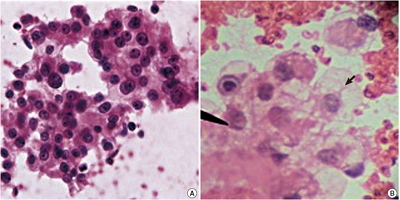
- 11,070 View
- 70 Download
- 5 Web of Science
- 6 Crossref
-
Abstract
PDF
- Background
Although using fine needle cytology with aspiration (FNC-A) for establishing diagnoses in the retroperitoneal region has shown promise, there is scant literature supporting a role of non-aspiration cytology (FNC-NA) for this region. We assessed the accuracy and reliability of FNC-A and FNC-NA as tools for preoperative diagnosis of retroperitoneal masses and compared the results of both techniques with each other and with histopathology. Methods: Fifty-seven patients with retroperitoneal masses were subjected to FNC-A and FNC-NA. Smears were stained with May-Grunwald Giemsa and hematoxylin and eosin stain. An individual slide was objectively analysed using a point scoring system to enable comparison between FNC-A and FNC-NA. Results: By FNC-A, 91.7% accuracy was obtained in cases of retroperitoneal lymph node lesions followed by renal masses (83.3%). The diagnostic accuracy of other sites by FNC-A varied from 75.0%–81.9%. By FNC-NA, 93.4% diagnostically accurate results were obtained in the kidney, followed by 75.0% in adrenal masses. The diagnostic accuracy of other sites by FNC-NA varied from 66.7%–72.8%. Conclusions: Although both techniques have their own advantages and disadvantages, FNC-NA may be a more efficient adjuvant method of sampling in retroperitoneal lesions. -
Citations
Citations to this article as recorded by- A comparison of cytologic quality in fine‐needle specimens obtained with and without aspiration from superficial lymph nodes in the dog
V. Karakitsou, M. M. Christopher, E. Meletis, P. Kostoulas, D. Pardali, C. K. Koutinas, M. E. Mylonakis
Journal of Small Animal Practice.2022; 63(1): 16. CrossRef - A comparison of cytological quality between fine‐needle aspiration and non‐aspiration techniques for obtaining ultrasound‐guided samples from canine and feline lymph nodes
James Whitlock, Olivier Taeymans, Paola Monti
Veterinary Record.2021;[Epub] CrossRef - A randomised controlled comparison of aspiration and non-aspiration fine-needle techniques for obtaining ultrasound-guided cytological samples from canine livers
K.L. Fleming, E.J. Howells, E.J. Villiers, T.W. Maddox
The Veterinary Journal.2019; 252: 105372. CrossRef - Minimally invasive biopsy in retroperitoneal tumors (Review)
Radu Marcu, Camelia Diaconu, Traian Constantin, Bogdan Socea, Florentina Ionita‑Radu, Dan Mischianu, Ovidiu Bratu
Experimental and Therapeutic Medicine.2019;[Epub] CrossRef - For diagnosis of liver masses, fine-needle aspiration versus needle core biopsy: which is better?
Liye Suo, Ruby Chang, Vijayalakshmi Padmanabhan, Shilpa Jain
Journal of the American Society of Cytopathology.2018; 7(1): 46. CrossRef - Fine needle aspiration in retroperitoneal lesions
Parikshaa Gupta, Arvind Rajwanshi, Raje Nijhawan, Radhika Srinivasan, Nalini Gupta, Uma Nahar Saikia, Pranab Dey
APMIS.2017; 125(1): 16. CrossRef
- A comparison of cytologic quality in fine‐needle specimens obtained with and without aspiration from superficial lymph nodes in the dog
- Diagnostic Accuracy of Endoscopic Ultrasound-Guided Fine Needle Aspiration Cytology of Pancreatic Lesions
- Hae Woon Baek, Min Jee Park, Ye-Young Rhee, Kyoung Bun Lee, Min A Kim, In Ae Park
- J Pathol Transl Med. 2015;49(1):52-60. Published online January 15, 2015
- DOI: https://doi.org/10.4132/jptm.2014.10.26
- 14,318 View
- 97 Download
- 35 Web of Science
- 34 Crossref
-
Abstract
PDF
- Background
Endoscopic ultrasound–guided fine needle aspiration cytology (EUS-FNAC) is currently the most commonly used procedure for obtaining cytologic specimens of the pancreas. It is accurate, minimally invasive, safe and cost-effective. However, there is discrepancy between cytological and surgical diagnoses. This study was aimed at evaluating the diagnostic accuracy of EUS-FNAC of the pancreas. Methods: We performed a retrospective review of 191 cases of pancreatic lesions initially diagnosed by EUS-FNAC with subsequent histological diagnosis between 2010 and 2012 in the Department of Pathology, Seoul National University Hospital. Cytologic and surgical diagnoses were categorized into five groups: negative, benign, atypical, malignant, and insufficient for diagnosis. Subsequently, 167 cases with satisfactory yield in both surgical and cytology specimens were statistically analyzed to determine correlations with diagnosis. Results: In comparison to surgical diagnoses, cytologic diagnoses were true-positive in 103 cases (61.7%), true-negative in 28 cases (16.8%), false-positive in 9 cases (5.4%), and false-negative in 27 cases (16.1%). The diagnostic accuracy was 78.4%, sensitivity was 79.2%, and specificity was 75.7%. The positive predictive value was 92.0%, and negative predictive value was 50.9%. Conclusions: EUS-FNAC has high accuracy, sensitivity, specificity and positive predictive value. Overcoming the limitations of EUS-FNAC will make it a useful and reliable diagnostic tool for accurate evaluation of pancreatic lesions. -
Citations
Citations to this article as recorded by- Diagnostic efficacy of endoscopic ultrasound-guided fine needle aspiration cytology in pancreatic lesions
Pranita Mohanty, Priyadarshini Dehuri, Jimmy Narayan
Scientific Reports.2026;[Epub] CrossRef - miR-6855-5p Enhances Radioresistance and Promotes Migration of Pancreatic Cancer by Inducing Epithelial-Mesenchymal Transition via Suppressing FOXA1: Potential of Plasma Exosomal miR-6855-5p as an Indicator of Radiosensitivity in Patients with Pancreatic
Hiroki Ueda, Hidenori Takahashi, Shogo Kobayashi, Masahiko Kubo, Kazuki Sasaki, Yoshifumi Iwagami, Daisaku Yamada, Yoshito Tomimaru, Tadafumi Asaoka, Takehiro Noda, Junzo Shimizu, Yuichiro Doki, Hidetoshi Eguchi
Annals of Surgical Oncology.2025; 32(2): 720. CrossRef - Combating false negatives in pancreatic cancer: A deep learning approach for aiding fine needle aspiration via accurate subregion identification
Ms Jasmine Chhikara, Nidhi Goel, Neeru Rathee
Engineering Applications of Artificial Intelligence.2025; 157: 111347. CrossRef - Grading pancreatic adenocarcinomas on fine needle aspiration cytology. The outstanding issues
Mrinmay Kumar Mallik, Laila Rafiq Qadan, Asit Kumar Mohanty, Ali Alali, Kusum Kapila
Cytopathology.2024; 35(2): 256. CrossRef - Reporting Pancreatic FNAC using the Papanicolaou System: Still a Diagnostic Challenge
Parul Verma, Saloni Goyal, Ruchita Tyagi, Mehar Ghuman, Ramit Mahajan, Arshneet Kaur Selhi, Harpreet Kaur, Pavneet Kaur Selhi
Journal of Cytology.2024; 41(2): 123. CrossRef - Preoperative treatment response prediction for pancreatic cancer by multiple microRNAs in plasma exosomes: Optimization using machine learning and network analysis
Hiroki Ueda, Hidenori Takahashi, Ryoto Sakaniwa, Tetsuhisa Kitamura, Shogo Kobayashi, Yoshito Tomimaru, Masahiko Kubo, Kazuki Sasaki, Yoshifumi Iwagami, Daisaku Yamada, Tadafumi Asaoka, Takehiro Noda, Junzo Shimizu, Yuichiro Doki, Hidetoshi Eguchi
Pancreatology.2024; 24(7): 1097. CrossRef - Comparison of Endoscopic Ultrasound-Guided Fine-Needle Aspiration with Fine-Needle Biopsy for Solid Gastrointestinal Lesions: A Randomized Crossover Single-Center study
Shivaraj Afzalpurkar, Vijay Kumar Rai, Nikhil Sonthalia, Gajanan Rodge, Awanesh Tewary, Mahesh Goenka
Journal of Digestive Endoscopy.2023; 14(01): 014. CrossRef - Diagnosing and monitoring pancreatic cancer through cell-free DNA methylation: progress and prospects
María Victoria García-Ortiz, Pablo Cano-Ramírez, Marta Toledano-Fonseca, Enrique Aranda, Antonio Rodríguez-Ariza
Biomarker Research.2023;[Epub] CrossRef - The impact of preoperative EUS-FNA for distal resectable pancreatic cancer: Is it really effective enough to take risks?
Jin-Seok Park, Jae Hoon Lee, Tae Jun Song, Joune Seup Lee, Seok Jung Jo, Dong Wook Oh, Ki Byung Song, Dae Wook Hwang, Do Hyun Park, Sang Soo Lee, Song Cheol Kim, Dong Wan Seo, Sung Koo Lee, Myung-Hwan Kim
Surgical Endoscopy.2022; 36(5): 3192. CrossRef - Magnetic Resonance Imaging Radiomics‐Based Nomogram From Primary Tumor for Pretreatment Prediction of Peripancreatic Lymph Node Metastasis in Pancreatic Ductal Adenocarcinoma: A Multicenter Study
Zhenshan Shi, Chengle Ma, Xinming Huang, Dairong Cao
Journal of Magnetic Resonance Imaging.2022; 55(3): 823. CrossRef - Role of Pathologist in the Era of Image-Guided and EUS-Guided Aspirations: A 10-Year Study at a Single Tertiary Care Oncology Institute in North India
Gurudutt Gupta, Anila Sharma, Meenakshi Kamboj, Anurag Sharma, Sunil Pasricha, Garima Durga, Anurag Mehta, Avinash Rao
Acta Cytologica.2022; 66(3): 187. CrossRef - Predicting Factors for Pancreatic Malignancy with Computed Tomography and Endoscopic Ultrasonography in Chronic Pancreatitis
Jian-Han Lai, Keng-Han Lee, Chen-Wang Chang, Ming-Jen Chen, Ching-Chung Lin
Diagnostics.2022; 12(4): 1004. CrossRef - Phenotypic profiling of pancreatic ductal adenocarcinoma plasma-derived small extracellular vesicles for cancer diagnosis and cancer stage prediction: a proof-of-concept study
Wei Zhang, Ling Wang, Dan Li, Douglas H. Campbell, Bradley J. Walsh, Nicolle H. Packer, Qing Dong, Erkang Wang, Yuling Wang
Analytical Methods.2022; 14(23): 2255. CrossRef - Cancer-derived small extracellular vesicles: emerging biomarkers and therapies for pancreatic ductal adenocarcinoma diagnosis/prognosis and treatment
Wei Zhang, Douglas H. Campbell, Bradley J. Walsh, Nicolle H. Packer, Dingbin Liu, Yuling Wang
Journal of Nanobiotechnology.2022;[Epub] CrossRef - Single Cell RNA Sequencing: A New Frontier in Pancreatic Ductal Adenocarcinoma
Maroun Bou Zerdan, Malek Shatila, Dhruv Sarwal, Youssef Bouferraa, Morgan Bou Zerdan, Sabine Allam, Merima Ramovic, Stephen Graziano
Cancers.2022; 14(19): 4589. CrossRef - Molecular Subtyping and Precision Medicine for Pancreatic Cancer
Fieke Froeling, Raffaella Casolino, Antonio Pea, Andrew Biankin, David Chang
Journal of Clinical Medicine.2021; 10(1): 149. CrossRef - Reshaping preoperative treatment of pancreatic cancer in the era of precision medicine
R. Casolino, C. Braconi, G. Malleo, S. Paiella, C. Bassi, M. Milella, S.B. Dreyer, F.E.M. Froeling, D.K. Chang, A.V. Biankin, T. Golan
Annals of Oncology.2021; 32(2): 183. CrossRef - Endoscopic Ultrasound-Guided Fine Needle Aspiration Cytology of Pancreatic Adenocarcinomas Revisited. A Detailed Cytological Analysis
Mrinmay Kumar Mallik, Kusum Kapila, Asit Kumar Mohanty, Shafi Ahmed Inamdar, Ali AlAli, Abdullah Al Naseer
Journal of Cytology.2021; 38(1): 31. CrossRef - Diagnostic value of various liquid biopsy methods for pancreatic cancer
Yuzhou Zhu, Hao Zhang, Nan Chen, Jianqi Hao, Hongyu Jin, Xuelei Ma
Medicine.2020; 99(3): e18581. CrossRef - Factors affecting cytological results of endoscopic ultrasound guided-fine needle aspiration during learning
Jian-Han Lai, Hsiang-Hung Lin, Ching-Chung Lin
Diagnostic Pathology.2020;[Epub] CrossRef - Comparison between Conventional Smear and Liquid-Based Preparation in Endoscopic Ultrasonography-Fine Needle Aspiration Cytology of Pancreatic Lesions
Soo Hee Ko, Jung-Soo Pyo, Byoung Kwan Son, Hyo Young Lee, Il Whan Oh, Kwang Hyun Chung
Diagnostics.2020; 10(5): 293. CrossRef - Evaluation of Pancreatic Lesions With Endoscopic Ultrasound and Fine Needle Aspiration
Yan Luk, Wong Hoi She, Felix Che Lok Chow, Ka Wing Ma, Simon Hing Yin Tsang, Wing Chiu Dai, Tan To Cheung, Chung Mau Lo
Surgical Innovation.2020; 27(5): 431. CrossRef - The applicability of Papanicolaou Society of Cytopathology system on reporting endoscopic ultrasound‐guided fine needle aspiration cytology specimens of pancreatic lesions in situations with limited availability of ancillary tests. Experience at a single
Mrinmay Kumar Mallik, Laila Rafiq Qadan, Abdullah Al Naseer, Ali AlAli, Taiba Al Ansari, Shafi Ahmed Inamdar Naquib, Dilip Kumar Das, Kusum Kapila
Cytopathology.2020; 31(6): 564. CrossRef - Circulating Cell-Free DNA-Based Liquid Biopsy Markers for the Non-Invasive Prognosis and Monitoring of Metastatic Pancreatic Cancer
Marta Toledano-Fonseca, M. Teresa Cano, Elizabeth Inga, Rosa Rodríguez-Alonso, M. Auxiliadora Gómez-España, Silvia Guil-Luna, Rafael Mena-Osuna, Juan R. de la Haba-Rodríguez, Antonio Rodríguez-Ariza, Enrique Aranda
Cancers.2020; 12(7): 1754. CrossRef - Risk of malignancy in the categories of the Papanicolaou Society of Cytopathology system for reporting pancreaticobiliary cytology
Raza S. Hoda, Elizabeth B. Finer, Ronald N. Arpin, Matthew Rosenbaum, Martha B. Pitman
Journal of the American Society of Cytopathology.2019; 8(3): 120. CrossRef - Endoscopic ultrasound guided fine‐needle aspiration vs core needle biopsy for solid pancreatic lesions: Comparison of diagnostic accuracy and procedural efficiency
Aslam Syed, Olivia Babich, Bharat Rao, Shailendra Singh, Neil Carleton, Abhishek Gulati, Archana Kulkarni, Mrinal Garg, Katie Farah, Gursimran Kochhar, Suzanne Morrissey, Marcia Mitre, Abhijit Kulkarni, Manish Dhawan, Jan F. Silverman, Majed Pharaon, Shya
Diagnostic Cytopathology.2019; 47(11): 1138. CrossRef - Liquid Biopsy as Surrogate for Tissue for Molecular Profiling in Pancreatic Cancer: A Meta-Analysis Towards Precision Medicine
Claudio Luchini, Nicola Veronese, Alessia Nottegar, Vera Cappelletti, Maria G. Daidone, Lee Smith, Christopher Parris, Lodewijk A. A. Brosens, Maria G. Caruso, Liang Cheng, Christopher L. Wolfgang, Laura D. Wood, Michele Milella, Roberto Salvia, Aldo Scar
Cancers.2019; 11(8): 1152. CrossRef - It is necessary to exam bottom and top slide smears of EUS-FNA for pancreatic cancer
Jong-chan Lee, Haeryoung Kim, Hyoung Woo Kim, Jongchan Lee, Kyu-hyun Paik, Jingu Kang, Jin-Hyeok Hwang, Jaihwan Kim
Hepatobiliary & Pancreatic Diseases International.2018; 17(6): 553. CrossRef - Preoperative EUS-guided FNA: effects on peritoneal recurrence and survival in patients with pancreatic cancer
Sun Hwa Kim, Young Sik Woo, Kwang Hyuck Lee, Jong Kyun Lee, Kyu Taek Lee, Joo Kyung Park, Soo Hoon Kang, Ji Won Kim, Jae Keun Park, Sung-Wook Park
Gastrointestinal Endoscopy.2018; 88(6): 926. CrossRef - Cytological characteristics of atypical cells in endoscopic ultrasound-guided fine-needle aspiration specimens obtained from the pancreas
Shikine ESAKA, Yoko MATSUDA, Yuri HAMASHIMA, Masayuki IMAIZUMI, Hiroya KOJIMA, Yuri KISO, Hiroto SHIRAHATA, Mayumi KINOSHITA, Akemi SUZUKI, Tomio ARAI
The Journal of the Japanese Society of Clinical Cytology.2018; 57(4): 199. CrossRef - Performance measures for endoscopic retrograde cholangiopancreatography and endoscopic ultrasound: A European Society of Gastrointestinal Endoscopy (ESGE) Quality Improvement Initiative
Dirk Domagk, Kofi W Oppong, Lars Aabakken, Laszlo Czakó, Tibor Gyökeres, Gianpiero Manes, Peter Meier, Jan-Werner Poley, Thierry Ponchon, Andrea Tringali, Cristina Bellisario, Silvia Minozzi, Carlo Senore, Cathy Bennett, Michael Bretthauer, Cesare Hassan,
United European Gastroenterology Journal.2018; 6(10): 1448. CrossRef - Performance measures for ERCP and endoscopic ultrasound: a European Society of Gastrointestinal Endoscopy (ESGE) Quality Improvement Initiative
Dirk Domagk, Kofi W. Oppong, Lars Aabakken, Laszlo Czakó, Tibor Gyökeres, Gianpiero Manes, Peter Meier, Jan-Werner Poley, Thierry Ponchon, Andrea Tringali, Cristina Bellisario, Silvia Minozzi, Carlo Senore, Cathy Bennett, Michael Bretthauer, Cesare Hassan
Endoscopy.2018; 50(11): 1116. CrossRef - Imaging modalities for characterising focal pancreatic lesions
Lawrence MJ Best, Vishal Rawji, Stephen P Pereira, Brian R Davidson, Kurinchi Selvan Gurusamy
Cochrane Database of Systematic Reviews.2017;[Epub] CrossRef - Percutaneous ultrasound‐guided core needle biopsy of solid pancreatic masses: Results in 250 patients
Guven Kahriman, Nevzat Ozcan, Serap Dogan, Soner Ozmen, Kemal Deniz
Journal of Clinical Ultrasound.2016; 44(8): 470. CrossRef
- Diagnostic efficacy of endoscopic ultrasound-guided fine needle aspiration cytology in pancreatic lesions
Brief Case Reports
- A Rare Case of Mesothelioma Showing Micropapillary and Small Cell Differentiation with Aggressive Behavior
- Yoon Jin Cha, Binnari Kim, Joungho Han, Chin A Yi, Jae Ill Zo
- Korean J Pathol. 2014;48(6):466-468. Published online December 31, 2014
- DOI: https://doi.org/10.4132/KoreanJPathol.2014.48.6.466
- 11,019 View
- 40 Download
- 2 Crossref
-
PDF
-
Citations
Citations to this article as recorded by- Small cell mesothelioma: A rare entity and diagnostic pitfall mimicking small cell lung carcinoma on fine‐needle aspiration
Yanhong Zhang, Alaa Afify, Regina F. Gandour‐Edwards, John W. Bishop, Eric C. Huang
Diagnostic Cytopathology.2016; 44(6): 526. CrossRef - A Biphasic Pleural Tumor with Features of an Epithelioid and Small Cell Mesothelioma: Morphologic and Molecular Findings
Sarah Hackman, Richard D. Hammer, Lester Layfield
Case Reports in Pathology.2016; 2016: 1. CrossRef
- Small cell mesothelioma: A rare entity and diagnostic pitfall mimicking small cell lung carcinoma on fine‐needle aspiration
- Supratentorial Hemangioblastoma with Unusual Features
- Yooju Shin, Seokhwi Kim, Hyun-Woo Lee, Heejin Bang, Yeon-Lim Suh
- Korean J Pathol. 2014;48(6):462-465. Published online December 31, 2014
- DOI: https://doi.org/10.4132/KoreanJPathol.2014.48.6.462
- 13,629 View
- 81 Download
- 6 Crossref
-
PDF
-
Citations
Citations to this article as recorded by- Supratentorial Hemangioblastoma in Adults: A Systematic Review and Comparison of Infratentorial and Spinal Cord Locations
Dragan Jankovic, Kyna Vuong, Bruno Splavski, Kresimir Rotim, Kenan I. Arnautovic
World Neurosurgery.2023; 173: 48. CrossRef - Supratentorial hemangioblastoma: correlation between phenotype, gender and vascular territory affected
Yosef Laviv, David Saraf, Liat Oxman, Ido Ben Zvi
Neurosurgical Review.2023;[Epub] CrossRef - Neuropathologic features of central nervous system hemangioblastoma
Rebecca A. Yoda, Patrick J. Cimino
Journal of Pathology and Translational Medicine.2022; 56(3): 115. CrossRef - The loss of succinate dehydrogenase B expression is frequently identified in hemangioblastoma of the central nervous system
Tae Hoon Roh, Hyunee Yim, Jin Roh, Kyi Beom Lee, So Hyun Park, Seon-Yong Jeong, Se-Hyuk Kim, Jang-Hee Kim
Scientific Reports.2019;[Epub] CrossRef - Supratentorial hemangioblastomas in von Hippel–Lindau wild-type patients – case series and literature review
Luís Rocha, Carolina Noronha, Ricardo Taipa, Joaquim Reis, Mário Gomes, Ernesto Carvalho
International Journal of Neuroscience.2018; 128(3): 295. CrossRef - MR Imaging Findings of Supratentorial Meningeal Hemangioblastoma: A Case Report
Gi Hong Kim, Ho Kyu Lee, Myeong Ju Koh, Young Hee Maeng
Journal of the Korean Society of Radiology.2016; 75(1): 26. CrossRef
- Supratentorial Hemangioblastoma in Adults: A Systematic Review and Comparison of Infratentorial and Spinal Cord Locations
- Malignant Perivascular Epithelioid Cell Tumor of the Uterus with Lung Metastasis
- Jeong Bae Kang, Jin Won Seo, Young-Han Park, Pong Rheem Jang
- Korean J Pathol. 2014;48(6):454-457. Published online December 31, 2014
- DOI: https://doi.org/10.4132/KoreanJPathol.2014.48.6.454
- 10,733 View
- 33 Download
- 3 Crossref
-
PDF
-
Citations
Citations to this article as recorded by- A rare case of uterine PEComa with pancreatic metastases – the role of endoscopic ultrasound-guided biopsy
I. Garrido, M. Pedro, M. Guilherme
Endoscopy.2023; 55(S 02): S335. CrossRef - A Retrospective Case Study of 13 Uterine Perivascular Epithelioid Cell Neoplasm (PEComa) Patients
Jiahui Gu, Wantong Wang, Shizhuo Wang
OncoTargets and Therapy.2021; Volume 14: 1783. CrossRef - Predicting the Behavior of Perivascular Epithelioid Cell Tumors of the Uterine Corpus
Andres Martin Acosta, Brian P. Adley
Archives of Pathology & Laboratory Medicine.2017; 141(3): 463. CrossRef
- A rare case of uterine PEComa with pancreatic metastases – the role of endoscopic ultrasound-guided biopsy
- Peritoneal Carcinosarcoma and Ovarian Papillary Serous Carcinoma Are the Same Origin: Analysis of TP53 Mutation and Microsatellite Suggests a Monoclonal Origin
- Chang Gok Woo, Dae Shik Suh, Joo Young Kim, Chang Ohk Sung, Jene Choi, Kyu-Rae Kim
- Korean J Pathol. 2014;48(6):449-453. Published online December 31, 2014
- DOI: https://doi.org/10.4132/KoreanJPathol.2014.48.6.449
- 14,541 View
- 56 Download
- 3 Crossref
-
PDF
-
Citations
Citations to this article as recorded by- A successfully treated primary peritoneal carcinosarcoma and serous carcinoma of stage IIIC rescued from hypovolemic shock due to tumor rupture
Che-Cheng Huang, Horng-Jyh Tsai, Shih Hsuan Huang, Victor C. Kok
Taiwanese Journal of Obstetrics and Gynecology.2019; 58(2): 296. CrossRef - Ovarian Carcinosarcoma and Concurrent Serous Tubal Intraepithelial Carcinoma With Next-Generation Sequencing Suggesting an Origin From the Fallopian Tube
Sharlene Helene C. See, Amir Behdad, Kruti P. Maniar, Luis Z. Blanco
International Journal of Surgical Pathology.2019; 27(5): 574. CrossRef - Progression inference for somatic mutations in cancer
Leif E. Peterson, Tatiana Kovyrshina
Heliyon.2017; 3(4): e00277. CrossRef
- A successfully treated primary peritoneal carcinosarcoma and serous carcinoma of stage IIIC rescued from hypovolemic shock due to tumor rupture
- A Case of Mixed Adenoneuroendocrine Carcinoma of the Common Bile Duct: Initially Diagnosed as Cholangiocarcinoma
- Soon Wook Lee, In Seok Lee, Yu Kyung Cho, Jae Myung Park, Sang Woo Kim, Myung-Gyu Choi, Kyu Yong Choi, Myung Ah Lee, Tae Ho Hong, Young Kyoung You, Eun-Sun Jung
- Korean J Pathol. 2014;48(6):445-448. Published online December 31, 2014
- DOI: https://doi.org/10.4132/KoreanJPathol.2014.48.6.445
- 11,080 View
- 44 Download
- 12 Crossref
-
PDF
-
Citations
Citations to this article as recorded by- Long-term survival after chemotherapy combined immunotherapy for recurrent mixed neuroendocrine–non-neuroendocrine neoplasms of the common bile duct
Jia Li, Chunyan Yuan, Yulin Pan, Yuanyuan Yang, Nannan Lai, Xia Sheng
Clinical Journal of Gastroenterology.2025; 18(4): 653. CrossRef - Neuroendocrine carcinoma of the common hepatic duct coexisting with distal cholangiocarcinoma: A case report and review of literature
Fei Chen, Wei-Wei Li, Juan-Fen Mo, Min-Jie Chen, Su-Hang Wang, Shu-Ying Yang, Zheng-Wei Song
World Journal of Gastrointestinal Surgery.2024; 16(5): 1449. CrossRef - Comparison of Metastatic Patterns Among Neuroendocrine Tumors, Neuroendocrine Carcinomas, and Nonneuroendocrine Carcinomas of Various Primary Organs
Hyung Kyu Park, Ghee Young Kwon
Journal of Korean Medical Science.2023;[Epub] CrossRef - Mixed adenoneuroendocrine carcinoma of the distal bile duct: a case report
Takashi Maeda, Kyohei Yugawa, Nao Kinjo, Hiroto Kayashima, Daisuke Imai, Koto Kawata, Shinichiro Ikeda, Keitaro Edahiro, Kazuki Takeishi, Tomohiro Iguchi, Noboru Harada, Mizuki Ninomiya, Shohei Yamaguchi, Kozo Konishi, Shinichi Tsutsui, Hiroyuki Matsuda
Surgical Case Reports.2020;[Epub] CrossRef - The clinical profiles, management, and prognostic factors of biliary mixed neuroendocrine nonneuroendocrine neoplasms
Li-Jia Wen, Jun-Hong Chen, Hong-Ji Xu, Qiong Yu, Yu Deng, Kai Liu
Medicine.2020; 99(50): e23271. CrossRef - Rapidly progressed neuroendocrine carcinoma in the extrahepatic bile duct: a case report and review of the literature
Mariko Kamiya, Naoto Yamamoto, Yuto Kamioka, Hirohide Inoue, Hirokazu Yotsumoto, Masaaki Murakawa, Toru Aoyama, Kota Washimi, Kae Kawachi, Takashi Oshima, Makoto Ueno, Norio Yukawa, Yasushi Rino, Munetaka Masuda, Soichiro Morinaga
Surgical Case Reports.2020;[Epub] CrossRef - Mixed adenoneuroendocrine carcinoma of the hepatic bile duct: a case report and review of the literature
Sulai Liu, Zhendong Zhong, Meng Xiao, Yinghui Song, Youye Zhu, Bo Hu, Zengpeng Sun, Weimin Yi, Chuang Peng
BMC Gastroenterology.2020;[Epub] CrossRef - Mixed adenoendocrine carcinoma in the extrahepatic biliary tract: A case report and literature review
Liang Zhang, Zhengtao Yang, Qing Chen, Mengxia Li, Xiaolu Zhu, Dalong Wan, Haiyang Xie, Shusen Zheng
Oncology Letters.2019;[Epub] CrossRef - Large Cell Neuroendocrine Carcinoma Coexisting with Adenocarcinoma in the Extrahepatic Bile Duct
Masami Yuda, Teruyuki Usuba, Shin Hagiwara, Masahisa Okuma, Hisatoshi Asano, Hitoshi Sakuda, Hiroaki Katagi, Yoshiyuki Furukawa
The Japanese Journal of Gastroenterological Surgery.2018; 51(3): 187. CrossRef - Mixed Adenoneuroendocrine Carcinoma of the Distal Bile Duct
Chiaki Uchida, Yoshikazu Toyoki, Keinosuke Ishido, Daisuke Kudo, Norihisa Kimura, Shinji Tsutsumi, Takuji Kagiya, Toshiro Kimura, Kenichi Hakamada
The Japanese Journal of Gastroenterological Surgery.2017; 50(1): 43. CrossRef - Mixed adenoneuroendocrine carcinoma of the distal bile duct: A case report
Toshiaki Komo, Toshihiko Kohashi, Akira Nakashima, Ichiro Ohmori, Jun Hihara, Hidenori Mukaida, Mayumi Kaneko, Naoki Hirabayashi
International Journal of Surgery Case Reports.2017; 39: 203. CrossRef - Common Hepatic Duct Mixed Adenoneuroendocrine Carcinoma Masquerading as Cholangiocarcinoma
Sali Priyanka Akhilesh, Yadav Kamal Sunder, Tampi Chandralekha, Parikh Samir, Wagle Prasad Kashinath
Case Reports in Gastrointestinal Medicine.2016; 2016: 1. CrossRef
- Long-term survival after chemotherapy combined immunotherapy for recurrent mixed neuroendocrine–non-neuroendocrine neoplasms of the common bile duct
- Periductal Stromal Tumor of Breast: A Case Report and A Review of Literature
- Salma L. Abbasi, Kate McNamara, Mohammed S. Absar, Alison Darlington, Francene Clucas, Sami Titi
- Korean J Pathol. 2014;48(6):442-444. Published online December 31, 2014
- DOI: https://doi.org/10.4132/KoreanJPathol.2014.48.6.442
- 14,526 View
- 89 Download
- 5 Crossref
-
PDF
-
Citations
Citations to this article as recorded by- Genetic Profiling of Mammary Periductal Stromal Tumors With Histologic Correlation Highlights High-Grade and Low-Grade Groups and Similarities to Phyllodes Tumors
Gregor Krings, Gregory R. Bean, Elizabeth M. Hosfield, J Jordi Rowe, Joseph Geradts, Yunn-Yi Chen
Modern Pathology.2026; 39(3): 100961. CrossRef - Survey of recurrent diagnostic challenges in breast phyllodes tumours
Benjamin Yongcheng Tan, Stephen B Fox, Sunil R Lakhani, Puay Hoon Tan
Histopathology.2023; 82(1): 95. CrossRef - Management of a periductal stromal tumor in a young woman: Our breast unit experience
Irene Valente, Adela Ristani, Cristina Mancini, Eugenia Martella, Leonardo Quartieri, Cecilia D'Aloia
The Breast Journal.2020; 26(7): 1375. CrossRef - A Diagnostic Approach to Fibroepithelial Breast Lesions
Benjamin Yongcheng Tan, Puay Hoon Tan
Surgical Pathology Clinics.2018; 11(1): 17. CrossRef - A case of local recurrence of periductal stromal sarcoma of the breast
Kana TERAMOTO, Yasuro DOI, Kayo YAMAMOTO, Kaname MATSUKAWA, Hisaka IWAIHARA, Rumi MOTOSHIMA, Noboru TAKATA, Ichiro YOSHINAKA, Kazunori HARADA
Choonpa Igaku.2018; 45(1): 61. CrossRef
- Genetic Profiling of Mammary Periductal Stromal Tumors With Histologic Correlation Highlights High-Grade and Low-Grade Groups and Similarities to Phyllodes Tumors
- Digital Papillary Carcinoma
- Sharon Lim, Inju Cho, Mi Ja Lee
- Korean J Pathol. 2014;48(6):438-441. Published online December 31, 2014
- DOI: https://doi.org/10.4132/KoreanJPathol.2014.48.6.438
- 11,133 View
- 50 Download
- 5 Crossref
-
PDF
-
Citations
Citations to this article as recorded by- Digital Papillary Carcinoma: A Literature Review of Epidemiology, Management Strategies, and Patient Outcomes
William Liu, Rahul Nanda, David Zloty
Dermatologic Surgery.2026; 52(7): 627. CrossRef - Digital Papillary Adenocarcinoma: Uncommon Malignancy of Sweat Glands - Two Rare Cases
Neeti Goyal, Pawan Dhaman, Jasvinder Kaur Bhatia, Pragya Sharma, Prabha Shankar Mishra, Vikram Singh, Anvesh Rathore
Journal of Marine Medical Society.2025; 27(1): 103. CrossRef - Digital papillary adenocarcinoma: A case report of a rare malignant tumour with recommendations on management and follow-up
Varanindu Mudduwa, Mohammad Goodarzi, Richard Chalmers, Haitham Khashaba
International Journal of Surgery Case Reports.2025;[Epub] CrossRef - Digital papillary adenocarcinoma: A case report
Betty A. Kasimo, Vivian Akello, James J. Yahaya
Clinical Case Reports.2021;[Epub] CrossRef - A rare case of a digital papillary carcinoma of the hand with secondary conservative management
Rabeet Khan, Renu Irri, Effie Katsarma
Journal of Surgical Case Reports.2020;[Epub] CrossRef
- Digital Papillary Carcinoma: A Literature Review of Epidemiology, Management Strategies, and Patient Outcomes
- A Case of Epithelioid Blue Nevus
- Chung Hun Lee, Hye Sook Min, Eon Sub Park, Kye Yong Song
- Korean J Pathol. 2014;48(6):434-437. Published online December 31, 2014
- DOI: https://doi.org/10.4132/KoreanJPathol.2014.48.6.434
- 13,081 View
- 80 Download
- 3 Crossref
-
PDF
-
Citations
Citations to this article as recorded by- A Curious Case of a Pigmented Lesion
Dilshad Sachedina, Marisa Taylor
Indian Journal of Dermatology.2024; 69(1): 104. CrossRef - Recurrence of a Cellular Blue Nevus with Satellitosis—A Diagnostic Pitfall with Clinical Consequences
Hermann Kneitz, Christian Rose, Valerie Glutsch, Matthias Goebeler
Dermatopathology.2022; 9(4): 361. CrossRef - Epithelioid Blue Nevus
Kai-Lv Sun, Jian-Min Chang
International Journal of Dermatology and Venereology.2020; 3(3): 186. CrossRef
- A Curious Case of a Pigmented Lesion
- Indolent CD56-Positive Clonal T-Cell Lymphoproliferative Disease of the Stomach Mimicking Lymphomatoid Gastropathy
- Mineui Hong, Won Seog Kim, Young Hyeh Ko
- Korean J Pathol. 2014;48(6):430-433. Published online December 31, 2014
- DOI: https://doi.org/10.4132/KoreanJPathol.2014.48.6.430
- 11,654 View
- 71 Download
- 3 Crossref
-
PDF
-
Citations
Citations to this article as recorded by- Indolent T- and Natural Killer-Cell Lymphomas and Lymphoproliferative Diseases—Entities in Evolution
Chi Sing Ng
Lymphatics.2025; 3(4): 41. CrossRef - Case Report: Primary Indolent Epstein-Barr Virus-Positive T-Cell Lymphoproliferative Disease Involving the Central Nervous System
Kun Wang, Jinjian Li, Xuehui Zhou, Junhui Lv, Yirong Wang, Xinwei Li
Frontiers in Surgery.2022;[Epub] CrossRef - Indolent NK cell proliferative lesion mimicking NK/T cell lymphoma in the gallbladder
Su Hyun Hwang, Joon Seong Park, Seong Hyun Jeong, Hyunee Yim, Jae Ho Han
Human Pathology: Case Reports.2016; 5: 39. CrossRef
- Indolent T- and Natural Killer-Cell Lymphomas and Lymphoproliferative Diseases—Entities in Evolution
- A Case of Type II Enteropathy-Associated T-Cell Lymphoma with Epstein-Barr Virus Positivity
- Min Jeong Song, Chan Sik Park, Hee Sang Hwang, Cheol Won Suh, Dok Hyun Yoon, Jooryung Huh
- Korean J Pathol. 2014;48(6):426-429. Published online December 31, 2014
- DOI: https://doi.org/10.4132/KoreanJPathol.2014.48.6.426
- 11,298 View
- 53 Download
- 6 Crossref
-
PDF
-
Citations
Citations to this article as recorded by- Various Endoscopic Features in Monomorphic Epitheliotropic Intestinal T-Cell Lymphoma
Yasuhiro Aoki, Tomohisa Sujino, Kaoru Takabayashi, Makoto Mutakuchi, Katsura Emoto, Naoki Hosoe, Haruhiko Ogata, Takanori Kanai
Case Reports in Gastroenterology.2021; 15(1): 312. CrossRef - A Case Report of Monomorphic Epithelial Intestinal T-Cell Lymphoma and Literature Review
Jing Deng, Yongguang Zou, Guangshu Li
International Journal of Clinical Medicine.2020; 11(02): 15. CrossRef - A viral map of gastrointestinal cancers
Natália R. Costa, Rui M. Gil da Costa, Rui Medeiros
Life Sciences.2018; 199: 188. CrossRef - Type II Enteropathy-Associated T-cell Lymphoma: A Rare Report from Iran
Neda Nozari
Middle East Journal of Digestive Diseases.2017; 9(1): 55. CrossRef - Unusual Cause of Dysphagia
Shahram Agah, Ramak Ghavam, Ahmad Darvishi Zeidabadi, Arash Sarveazad
Middle East Journal of Digestive Diseases.2017; 9(1): 58. CrossRef - Multiple lesions of gastrointestinal tract invasion by monomorphic epitheliotropic intestinal T-cell lymphoma, accompanied by duodenal and intestinal enteropathy-like lesions and microscopic lymphocytic proctocolitis: a case series
Hideki Ishibashi, Satoshi Nimura, Yoshiyuki Kayashima, Yasushi Takamatsu, Kunihiko Aoyagi, Naohiko Harada, Masanori Kadowaki, Takihiko Kamio, Shotaro Sakisaka, Morishige Takeshita
Diagnostic Pathology.2016;[Epub] CrossRef
- Various Endoscopic Features in Monomorphic Epitheliotropic Intestinal T-Cell Lymphoma
- Primary Leiomyosarcoma of Adrenal Gland with Tissue Eosinophilic Infiltration
- Seungkoo Lee, Gail Domecq C. Tanawit, Rolando A. Lopez, Jaime T. Zamuco, Betsy Grace G. Cheng, Menandro V. Siozon
- Korean J Pathol. 2014;48(6):423-425. Published online December 31, 2014
- DOI: https://doi.org/10.4132/KoreanJPathol.2014.48.6.423
- 10,758 View
- 48 Download
- 8 Crossref
-
PDF
-
Citations
Citations to this article as recorded by- Primary adrenal leiomyosarcoma
Syed Muhammad Nazim, Muhammad Hummam Siddique, Imran Khan Jalbani, Ayesha Nusrat
BMJ Case Reports.2025; 18(10): e266476. CrossRef - Outcomes and Follow-Up Trends in Adrenal Leiomyosarcoma: A Comprehensive Literature Review and Case Report
Federico Maria Mongardini, Maddalena Paolicelli, Antonio Catauro, Alessandra Conzo, Luigi Flagiello, Giusiana Nesta, Rosetta Esposito, Andrea Ronchi, Alessandro Romano, Renato Patrone, Ludovico Docimo, Giovanni Conzo
Journal of Clinical Medicine.2024; 13(12): 3499. CrossRef - Challenges in the diagnosis of the enigmatic primary adrenal leiomyosarcoma: two case reports and review of the literature
Sawako Suzuki, Naoya Takahashi, Masafumi Sugo, Kazuki Ishiwata, Akiko Ishida, Suzuka Watanabe, Katsushi Igarashi, Yutaro Ruike, Kumiko Naito, Masanori Fujimoto, Hisashi Koide, Yusuke Imamura, Shinichi Sakamoto, Tomohiko Ichikawa, Yoshihiro Kubota, Takeshi
BMC Endocrine Disorders.2023;[Epub] CrossRef - Primary adrenal leiomyosarcoma: clinical case and literature review
S. V. Lukyanov, K. M. Blikyan, S. S. Todorov, V. Y. Deribas, N. S. Lukyanov
Endocrine Surgery.2021; 15(1): 36. CrossRef Pleomorphic Leiomyosarcoma of the Adrenal Gland in a Young Woman: A Case Report and Review of the Literature
Yuanyuan Wang, Yongliang Teng, Shibo Na, Ye Yuan
OncoTargets and Therapy.2020; Volume 13: 4705. CrossRef- Primary Adrenal Leiomyosarcoma: Clinical, Radiological, and Histopathological Characteristics
Fatema Jabarkhel, Henri Puttonen, Lina Hansson, Andreas Muth, Oskar Ragnarsson
Journal of the Endocrine Society.2020;[Epub] CrossRef - Primary adrenal leiomyosarcoma with inferior vena cava extension in a 70-year-old man
Sai K Doppalapudi, Tejash Shah, Valerie A Fitzhugh, Vladislav Bargman
BMJ Case Reports.2019; 12(3): e227670. CrossRef - Primary Adrenal Leiomyosarcoma: An Extremely Rare Mesenchymal Tumor
D Lokanatha, Linu Abraham Jacob, MC Suresh Babu, KN Lokesh, Ram Krishna Sai, AH Rudresha, LK Rajeev, Smitha Saldanha, MN Suma, A Usha
Indian Journal of Medical and Paediatric Oncology.2019; 40(04): 559. CrossRef
- Primary adrenal leiomyosarcoma
- Combined Squamous Cell Carcinoma and Follicular Carcinoma of the Thyroid
- Da Hye Son, Jong-Lyel Roh, Kyung-Ja Cho
- Korean J Pathol. 2014;48(6):418-422. Published online December 31, 2014
- DOI: https://doi.org/10.4132/KoreanJPathol.2014.48.6.418
- 10,832 View
- 59 Download
- 4 Crossref
-
PDF
-
Citations
Citations to this article as recorded by- Efficacy of combined immunotherapy and targeted therapy in overcoming barriers to postoperative recurrence in squamous subtype anaplastic thyroid carcinoma with abscess: a case report and literature review
Shuyun Jiang, Xiaowu Wang, Zhijun Ma
Frontiers in Oncology.2025;[Epub] CrossRef - State of Knowledge About Thyroid Cancers in the Era of COVID-19—A Narrative Review
Agnieszka Bronowicka-Szydełko, Maciej Rabczyński, Ilias Dumas, Żanna Fiodorenko-Dumas, Beata Wojtczak, Łukasz Kotyra, Irena Kustrzeba-Wójcicka, Łukasz Lewandowski, Beata Ponikowska, Aleksandra Kuzan, Joanna Kluz, Andrzej Gamian, Katarzyna Madziarska
Biomedicines.2024; 12(12): 2829. CrossRef - Primary and metastatic squamous cell carcinoma of the thyroid gland: Two case reports
Xing Zhao, Pengyu Hao, Jiangbei Tian, Jirun Sun, Dawei Chen, Zhehui Cui, Libo Xin, Yanmin Song, Gang Zhang
Open Life Sciences.2022; 17(1): 1148. CrossRef - Aggressive Thyroid Gland Carcinoma: A Case Series
JP Dworkin-Valenti
Archives of Otolaryngology and Rhinology.2017; : 129. CrossRef
- Efficacy of combined immunotherapy and targeted therapy in overcoming barriers to postoperative recurrence in squamous subtype anaplastic thyroid carcinoma with abscess: a case report and literature review
- Myoepithelial Carcinoma of Soft Tissue: A Case Report and Review of the Literature
- Chang Hwan Choi, Young Chae Chu, Lucia Kim, Suk Jin Choi, In Suh Park, Jee Young Han, Joon Mee Kim
- Korean J Pathol. 2014;48(6):413-417. Published online December 31, 2014
- DOI: https://doi.org/10.4132/KoreanJPathol.2014.48.6.413
- 15,289 View
- 134 Download
- 9 Crossref
-
PDF
-
Citations
Citations to this article as recorded by- Hyalinizing Clear Cell Carcinoma of the Lung With Uncommon Distant Cutaneous Metastasis and Aggressive Clinical Course
David M. Gustafson, Sansar Tiwari, Catherine G. Chung
Journal of Cutaneous Pathology.2026;[Epub] CrossRef - Myoepithelial Carcinoma Mimicking Basal Cell Carcinoma: A Case Report
Farlin Asharaff, Neena Nayak, Roger Webb, Karwan Moutasim, Soogan Lalla
Cureus.2025;[Epub] CrossRef - Fine‐needle aspiration cytology of retroperitoneal myoepithelial carcinoma: A rare encounter with diagnostic dilemmas
Aadya Kerkar, Ajay Savlania, Reetu Kundu, Suvradeep Mitra, Manish Rohilla, Harmandeep Singh, Harish Bhujade
Diagnostic Cytopathology.2024;[Epub] CrossRef - EWSR1::NR4A3 gene fusion in a cutaneous atypical myoepithelial neoplasm
Ashley Rose Scholl, Evelyna Kliassov, Diana M. Cardona, Rex Bentley, Rami N. Al‐Rohil
Journal of Cutaneous Pathology.2023; 50(7): 601. CrossRef - Abdominal myoepithelial carcinoma: A rare abdominal wall entity of an uncommon tumor
Daania Shoaib, Saqib Raza Khan, Yasmin Abdul Rashid, Muhammad Nauman Zahir
International Journal of Surgery Case Reports.2022; 99: 107618. CrossRef - Adult soft tissue myoepithelial carcinoma: treatment outcomes and efficacy of chemotherapy
Florence Chamberlain, Elena Cojocaru, Mariana Scaranti, Jonathan Noujaim, Anastasia Constantinou, Khin Thway, Cyril Fisher, Christina Messiou, Dirk C. Strauss, Aisha Miah, Shane Zaidi, Charlotte Benson, Spyridon Gennatas, Robin L. Jones
Medical Oncology.2020;[Epub] CrossRef - Foot plantar soft tissue malignant myoepithelioma tumor: Case report and review of the literature
Manuel Trevino, Chetan Moorthy, Lisa Kafchinski, Daniel Bustamante
Clinical Imaging.2020; 61: 90. CrossRef - Presumed choroidal metastasis from soft tissue myoepithelial carcinoma
Michelle M. Hui, Rohan Merani, Fiona Bonar, Angela M. Hong, Adrian T. Fung
American Journal of Ophthalmology Case Reports.2019; 14: 55. CrossRef - Myoepithelial carcinoma of the elbow diagnosed by immunohistochemistry: Case report of an uncommon neoplasm with metastatic recurrence
Madhura Mahapatra, Travis Lambert, Abdal Rahman El-Mallah, Andressa Balbi, Mohamad Aziz
Case Reports International.2019; 8(2): 1. CrossRef
- Hyalinizing Clear Cell Carcinoma of the Lung With Uncommon Distant Cutaneous Metastasis and Aggressive Clinical Course
- Solid Form of Epithelioid Hemangioma: A Case Report
- Jin Roh, Min Jeong Song, Mi Woo Lee, Chan-Sik Park
- Korean J Pathol. 2014;48(5):394-397. Published online October 27, 2014
- DOI: https://doi.org/10.4132/KoreanJPathol.2014.48.5.394
- 12,922 View
- 93 Download
- 4 Crossref
-
PDF
-
Citations
Citations to this article as recorded by- Clinicopathologic and radio-genomic insights into hepatic epithelioid hemangioma: An illustrated review
Elijah Brown, Karen Schoedel, Adarsh Barwad, Alessandro Furlan, Paul J.L. Zhang, Nigar Anjuman Khurram
Human Pathology.2026; 170: 105955. CrossRef - Epithelioid Haemangioma of Bone: A Case Series and Comprehensive Literature Review Reappraising the Diagnostic Classification of All Epithelioid Vascular Neoplasms of Bone
Subramaniam Ramkumar
Cureus.2021;[Epub] CrossRef - Update on cutaneous epithelioid vascular tumours
Boštjan Luzar, Eduardo Calonje
Diagnostic Histopathology.2018; 24(8): 273. CrossRef - Epithelioid Hemangioma (Angiolymphoid Hyperplasia With Eosinophilia) of the Heart With Peripheral Eosinophilia and Nephrotic Syndrome
Isidro Machado, Agustín Chong, Anisia Serrano, Alfredo Mario Naranjo Ugalde, Damian Pineda, Laynes Savón, Ever Olivera, Antonio Llombart-Bosch
International Journal of Surgical Pathology.2016; 24(1): 59. CrossRef
- Clinicopathologic and radio-genomic insights into hepatic epithelioid hemangioma: An illustrated review
- Cytotoxic Variant of Mycosis Fungoides with CD8+ CD56+ Phenotype: A Case Report and Review of Literature
- Meeran Kim, Moon Il Park, Myung Lim, Jinman Kim
- Korean J Pathol. 2014;48(5):390-393. Published online October 27, 2014
- DOI: https://doi.org/10.4132/KoreanJPathol.2014.48.5.390
- 9,423 View
- 63 Download
- 8 Crossref
-
PDF
-
Citations
Citations to this article as recorded by- Chronic Radiation Dermatitis Secondary to Narrow-Band Ultraviolet B Therapy in a Patient With Primary Cutaneous CD8+ T-Cell Lymphoma With Cytotoxic Granules
Mia P. Edelson, Jane J. Gay, Robert W. Thiel, Douglas J. Grider
The American Journal of Dermatopathology.2024; 46(5): 312. CrossRef - Null T‐cell phenotype mycosis fungoides with aberrant CD20 and CD56 expression: A diagnostic dilemma
Brenna M. Aran, Regina Burton, Whitney A. High, Alejandro A. Gru
Journal of Cutaneous Pathology.2024; 51(8): 614. CrossRef - Primary cutaneous CD8+ cytotoxic T‐cell lymphoma of the face with intraoral involvement, resulting in facial nerve palsy after chemotherapy
Daphine Caxias Travassos, Heitor Albergoni Silveira, Evânio Vilela Silva, Beatriz Zamboni Martins Panucci, Nilson Coelho da Silva Filho, Paula Verona Ragusa Silva, Andreia Bufalino, Jorge Esquiche León
Journal of Cutaneous Pathology.2022; 49(6): 560. CrossRef - A Study of Antimicrobial Activity of Herbal Extracts on Clostridium difficile
Eunhak Seong, Sookyoung Lim, Myeongjong Lee, Hojun Kim
Journal of Korean Medicine Rehabilitation.2021; 31(1): 47. CrossRef - Rare case of CD8+ CD56+ cytotoxic variant of mycosis fungoides clinically presenting with a combination of hypopigmentation and poikiloderma
Min‐Young Park, Shinwon Hwang, Jemin Kim, Abdurrahman I. Almurayshid, Sun Och Yoon, Sang Ho Oh
International Journal of Dermatology.2020;[Epub] CrossRef - Mycosis fungoides in Taiwan shows a relatively high frequency of large cell transformation and CD56 expression
Ren Ching Wang, Seiji Sakata, Bo-Jung Chen, Sheng-Tsung Chang, Pin-Pen Hsieh, Chi-Shun Yang, Satoko Baba, Kengo Takeuchi, Shih-Sung Chuang
Pathology.2018; 50(7): 718. CrossRef - CD8+ mycosis fungoides: A low-grade lymphoproliferative disorder
Maria Estela Martinez-Escala, Robert W. Kantor, Ahuva Cices, Xiaolong A. Zhou, Jason B. Kaplan, Barbara Pro, Jaehyuk Choi, Joan Guitart
Journal of the American Academy of Dermatology.2017; 77(3): 489. CrossRef - Phenotypic Variation in Different Lesions of Mycosis Fungoides Biopsied Within a Short Period of Time From the Same Patient
Natalie Kash, Cesare Massone, Regina Fink-Puches, Lorenzo Cerroni
The American Journal of Dermatopathology.2016; 38(7): 541. CrossRef
- Chronic Radiation Dermatitis Secondary to Narrow-Band Ultraviolet B Therapy in a Patient With Primary Cutaneous CD8+ T-Cell Lymphoma With Cytotoxic Granules
- Pulmonary Hodgkin Lymphoma in a Patient with Crohn’s Disease
- Jae-Young Park, Juhie Lee
- Korean J Pathol. 2014;48(5):387-389. Published online October 27, 2014
- DOI: https://doi.org/10.4132/KoreanJPathol.2014.48.5.387
- 8,683 View
- 35 Download
- 2 Crossref
-
PDF
-
Citations
Citations to this article as recorded by- The Complex Relationship between Mechanisms Underlying Inflammatory Bowel Disease, Its Treatment, and the Risk of Lymphomas: A Comprehensive Review
Katarzyna Stasik, Rafał Filip
International Journal of Molecular Sciences.2024; 25(8): 4241. CrossRef - Extra-intestinal malignancies in inflammatory bowel diseases: An update with emphasis on MDCT and MR imaging features
A. Dohan, S.A. Faraoun, M. Barral, Y. Guerrache, M. Boudiaf, X. Dray, C. Hoeffel, M. Allez, O. Farges, L. Beaugerie, T. Aparicio, P. Marteau, E.K. Fishman, O. Lucidarme, C. Eveno, M. Pocard, R. Dautry, P. Soyer
Diagnostic and Interventional Imaging.2015; 96(9): 871. CrossRef
- The Complex Relationship between Mechanisms Underlying Inflammatory Bowel Disease, Its Treatment, and the Risk of Lymphomas: A Comprehensive Review
- One Case of a BRCA1 Germ Line Mutation Ovarian Carcinoma Patient Based on Abnormal Immunohistochemistry Finding
- Hyun Joo Kim, Jung Min Park, Hyoun Wook Lee, Eun Hee Lee, Min Kyu Kim
- Korean J Pathol. 2014;48(5):379-381. Published online October 27, 2014
- DOI: https://doi.org/10.4132/KoreanJPathol.2014.48.5.379
- 8,883 View
- 34 Download
- Cytokeratin-Positive Gastrointestinal Stromal Tumor of Biphasic Morphology: A Case Report
- Sung Sun Kim, Yoo Duk Choi, Jae Hyuk Lee, Chan Choi
- Korean J Pathol. 2014;48(5):375-378. Published online October 27, 2014
- DOI: https://doi.org/10.4132/KoreanJPathol.2014.48.5.375
- 9,714 View
- 39 Download
- 2 Crossref
-
PDF
-
Citations
Citations to this article as recorded by- CYTOKERATINS: NOT AN EPITHELIAL ENTITY ANYMORE?
Geetpriya Kaur, Devicharan Shetty, Seema Sikka, Aparna Pathak
INTERNATIONAL JOURNAL OF SCIENTIFIC RESEARCH.2022; : 15. CrossRef - Gastrointestinal stromal tumors of the stomach in a 10-year-old child
Saeed Nasher, Fayed Al-Yousofy, Faisal Ahmed
Journal of Pediatric Surgery Case Reports.2021; 74: 102044. CrossRef
- CYTOKERATINS: NOT AN EPITHELIAL ENTITY ANYMORE?
- Cystic Brunner’s Gland Hamartoma in the Gastric Body: A Case Report
- Dong Hae Chung, Na Rae Kim, Hyun Yee Cho, Yoon Jae Kim
- Korean J Pathol. 2014;48(5):371-374. Published online October 27, 2014
- DOI: https://doi.org/10.4132/KoreanJPathol.2014.48.5.371
- 12,158 View
- 64 Download
- 1 Crossref
Case Study
- Nodular Fasciitis of the Parotid Gland, Masquerading as Pleomorphic Adenoma
- Chung Su Hwang, Chang Hun Lee, Ahrong Kim, Nari Shin, Won Young Park, Min Gyoung Park, Do Youn Park
- Korean J Pathol. 2014;48(5):366-370. Published online October 27, 2014
- DOI: https://doi.org/10.4132/KoreanJPathol.2014.48.5.366
- 10,020 View
- 56 Download
- 10 Crossref
-
Abstract
PDF
- It is difficult to distinguish nodular fasciitis (NF) from other neoplasm of the parotid gland, especially pleomorphic adenoma (PA) by fine needle aspiration cytology. A 39-year-old female noticed a mass in the parotid region. The aspirate material showed cohesive parts composed of the cells that had oval or spindle-shaped nuclei and relatively abundant cytoplasm and some cells with plasmacytoid features. The background substance was fibromyxoid. PA was diagnosed based on the cytologic findings. Subsequently, parotidectomy was performed and NF was diagnosed based on histologic and immunohistochemical findings. NF in the parotid region is rare and may be misdiagnosed as other benign or malignant tumors of the parotid gland. The clinical history of rapid growth and the presence of mitoses and inflammatory cells help to distinguish NF from PA. In addition, immunohistochemical stains for smooth muscle actin and CD68 are useful to confirm the diagnosis of NF.
-
Citations
Citations to this article as recorded by- Nodular Fasciitis of the Parotid Gland Mimicking Pleomorphic Adenoma: Cytological Features of a Diagnostic Pitfall
Xiaobing Jin, Steven Hrycaj, Stephanie L. Skala, Jonathan B. McHugh, Richard L. Cantley
Diagnostic Cytopathology.2026;[Epub] CrossRef - A case report of nodular fasciitis of the parotid gland: An entity of concern
Andrea Varazzani, Laura Tognin, Silvia Eleonora Gazzani, Luigi Corcione, Tito Poli
Journal of Oral and Maxillofacial Surgery, Medicine, and Pathology.2024; 36(3): 422. CrossRef - Cytologic findings of nodular fasciitis in the parotid region
Yoshihiro KATO, Keiko TSUCHIDO, Makoto YAMADA, Yasuhiro AKAZAWA, Shogo MIZUNO, Yoshiro OTSUKI, Shin-ichi SHIMIZU, Hiroshi KOBAYASHI
The Journal of the Japanese Society of Clinical Cytology.2024; 63(3): 129. CrossRef - A Case of Parotid Nodular Fasciitis in Children and Literature Review
豆豆 屈
Advances in Clinical Medicine.2024; 14(10): 854. CrossRef - A Rare Case of Parotid Nodular Fasciitis in a Six-Month-Old Female
Mazin Merdad, Linah Qasim, Mohammed Nujoom, Hani Z Marzouki, Abdulaziz Neazy
Cureus.2023;[Epub] CrossRef - A rare case of nodular fasciitis presenting as a parotid tumor: Clues to cytodiagnosis
Seetu Palo, Chitrawati Bal Gargade
Journal of Laboratory Physicians.2023; 16: 124. CrossRef - Condylar Reshape in Orthognathic Surgery: Morphovolumetric and Densitometric Analysis Based on 3D Imaging and Digital Workflow
Vincenzo Abbate, Giovanni Audino, Giovanni Dell’Aversana Orabona, Marco Friscia, Paola Bonavolontà, Carmelo Lo Faro, Umberto Committeri, Carlos Navarro Cuéllar, Giorgio Iaconetta, Luigi Califano
Journal of Maxillofacial and Oral Surgery.2022; 21(2): 501. CrossRef - Nodular fasciitis of the submandibular gland
Ting Suen Wong, Richard Wei Chern Gan, Laszlo Karsai, Bun Yin Winson Wong
BMJ Case Reports.2022; 15(4): e245584. CrossRef - Nodular fasciitis in cervicofacial region: a rare case description and literature review
Vincenzo Abbate, Giovanni Dell’Aversana Orabona, Giovanni Audino, Antonio Romano, Paola Bonavolontà, Daniela Russo, Silvia Varricchio, Roberto Ferrigno, Giorgio Iaconetta, Luigi Califano
Oral Surgery.2022; 15(4): 550. CrossRef - Nodular fasciitis of the parotid gland engulfing the facial nerve: a conservative approach
Stephen Bennett, Kristian Hutson, Olakunle Ajayi, Andreas Hilger
BMJ Case Reports.2019; 12(10): e231203. CrossRef
- Nodular Fasciitis of the Parotid Gland Mimicking Pleomorphic Adenoma: Cytological Features of a Diagnostic Pitfall
Original Article
- Preparation of Compact Agarose Cell Blocks from the Residues of Liquid-Based Cytology Samples
- Suk Jin Choi, Yeon Il Choi, Lucia Kim, In Suh Park, Jee Young Han, Joon Mee Kim, Young Chae Chu
- Korean J Pathol. 2014;48(5):351-360. Published online October 27, 2014
- DOI: https://doi.org/10.4132/KoreanJPathol.2014.48.5.351
- 19,288 View
- 396 Download
- 25 Crossref
-
Abstract
PDF
- Background: Inevitable loss of diagnostic material should be minimized during cell block preparation. We introduce a modified agarose cell block technique that enables the synthesis of compact cell blocks by using the entirety of a cell pellet without the loss of diagnostic material during cell block preparations. The feasibility of this technique is illustrated by high-throughput immunocytochemistry using high-density cell block microarray (CMA). Methods: The cell pellets of Sure- Path residues were pre-embedded in ultra-low gelling temperature agarose gel and re-embedded in standard agarose gel. They were fixed, processed, and embedded in paraffin using the same method as tissue sample processing. The resulting agarose cell blocks were trimmed and represented on a CMA for high-throughput analysis using immunocytochemical staining. Results: The SurePath residues were effectively and entirely incorporated into compact agarose cell buttons and embedded in paraffin. Sections of the agarose cell blocks revealed cellularities that correlated well with corresponding SurePath smears and had immunocytochemical features that were sufficient for diagnosis of difficult cases. Conclusions: This agarose-based compact cell block technique enables preparation of high-quality cell blocks by using up the residual SurePath samples without loss of diagnostic material during cell block preparation.
-
Citations
Citations to this article as recorded by- Assessment of an innovative synthetic matrix for enhanced cell preservation: evaluating its clinical utility and impact on diagnostic precision in thyroid fine-needle aspiration cytology
Diana-Raluca Streinu, Octavian Constantin Neagoe, Andreea Bena, Dana-Liana Stoian
BMC Endocrine Disorders.2026;[Epub] CrossRef - Establishing 3D organoid models from patient-derived conditionally reprogrammed cells to bridge preclinical and clinical insights in pancreatic cancer
Jin Su Kim, Chan Hee Park, Eunyoung Kim, Hee Seung Lee, Jinyoung Lee, Jeehoon Kim, Eun Hee Kam, Sanghee Nam, Moon Jae Chung, Jeong Youp Park, Seung Woo Park, Sangwoo Kim, Galam Leem, Seungmin Bang
Molecular Cancer.2025;[Epub] CrossRef - Assessing Determinants of Response to PARP Inhibition in Germline ATM Mutant Melanoma
Eleonora Allavena, Michela Croce, Bruna Dalmasso, Cecilia Profumo, Valentina Rigo, Virginia Andreotti, Irene Vanni, Benedetta Pellegrino, Antonino Musolino, Nicoletta Campanini, William Bruno, Luca Mastracci, Gabriele Zoppoli, Enrica Teresa Tanda, Frances
International Journal of Molecular Sciences.2025; 26(15): 7420. CrossRef - Immunocytochemistry on frozen-embedded cell block for the diagnosis of hematolymphoid cytology specimen: a straightforward alternative to the conventional cell block
Youjeong Seo, Sanzida Alam Prome, Lucia Kim, Jee Young Han, Joon Mee Kim, Suk Jin Choi
Journal of Hematopathology.2024; 17(1): 1. CrossRef - Comparison of liquid-based cytology and cell blocks prepared from cell remnants for diagnosis of cervical pathology
Elif Kuzucular, Ferhat Ozden, Bahar Muezzinoglu
Annals of Diagnostic Pathology.2024; 69: 152265. CrossRef - Advances in diagnostic liquid‐based cytology
Hideyuki Abe, Akihiko Kawahara, Jun Akiba, Rin Yamaguchi
Cytopathology.2024; 35(6): 682. CrossRef - Enhancing diagnostic precision in thyroid nodule assessment: evaluating the efficacy of a novel cell preservation technique in fine-needle aspiration cytology
Diana-Raluca Streinu, Octavian Constantin Neagoe, Andreea Borlea, Ion Icma, Mihnea Derban, Dana Stoian
Frontiers in Endocrinology.2024;[Epub] CrossRef - Cell blocks in cytology: review of preparation methods, advantages, and limitations
Vanda F. Torous, Jacqueline M. Cuda, Varsha Manucha, Melissa L. Randolph, Qiuying Shi, Christopher J. VandenBussche
Journal of the American Society of Cytopathology.2023; 12(2): 77. CrossRef - Cerebral Organoid Arrays for Batch Phenotypic Analysis in Sections and Three Dimensions
Juan Chen, Haihua Ma, Zhiyu Deng, Qingming Luo, Hui Gong, Ben Long, Xiangning Li
International Journal of Molecular Sciences.2023; 24(18): 13903. CrossRef - Diagnosis of pancreatic solid pseudopapillary neoplasms using cell‐blocks and immunohistochemical evaluation of endoscopic ultrasound‐guided fine needle aspiration biopsy specimens
José Celso Ardengh, César Vivian Lopes, Filadélfio Euclides Venco, Marcel Autran Machado
Cytopathology.2021; 32(1): 50. CrossRef - Somatostatin receptor 2 expression in nasopharyngeal cancer is induced by Epstein Barr virus infection: impact on prognosis, imaging and therapy
Matt Lechner, Volker H. Schartinger, Christopher D. Steele, Wen Long Nei, Marc Lucas Ooft, Liesa-Marie Schreiber, Christodoulos P. Pipinikas, Grace Tin-Yun Chung, Yuk Yu Chan, Feng Wu, Ka-Fai To, Chi Man Tsang, Wayne Pearce, Daniele Morelli, Martin Philpo
Nature Communications.2021;[Epub] CrossRef - Utility of PD‐L1 immunocytochemistry using body‐fluid cell blocks in patients with non‐small‐cell lung cancer
Seung Geun Song, Jonghoon Lee, Jaemoon Koh, Sehui Kim, Doo Hyun Chung, Yoon Kyung Jeon
Diagnostic Cytopathology.2020; 48(4): 291. CrossRef - Agarose cell block and ancillary molecular tests enhance diagnostic efficacy of liquid-based cytology samples
EmanS Abusinna, MervatM El-Deftar, YasmineF El-Esawy
Egyptian Journal of Pathology.2020; 40(2): 169. CrossRef - Preparation of frozen cell-block sections amenableto diagnostic immunocytochemistry: a technical report on cryo-embedded cell-block method
Joo Sung Lee, Suk Jin Choi, Lucia Kim, In Suh Park, Jee Young Han, Joon Mee Kim
Polish Journal of Pathology.2020; 71(2): 146. CrossRef - Brain-Derived Neurotrophin and TrkB in Head and Neck Squamous Cell Carcinoma
József Dudás, Anna Riml, Raphaela Tuertscher, Christian Pritz, Teresa Bernadette Steinbichler, Volker Hans Schartinger, Susanne Sprung, Rudolf Glueckert, Anneliese Schrott-Fischer, Lejo Johnson Chacko, Herbert Riechelmann
International Journal of Molecular Sciences.2019; 20(2): 272. CrossRef - Pleiotropic Effects of Epithelial Mesenchymal Crosstalk on Head and Neck Cancer: EMT and beyond
T. B. Steinbichler, D. Savic, D. Dejaco, A. Romani, B. Kofler, I. I. Skvortsova, H. Riechelmann, J. Dudas
Cancer Microenvironment.2019; 12(2-3): 67. CrossRef - Microarray Embedding/Sectioning for Parallel Analysis of 3D Cell Spheroids
Jonathan Gabriel, David Brennan, Jennifer H. Elisseeff, Vince Beachley
Scientific Reports.2019;[Epub] CrossRef - Photodynamic Effect of Methylene Blue and Low Level Laser Radiation in Head and Neck Squamous Cell Carcinoma Cell Lines
Barbara Kofler, Angela Romani, Christian Pritz, Teresa Steinbichler, Volker Schartinger, Herbert Riechelmann, Jozsef Dudas
International Journal of Molecular Sciences.2018; 19(4): 1107. CrossRef - Predictors of Response to Autologous Dendritic Cell Therapy in Glioblastoma Multiforme
Chia-Ing Jan, Wan-Chen Tsai, Horng-Jyh Harn, Woei-Cherng Shyu, Ming-Chao Liu, Hsin-Man Lu, Shao-Chih Chiu, Der-Yang Cho
Frontiers in Immunology.2018;[Epub] CrossRef - Nerve Growth Factor (NGF)—Receptor Survival Axis in Head and Neck Squamous Cell Carcinoma
József Dudás, Wolfgang Dietl, Angela Romani, Susanne Reinold, Rudolf Glueckert, Anneliese Schrott-Fischer, Daniel Dejaco, Lejo Johnson Chacko, Raphaela Tuertscher, Volker Hans Schartinger, Herbert Riechelmann
International Journal of Molecular Sciences.2018; 19(6): 1771. CrossRef - Cell blocks in cytopathology: An update
Aruna Nambirajan, Deepali Jain
Cytopathology.2018; 29(6): 505. CrossRef - Cell transfer technique for constructing cytological microarrays for immunocytochemical analysis
C.‐H. Wen, C.‐H. Lin, P.‐L. Ko, Y.‐F. Kuo, Y.‐J. Chen, C.‐Y. Chai
Cytopathology.2017; 28(2): 157. CrossRef - Cell and Tissue Display
Nicholas Theodosakis, Goran Micevic, Marcus W. Bosenberg, Nemanja Rodić
Journal of Histochemistry & Cytochemistry.2016; 64(7): 403. CrossRef - Diagnostic Usefulness of Claudin-3 and Claudin-4 for Immunocytochemical Differentiation between Metastatic Adenocarcinoma Cells and Reactive Mesothelial Cells in Effusion Cell Blocks
Nah Ihm Kim, Ga-Eon Kim, Ji Shin Lee
Acta Cytologica.2016; 60(3): 232. CrossRef - Do We Know What Is in Our Samples?
Louise Gilroy, Kathy Walsh, Anca Oniscu
Journal of Thoracic Oncology.2015; 10(12): e122. CrossRef
- Assessment of an innovative synthetic matrix for enhanced cell preservation: evaluating its clinical utility and impact on diagnostic precision in thyroid fine-needle aspiration cytology
 First
First Prev
Prev


